
Synagelides Strand, 1906
|
publication ID |
https://doi.org/10.11646/zootaxa.4790.1.2 |
|
publication LSID |
lsid:zoobank.org:pub:9BA7A89F-B54F-4EED-9040-871E63CB75DC |
|
DOI |
https://doi.org/10.5281/zenodo.10564078 |
|
persistent identifier |
https://treatment.plazi.org/id/03B887D6-235B-FFD3-FF27-FC94526D03D4 |
|
treatment provided by |
Plazi (2020-06-10 07:42:19, last updated 2024-01-24 18:06:59) |
|
scientific name |
Synagelides Strand, 1906 |
| status |
|
Synagelides Strand, 1906 View in CoL View at ENA
Synagelides Strand in Bosenberg & Strand 1906: 330 View in CoL (type species: Synagelides agoriformis Strand, 1906 View in CoL ).
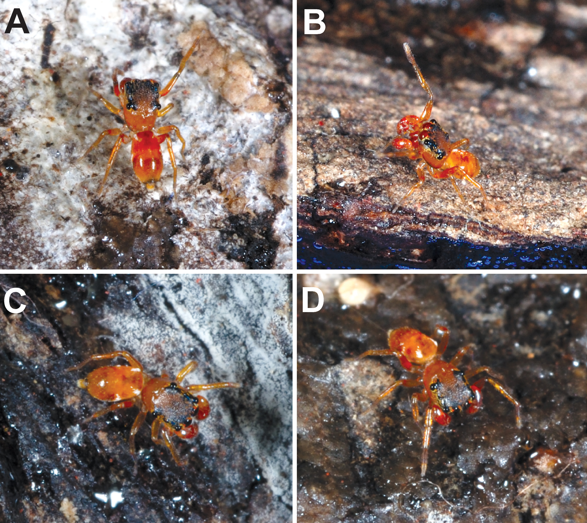
Diagnosis: Synagelides can be recognized by the strikingly modified male palps with enlarged patella with a femur connected at 90°, and presence of femoral apophysis ( Figs 7 View FIGURE 7 E–F, 8D, 8F), several apical tibial apophysis, spirally arranged embolus ( Figs 1 View FIGURE 1 D–E, 2A–C, 4D, 5B–C, 7D–F, 8A–F, 9C–E) and epigyne with arcuated rims and pockets ( Figs 2 View FIGURE 2 D–E, 3C–E, H–J, 5D–E). Further, the ant-like sandy brown habitus ( Figs 1A, 1C View FIGURE 1 , 3A, F View FIGURE 3 , 4A View FIGURE 4 , 6 View FIGURE 6 A–D, 7A, 9A), patella I as long as femur I, elongated front legs with long and stout spines on metatarsi and tibiae ( Prószyński 2009) separate Synagelides from most other jumping spiders.
Synagelides are similar to Agorius Thorell, 1877 , but differ by the more compact body (elongate with abdomen constricted in the middle in Agorius ). However, some Synagelides also seem to possess the above characters and cannot be unambiguously separated from Agorius .
Description: Small, ant-like spiders with size ranging from 2–4 mm in length ( Bohdanowicz 1987; Logunov & Hereward 2006). Flattened and stippled prosoma with distinct fovea ( Liu et al. 2017). Square-shaped ocular quadrangle. Cervical groove in between PMEs. Chelicerae with 2 promarginal teeth and one large retromarginal tooth with a bifurcated tip ( Liu et al. 2017). Leg formula: I, IV, III and II. Front legs elongated with massive femur and long and rigid spines on metatarsi and tibiae I ( Prószyński 2009). Other legs lack spines ( Logunov 2017). Oval abdomen with ‘herringbone-like’ dorsal patterns at posterior region ( Bohdanowicz 1987; Liu et al. 2017). Male palp with massive patella, prolateral femoral apophysis articulates on bulges of patella and tibia, long and slender RTA lies in alveolus of cymbium, posterior apophysis on dorsal cymbium, massive bulb with distal hook-shaped apophyses on prolateral side, triangular dorsal cymbium, short embolus broad at spiral base ( Figs 1 View FIGURE 1 D–E, 2A–C, 4D, 5B–C, 7D–F, 8A–F, 9C–E) and developed median apophysis ( Figs 1 View FIGURE 1 D–E, 2B) in some species ( Bohdanowicz 1987). Epigyne characterized with fossae ( Figs 2 View FIGURE 2 D–E, 3C–E, H–J, 5D–E), sculptures and pockets, arcuated rims separated by median septum, inconspicuous copulatory openings, developed copulatory duct and oval spermathecae ( Bohdanowicz 1987; Liu et al. 2017).
Bohdanowicz, A. (1987) Salticidae from the Nepal Himalayas. The genus Synagelides Bosenberg & Strand 1906. Courier Forschungsinstitut Senckenberg, 93, 65 - 87.
Bosenberg, W. & Strand, E. (1906) Japanische Spinnen. Abhandlungen der Senckenbergischen Naturforschenden Gesellschaft, 30, 93 - 422.
Liu, K., Chen, Z. W., Xu, X. & Peng, X. J. (2017) Three new species of Synagelides Strand, 1906 from China (Araneae: Salticidae). Zootaxa, 4350 (2), 291 - 300. https: // doi. org / 10.11646 / zootaxa. 4350.2.5
Logunov, D. V. & Hereward, J. (2006) New species and synonymies in the genus Synagelides Strand in Bosenberg & Strand, 1906 (Araneae: Salticidae). Bulletin of the British Arachnological Society, 13, 281 - 292.
Logunov, D. V. (2017). New species and records in the genus Synagelides Strand in Bosenberg et Strand, 1906 (Aranei: Salticidae) from the Oriental region. Arthropoda Selecta, 26, 315 - 322. https: // doi. org / 10.15298 / arthsel. 26.4.06
Proszynski, J. (2009) Comments on the Oriental genera Agorius and Synagelides (Araneae: Salticidae). In: Makarov, S. E. & Dimitrijevic, R. N. (Eds.), Advances in Arachnology and Developmental Biology. Institute of Zoology, Bulgarian Academy of Sciences Monographs, 12, pp. 311 - 325.

FIGURE 1. Synagelides hortonensis sp. nov. A, B, D, F, G, Male holotype from Horton plains (A, dorsal view; B, ventral view; D, left palp, ventral view; F, prolateral view; G, retrolateral view). C, E, male from Piduruthalagala (C, dorsal view; E, left palp, ventral view). Scale bars: A–C = 0.5 mm; D–G = 0.1mm.

FIGURE 2. Synagelides hortonensis sp. nov. A–C, left male palp (A, prolateral view; B, ventral view; C, retrolateral view). D–E, epigyne (D, ventral view; E, vulva, dorsal view). Abbreviations: AR, arcuated rim; CD: copulatory ducts; E, embolus; F, fossa; FD, fertilization duct; MA, median apophysis; MS: median septum; PL, proximal lobe of bulb; PT, patella; RTA, retrolateral tibial apophysis; S, spermatheca; T, tegulum; VTA, ventral tibial apophysis. Scale bars: A–C = 0.2 mm; D–E = 0.1mm.

FIGURE 3. Synagelides hortonensis sp. nov. A–B, female (A, dorsal view; B, ventral view); C–E, epigyne (C, ventral view; D, cleared, ventral view; E, vulva, dorsal view). Synagelides lakmalii sp. nov. F–G, female (F, dorsal view; G, ventral view); H–J (H, ventral view; I, cleared, ventral view; J, vulva, dorsal view). Scale bars: A–B, F–G = 1 mm; C–E, H–J = 0.1 mm.
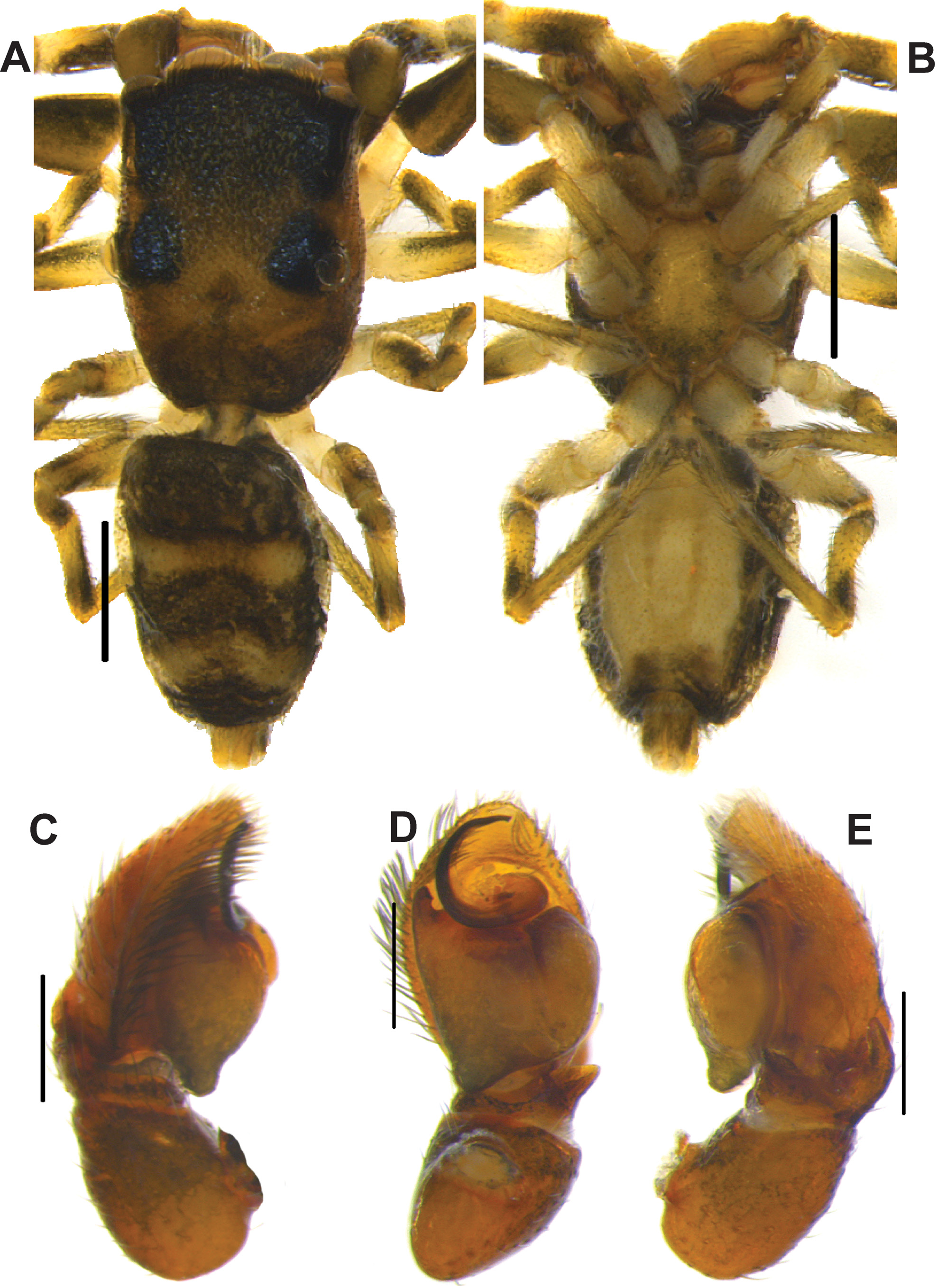
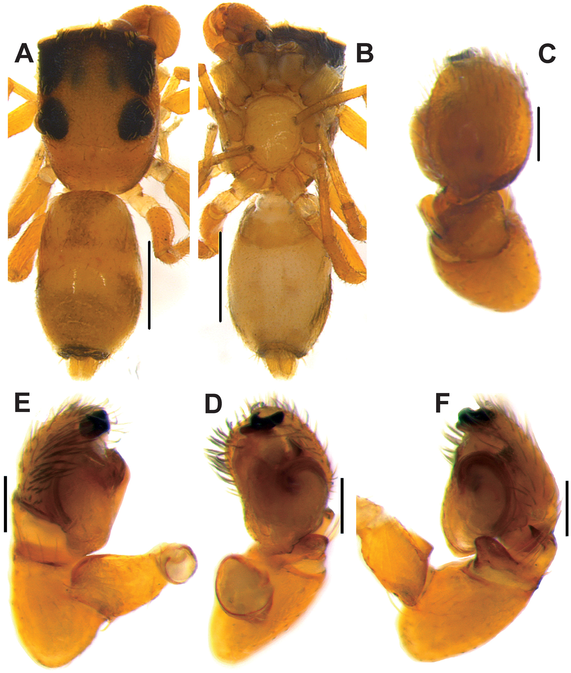
FIGURE 4. Synagelides lakmalii sp. nov. A–B, male (A, dorsal view; B, ventral view); C–E, left palp (C, prolateral view; D, ventral view; E, retrolateral view). Scale bars: A–B = 0.5 mm; C–E = 0.2 mm.
No known copyright restrictions apply. See Agosti, D., Egloff, W., 2009. Taxonomic information exchange and copyright: the Plazi approach. BMC Research Notes 2009, 2:53 for further explanation.
|
Kingdom |
|
|
Phylum |
|
|
Class |
|
|
Order |
|
|
Family |
Synagelides Strand, 1906
| Kanesharatnam, Nilani & Benjamin, Suresh P. 2020 |
Synagelides Strand in Bosenberg & Strand 1906: 330
| Bosenberg, W. & Strand, E. 1906: 330 |