
Ascidiidae
|
publication ID |
https://doi.org/10.5281/zenodo.277174 |
|
DOI |
https://doi.org/10.5281/zenodo.6187340 |
|
persistent identifier |
https://treatment.plazi.org/id/03B887B6-FFBF-FFCB-FF62-162DFA63FD30 |
|
treatment provided by |
Plazi (2016-04-10 08:17:19, last updated 2024-11-27 00:17:51) |
|
scientific name |
Ascidiidae |
| status |
|
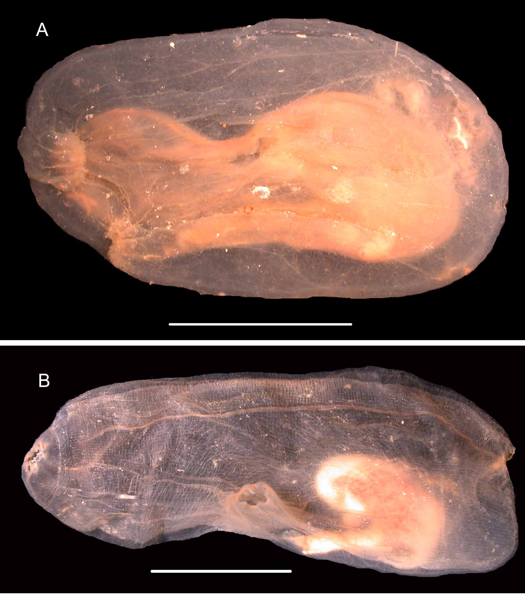
Ascidia challengeri Herdman, 1882 ( Figures 12A View FIGURE 12. A , 13 View FIGURE 13 A, 14)
Herdman, 1882: 202 pl. 30. Kott 1969: 90 and synonymy. Monniot & Monniot 1983: 64 Fig12 a View FIGURE 12. A –e; 1994: 28. Tatian et al. 1998: 149. Primo & Vazquez 2007: 1796.
Stations (events when several trawling operations per station): 2-3- 5-9-12-13 A-17-30(66)-35-42.
The largest specimens of the collection reach 20 cm in length. The oral siphon is apical; the atrial siphon opens at 1/4 to 1/3 of the body length ( Fig. 12A View FIGURE 12. A ). Around both siphons the tunic is raised in irregular finger-like papillae. Elsewhere the surface of the tunic is smooth. A trace of red pigment remained on some specimens. A thin vascular web in polygonal meshes can be seen in the transparent surface of the tunic. On the right side the muscular fibres are thin and regularly crossed. On the left side the musculature is limited to the anterior part of the body. The oral tentacles are not numerous: 16 to 20 large ones are distributed in two rings. The dorsal tubercle opens in a U near the neural ganglion ( Fig. 14). The dorsal lamina, in a plain membrane, does not overpass the oesophagus entrance. In a specimen 65 mm long, 38 longitudinal vessels were counted on the right side of the branchial sac. High and thick crooked papillae protrude at the intersection of the longitudinal and transverse vessels, others, with almost the same size, arise on the longitudinal vessels between each transverse vessel ( Fig. 13 View FIGURE 13 A). The branchial sac does extend below the level of the gut. The digestive tract, in a double loop, occupies 1/3 of the left body side. The stomach is grooved. The long rectum obviously overpasses the level of the top of the primary gut loop. The gonads lie inside and over the intestinal loop.
A. challengeri may be easily confused at first look with A. meridionalis of a same size and general aspect ( Fig. 12 View FIGURE 12. A ). The differences concern the fine structure of the tunic, the number of oral tentacles and the presence of secondary branchial papillae ( Fig. 13 View FIGURE 13 ). The synonymy of A. challengeri with other Antarctic Ascidia species is discussed in Monniot and Monniot (1983).
A. challengeri is widely distributed and bathyal in the Antarctic and Sub-Antarctic seas.
Ascidia meridionalis Herdman, 1880 ( Figures 12 View FIGURE 12. A B, 13B)
Herdman, 1880:465. Monniot & Monniot. 1983: 61 and synonymy. Kott 1969: 72 part, and synonymy. Tatian et al. 2005: 210. Primo & Vazquez 2007: 1803.
Stations (events when several trawling operations per station): 2-5-8-9-10 -20-26A-27(45)-27(46)-30(66)-31-34- 36(297)-41-42-50A-51-52-55-62-79.
The specimens are 3 to 15 cm in length. They differ from A.challengeri by a tunic without papillae at the siphons. The body surface has a velvet-like aspect due to the thin granular surface of the tunic. The oral aperture is terminal with 4 lobes. The atrial siphon is not protruding with 6 lobes, located at 1/3 or 1/2 of the body length ( Fig. 12 View FIGURE 12. A B). Both siphons possess ocelli. More than 70 oral tentacles are inserted along a wide crest. The dorsal tubercle is far from the neural ganglion, at mid distance from the siphons. The dorsal tubercle opens in a U with slightly rolled horns. The branchial sac extends down to the body end, far behind the gut. The dorsal lamina reaches the bottom of the branchial sac below the oesophageal aperture. Sixty longitudinal vessels were counted on the right side of the branchial sac of a specimen 12cm long. The crooked branchial papillae are regularly placed at the crossings of the longitudinal and transverse vessels ( Fig. 13 View FIGURE 13 B), rare intermediate papillae were found. The digestive tract draws a double loop. The stomach is plicated. The anus is at the level of the top of the primary intestinal loop. The gonads have the common structure of the genus; the gonoducts follow the rectum and open against the anus.
No close hit in BOLD (best: 76.49%) for the sequence of specimen P5 ASC.Aa 405 (BOLD: ASCAN023-10). The abundance of this species in Terre Adélie allows to confirm the differences with the closely allied A. challengeri . Both species are widely distributed in the Antarctic and Sub-Antarctic regions in the same bathyal habitats.
Herdman, W. A. (1880) Preliminary report on the Tunicata of the Challenger Expedition. Part II. Proceedings of the Royal Society of Edinburgh, 10, 714 - 726.
Herdman, W. A. (1882) Report on the Tunicata collected during the Voyage of H. M. S. Challenger during the years 1873 - 76; Part I. Ascidiae simplices. Report of the Scientific Results of the Voyage of H. M. S. Challenger during the years 1873 - 76, 6 (17), 1 - 296.
Kott, P. (1969) Antarctic Ascidiacea. Antarctic Research Series, 13, 1 - 239.
Monniot, C. & Monniot, F. (1983) Ascidies antarctiques et subantarctiques: Morphologie et Biogeographie. Memoires du Museum National d'Histoire Naturelle, Paris, 125, 1 - 168.
Primo, C. & Vazquez, E. (2007) Ascidians collected during the Spanish Antarctic expedition CIEMAR 99 / 00 in the Bransfield and Gerlache Straits. Journal of natural History, 41 (29 - 32), 1775 - 1810.
Tatian, M., Sahade, R. J., Doucet, M. E. & Esnal, G. I. (1998) Ascidians (Tunicata, Ascidiacea) of Potter Cove, South Shetland Islands, Antarctica. Antarctic Science, 10 (2) 147 - 152.
Tatian, M., Antacli, J. C. & Sahade, R. (2005) Ascidians (Tunicata, Ascidiacea): species distribution along the Scotia Arc. Scientia Marina, 69 (2), 205 - 214.

No known copyright restrictions apply. See Agosti, D., Egloff, W., 2009. Taxonomic information exchange and copyright: the Plazi approach. BMC Research Notes 2009, 2:53 for further explanation.
|
Kingdom |
|
|
Phylum |
|
|
SubPhylum |
Tunicata |
|
Class |
|
|
Order |
|
|
Family |
Ascidiidae
| Monniot, Françoise, Dettai, Agnès, Eleaume, Marc, Cruaud, Corinne & Ameziane, Nadia 2011 |
Ascidia challengeri
| Herdman 1882 |
Ascidia meridionalis
| Herdman 1880 |