
Sperchon insignis (Walter, 1906)
|
publication ID |
https://doi.org/10.5281/zenodo.172007 |
|
DOI |
https://doi.org/10.5281/zenodo.5679273 |
|
persistent identifier |
https://treatment.plazi.org/id/03B187E0-FFCC-574C-CD09-FA350F8FCD39 |
|
treatment provided by |
Plazi (2016-04-04 09:30:58, last updated 2021-11-12 09:02:13) |
|
scientific name |
Sperchon insignis (Walter, 1906) |
| status |
|
Sperchon insignis (Walter, 1906)
Records ( Table 2): Benthos samples at three spring sites in Gutland, 5 individuals. E 7 22 larvae, parasitic on dixids. Attribution to adults by cooccurrence. Rearing of larvae from female adults failed.
Description (n = 5; unless otherwise indicated): Since all larvae were in a poor condition and had engorged to different extents, an illustration of the larval morphology was not possible and not all characters could be measured. Idiosoma moderately ovate; in some specimens, a transversefolded field at posterior dorsal idiosoma was visible, similar to that in S. setiger ( Figs. 17, 18 View FIGURES 15 – 20 ). Size of idiosoma strongly variable depending on engorgement. Length of five engorged specimens: 219–252 (236), width 198–246 (218).
Dorsal idiosoma: Dp relatively small and finely striated. Anterior margin nearly straight or slightly concave, posteriorly slightly confined. Length/width Dp (n= 1) 108 / 68, Mp 2 Amdp (n= 2) 40–46 (43), Mp 1 Mp 1 43–47 (45), Mp 2 Mp 2 30–32 (31), Lp 1 Lp 1 38– 42 (40), Lp 2 Lp 2 50 – 50 (50), Mp 1 Lp 1 12 – 12 (12), Mp 2 Lp 2 14–17 (16), Mp 1 Mp 2 28– 32 (30), Lp 1 Lp 2 25–31 (28), Mp 1 (n= 1) 36, Mp 2 38, Lp 1 28, Lp 2 52.
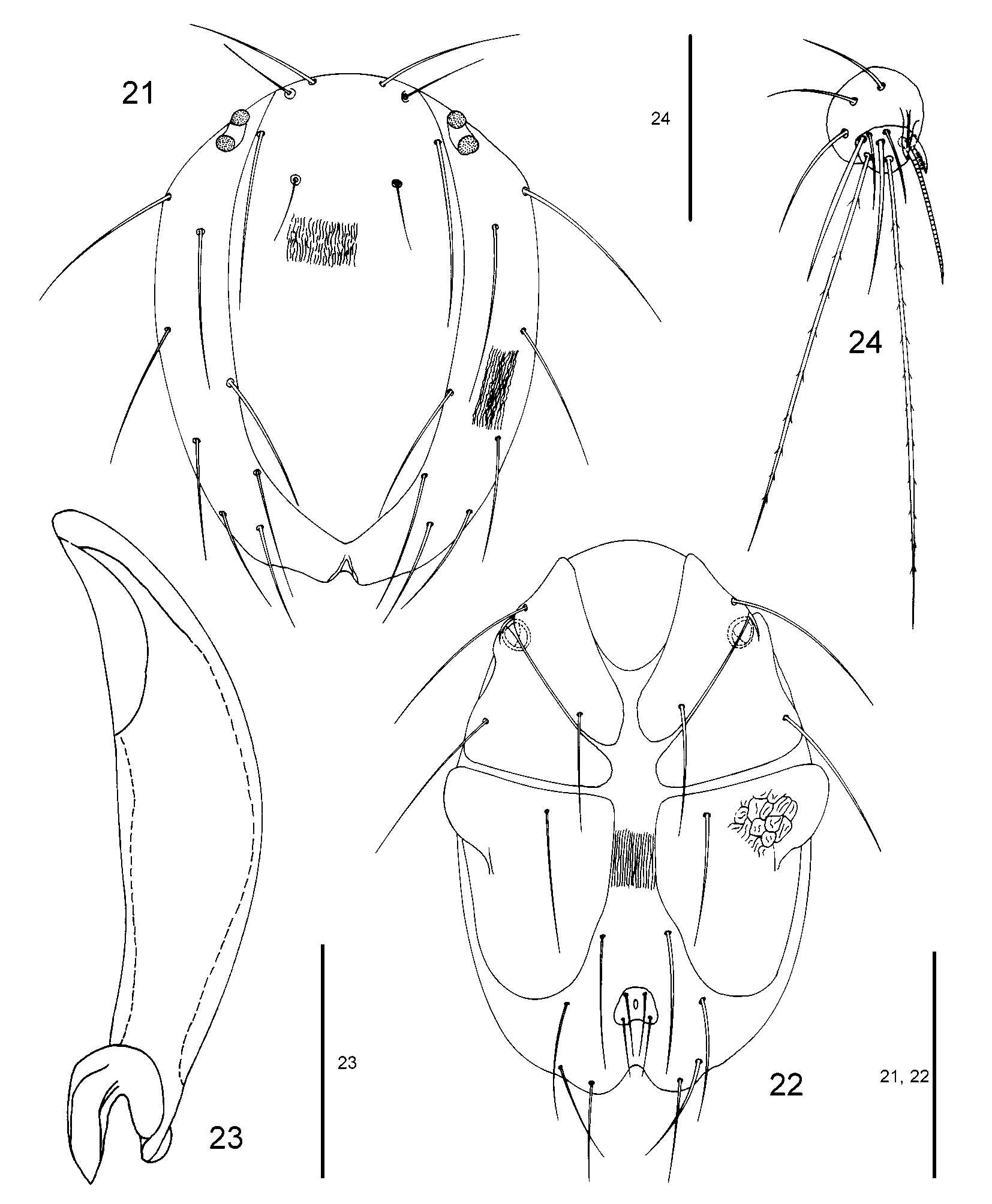
Ventral idiosoma: Length/width CXI (n= 5) 71–77 (74)/ 37–41 (39), CXII 64–72 (67)/ 42–47 (44), CXIII 78–83 (80)/ 64–70 (66), the coxal plates were sculptured in some specimens with a characteristic pattern, similar to that of S. longissimus (see Fig. 22 View FIGURES 21 – 24 ), urstigma slightly elongated, maximum diameter 12–13 (13), C 1 C 2 44–46 (45), C 1 posterior margin of CXI 19–23 (21), C 1 Mmcp 19–23 (21), C 4 Pmcp 64–72 (68), C 1 54– 59 (56), C 2 58–64 (62), C 4 (n= 2) 58–66 (62), length/width Expp (n= 5) 14–17 (15)/ 16–19 (17), E 1 E 1 6 –10 (8), E 2 E 2 10 –13 (12).
Gnathosoma: Length/height chelicera (n= 2) 72 – 72 (72)/ 22 – 22 (22), length chela (n= 1) 19, length/height P 2 41 / 34, length/height P 3 25 / 29, claw 20.
Legs (n= 5): All leg segments with the exception of first segments clearly striated.
Leg I: Total length 269–288 (279), length/height IL 1 32–37 (35)/ 25–28 (26), IL 2 42–47 (45)/ 26–29 (28), IL 3 54–58 (56)/ 24–28 (26), IL 4 61–62 (61)/ 24 – 24 (24), IL 5 79–84 (81)/ 22 – 22 (22).
Leg II: Total length 295–312 (303), length/height IIL 1 41–42 (42)/ 25–26 (26), IIL 2 44–48 (46)/ 26–28 (27), IIL 3 58–61 (60)/ 24–26 (25), IIL 4 68–72 (70)/ 22–24 (23), IIL 5 84–89 (86)/ 20–22 (21).
Leg III: Total length 349–374 (361), length/height IIIL 1 50–56 (53)/ 24–26 (25), III L 2 52–60 (55)/ 24–28 (26), IIIL 3 70–74 (72)/ 20–24 (23), IIIL 4 85–89 (87)/ 22 – 22 (22), IIIL 5 92–95 (93)/ 19–20 (20).
Diagnostic characters of larvae: Lack of C 3 and short legs. In general, the larval morphology of S. insignis is similar to that of S. setiger (e.g. related to the lack of seta C 3, the field of the folded integument located at the distal dorsal idiosoma). However, some morphological differences enable a separation between the two species. All legs and Dp in S. insignis are longer than those in S. setiger .
Remarks: The larva of Sperchon insignis was previously unknown. The species was formerly placed as a synonym or as a subspecies of S. setiger (e.g. Viets & Viets 1959; Viets 1987). Our morphological data of the larvae support recognition of S. insignis as a separate species (Gerecke, pers. com.). Since the state of the studied animals did not allow figures to be drawn, the description above is incomplete. Differences in size of legs, sclerotized structures of the idiosoma and setae are often found in sibling species exhibiting parasitic and nonparasitic lifehistory traits ( Smith 1998). A more thorough investigation of larval morphology of egg larvae from different populations of S. setiger and S. insignis should show to which extent larval morphology in S. setiger from different habitats is stable.
Smith, B. P. (1998) Loss of larval parasitism in parasitengonine mites. Experimental & Applied Acarology, 22, 187 - 200.
Viets, K., & Viets, K. O. (1959) Wassermilben, Hydracarina. In: Brohmer, P., Ehrmann, P. & Ulmer, G. (E d s.) Die Tierwelt Mitteleuropas. Quelle & Meyer, Leipzig, pp. 1 - 44.
Viets, K. O. (1987) Die Milben des Sußwassers (Hydrachnellae und Halacaridae [part.], Acari). II. Katalog. Sonderbande des Naturwissenschaftlichen Vereins in Hamburg, 8, 1 - 1012.
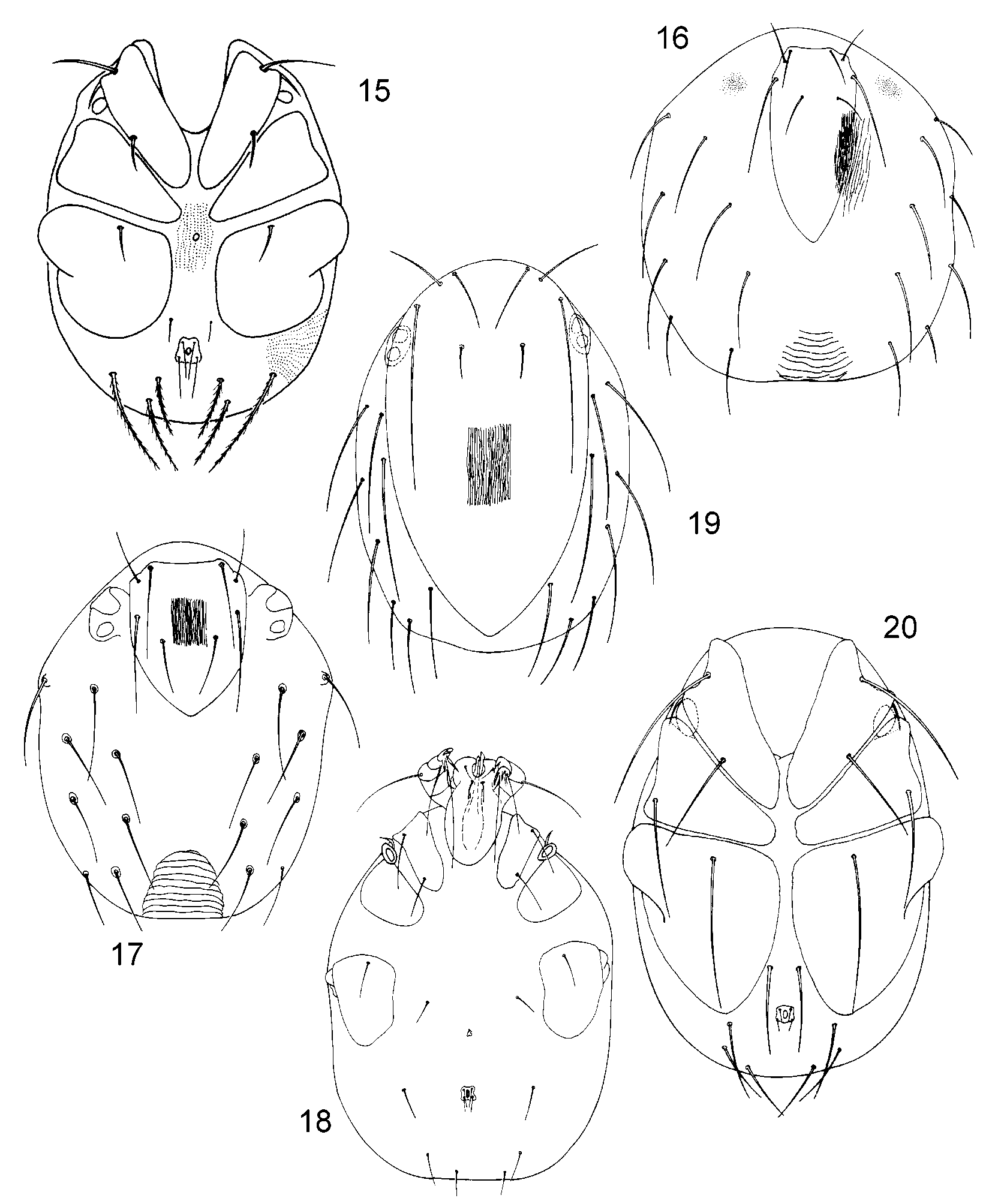
FIGURES 15 – 20. Fig. 15: Sperchonopsis verrucosa, ventral idiosoma (from Martin 2000), Fig. 16: Sperchon clupeifer dorsal idiosoma (redrawn and modified after Ullrich 1976), Fig. 17: Sperchon setiger, dorsal idiosoma, Fig. 18: Sperchon setiger, ventral idiosoma and gnathosoma (from Renz et al. 2004), Fig. 19: Sperchon thienemanni, dorsal idiosoma, Fig. 20: Sperchon thienemanni, ventral idiosoma (Figs. 19 – 20 from Martin 2003).
No known copyright restrictions apply. See Agosti, D., Egloff, W., 2009. Taxonomic information exchange and copyright: the Plazi approach. BMC Research Notes 2009, 2:53 for further explanation.
|
Kingdom |
|
|
Phylum |
|
|
Class |
|
|
Order |
|
|
Family |
|
|
Genus |
1 (by plazi, 2016-04-04 09:30:58)
2 (by ImsDioSync, 2016-11-27 03:28:29)
3 (by ImsDioSync, 2016-11-27 03:33:13)
4 (by ImsDioSync, 2018-06-29 13:35:29)
5 (by ExternalLinkService, 2019-09-26 22:29:22)
6 (by ExternalLinkService, 2021-11-09 17:19:20)
7 (by ExternalLinkService, 2021-11-10 06:04:56)
8 (by ExternalLinkService, 2021-11-12 09:02:13)
9 (by plazi, 2023-10-25 06:29:53)