
Scolelepis alisonae, Williams, Jason D., 2007
|
publication ID |
https://doi.org/10.5281/zenodo.176375 |
|
DOI |
https://doi.org/10.5281/zenodo.6242415 |
|
persistent identifier |
https://treatment.plazi.org/id/03AF87BA-6E3C-4D7A-FF7A-F96721CEFB11 |
|
treatment provided by |
Plazi (2016-04-05 05:04:00, last updated 2024-11-27 07:22:05) |
|
scientific name |
Scolelepis alisonae |
| status |
sp. nov. |
Scolelepis alisonae View in CoL sp. n.
( Figs. 8–10)
Material examined. Holotype Philippines, Morong, Bataan, sandy beach, 28 Feb 1999 ( USNM 1096804).— Paratypes, same data as holotype (16 anterior ends, two posterior ends, one middle piece in alcohol, USNM 1096805; one complete specimen on SEM stub, four anterior ends on SEM stubs, one set of palps on SEM stub, USNM 1096806); same location as holotype, 1 Mar 1999 (one anterior end, USNM 1096807); same location as holotype, 25 April 1999 (one complete specimen, eight anterior ends, ZRC 2006.0222).
Etymology. The species is named in honor of my wife, Dr. Alison S. Carson, for her help in collection of these specimens (sometimes under quite adverse conditions). Her cultural psychology studies in the Philippines (supported by the National Science Foundation and a Fulbright Scholarship) allowed for travel to make these collections.
Diagnosis. A species of Scolelepis with notosetae on setiger 1, notopodial hooded hooks and lacking an occipital tentacle. Palps short, palp sheaths absent, palps with two weakly separated transverse rows of cilia. Prostomium conical, with two pairs of eyes. Caruncle extending to middle of setiger 1, nuchal cilia in Ushaped pattern on sides of caruncle. Postsetal notopodial lamellae fused with branchiae from setiger 2, lamellae composed of up to nine conical lobes by setiger 5, number of lobes gradually decreasing posteriorly. Bidentate notopodial hooded hooks from setiger 68–99, with up to three per fascicle. Postsetal neuropodial lamellae with up to three lobes in anterior setigers. Bidentate neuropodial hooded hooks from setiger 25–33, with up to 11 hooks in middle and posterior fascicles. Pygidium broadly rounded, with short conical papillae surrounding anus.
A B C Pa C-G D E F G H I J K L M J-M Description. Largest specimen of 136 setigers, 47.6-mm long and 0.9-mm wide by setiger 8; holotype of 125 setigers, 37.5-mm long, 0.52-mm wide by setiger 8. Body widest anteriorly, gradually tapering to posterior end; body nearly rectangular in cross section. Color in alcohol opaque white, no pigmentation present.
Prostomium conical, extending anteriorly to long, tapering point, posteriorly extending to short caruncle with rounded posterior margin, ending in middle of setiger 1, caruncle not free on posterior margin ( Figs. 8 A– B, 9A–C). Two pairs of eyes, one pair of kidney-shaped eyes laterally, one pair of round eyes medially, all eyes in nearly straight row between base of palps ( Fig. 8 B); occipital tentacle absent. Palps short, extending to setigers 6–10 ( Figs. 8 B, 9C), with two weakly separated transverse rows of cilia along the ventral surface, long rows of cilia approximately 45 µm long, short rows approximately 24 µm long (short rows on medial side), long and short rows in approximately 1:1 ratio, rows of mucus secreting cells represented by tubular necks present proximal to transverse ciliary rows; ciliary rows extending to distal ends of palps, median ciliated groove lacking ( Figs. 10 View FIGURE 10 A–B, 18C). Palpal sheaths absent. Nuchal cilia in U-shaped pattern on both sides of caruncle, posterior to base of palps ( Fig. 9 View FIGURE 9 B).
Setiger 1 well developed with bluntly pointed, triangular notopodial and neuropodial postsetal lamellae, notosetae and neurosetae present ( Figs. 8 C, 9D). Postsetal notopodial lamellae fused with branchiae from setiger 2 ( Figs. 8 D, 9F), lamella composed of 2–6 conical lobes, number of lobes increasing to 5–9 on lamellae by setiger 5 ( Figs. 8 E, 9A, C), then gradually decreasing to two conical lobes distally and rounded basal portion by setigers 20–30 ( Figs. 8 F, 9G), posteriormost setigers with one conical lobe distally, with rounded basal portion free from branchiae ( Figs. 8 G, 9H, 10C). Neuropodial postsetal lamellae of setiger 1 rounded or conical ( Figs. 8 C, 9D), lamellae of setiger 2 with rounded base extending into short, bluntly pointed lobe ( Figs. 8 D, 9F), lamellae of setigers 3–5 sometimes with 2–3 bluntly pointed lobes ( Fig. 9 View FIGURE 9 C), lamellae from setigers 6-20 with broadly rounded lobe ( Fig. 8 F, 9G), developing notch dividing lamellae by setiger 30, on posterior setigers ventral lobe small, triangular, dorsal lobe elongate with dorsal end free and pointed, ventral end rounded ( Figs. 8 I, 9H), by setiger 65 ventral lobe reduced, dorsal lobe digitate ( Fig. 10 View FIGURE 10 C). Lateral organs between notopodial and neuropodial postsetal lamellae present from setiger 1 to posterior setigers ( Fig. 9 View FIGURE 9 G).
Notosetae of setiger 1 and subsequent setigers arranged in two vertical rows of bilimbate capillaries, the dorsal notosetae of these rows longer than ventral ones, often loosely coiled along their distal ends ( Figs. 8 L, 9A, C, F), one notopodial hooded hook beginning on setiger 68–99 ( Figs. 8 J, K, 10C), with up to three notopodial hooded hooks in posterior most setigers; notopodial hooded hooks with acute main fang and single accessory tooth. Neurosetae of setiger 1 and subsequent setigers arranged in two vertical rows of bilimbate capillaries ( Fig. 9 View FIGURE 9 F–G), 1–3 neuropodial hooded hooks from setiger 25–33, up to 5–11 neuropodial hooded hooks in middle to posterior setigers ( Fig. 8 G); neuropodial hooded hooks with acute main fang and single accessory tooth ( Fig. 8 M). Notosetae and neurosetae with minute spinelets ( Fig. 9 View FIGURE 9 E).
Branchiae present from setiger 2 to end of body, fused with postsetal notopodial lamellae but with tips free in anterior setigers ( Fig. 9 View FIGURE 9 F), separated from notopodial lamellae in posterior setigers, with band of cilia along inner edge and joined to corresponding branchiae on opposite side by a single band of cilia across dorsum ( Fig. 9 View FIGURE 9 B).
Pygidium broadly rounded, with short conical papillae surrounding dorsal anal opening ( Figs. 8 H–I, 10D).
Remarks. The genus Scolelepis was reviewed by Maciolek (1987) and more recently by Hutchings et al. (1998), both studies provided summary tables for the known members of the genus. Since then, nine species of Scolelepis , including those treated in this paper, have been described, and there are now 58–59 species within this genus ( Table 1). Confusion exists in the total number of species because MacCord & Amaral (2005) resurrected Scolelepis goodbodyi ( Jones, 1962) , a species that was previously considered a synonym of S. squamata ( Müller, 1806) ( Pettibone 1963; Foster 1971a; Light 1978; Maciolek 1987). However, MacCord & Amaral (2005) provided no taxonomic evidence to support the conclusion that these species are distinct (their identification was based on the authority of Radashevsky; pg. 829).
Scolelepis alisonae View in CoL sp. n. belongs to a group of six species including S. blakei Hartmann-Schröder, 1980 View in CoL ; S. carunculata Blake & Kudenov, 1978 View in CoL ; S. chilensis ( Hartmann-Schröder, 1962) View in CoL ; S. hutchingsae Dauer, 1985 View in CoL ; and S. kudenovi Hartmann-Schröder, 1981 View in CoL , that possess notosetae on setiger 1, notopodial hooded hooks, bidentate neuropodial hooded hooks and that lack an occipital tentacle. Scolelepis alisonae View in CoL sp. n. is distinguished from all these species in the possession of notopodial postsetal lamellae with up to nine lobes in anterior setigers and neuropodial postsetal lamellae with up to 3 lobes in anterior setigers. Scolelepis alisonae View in CoL sp. n. and S. laciniata Eibye-Jacobsen, 1997 View in CoL are similar in the possession of notopodial postsetal lamellae with conical lobes; however, Scolelepis alisonae View in CoL sp. n. is easily distinguished by the presence of notosetae on setiger 1 and notopodial hooded hooks (both absent in S. laciniata View in CoL ).
The palp morphology of S. alisonae View in CoL sp. n., is uniquely different from previously described species in exhibiting weakly separated long and short transverse rows of cilia in a 1:1 ratio. See Discussion for a comparative analysis of the palp morphology in members of the genus Scolelepis View in CoL .
Distribution. Sandy beach in Morong of the Bataan province in the Philippines; shallow subtidal (<5 m).
Blake, J. A. & Kudenov, J. D. (1978) The Spionidae (Polychaeta) from southeastern Australia and adjacent areas with a revision of the genera. Memoirs of the National Museum of Victoria, 39, 171 - 280.
Dauer, D. M. (1985) A new species of Scolelepis (Polychaeta: Spionidae) from Lizard Island, Australia. Proceedings of the Biological Society of Washington, 98, 678 - 681.
Eibye-Jacobsen, D. (1997) A new species of Scolelepis (Polychaeta: Spionidae), highly abundant on the sand beaches of western Phuket Island, Thailand. Bulletin of Marine Science, 60, 240 - 251.
Foster, N. M. (1971 a) Spionidae (Polychaeta) of the Gulf of Mexico and the Caribbean Sea. Studies on the Fauna of Curacao and other Caribbean Islands, 36, 1 - 183.
Hartmann-Schroder, G. (1962) Die Polychaeten des Eulittorals. In: Hartmann-Schroder, G. & Hartman, G. (Eds.), Zur Kenntnis des Eulitorals der chilenischen Pazifikkuste und argentinischen Kuste Sudpatagoniens unter besonderer Berucksichtigung der Polychaeten und Ostracoden. Mitteilungen aus dem Hamburgischen Zoologischen Museum und Institut, Supplement to 60, 57 - 167.
Hartmann-Schroder, G. (1980) Teil 4. Die Polychaeten der tropischen Nordwestkuste Australiens (zwischen Port Samson im Norden und Exmouth im Suden). In: Hartmann-Schroder, G. & Hartman, G. (Eds.), Zur Kenntnis des Eulitorals der australischen Kusten unter besonderer Berucksichtigung der Polychaeten und Ostracoden. Mitteilungen aus dem Hamburgischen Zoologischen Museum und Institut, 77, 41 - 110.
Hartmann-Schroder, G. (1981) Teil 6. Die Polychaeten der tropischen-subtropischen Westkuste Australiens (zwischen Exmouth im Norden und Cervantes im Suden). In: Hartmann-Schroder, G. & Hartman, G. (Eds.), Zur Kenntnis des Eulitorals der australischen Kusten unter besonderer Berucksichtigung der Polychaeten und Ostracoden. Mitteilungen aus dem Hamburgischen Zoologischen Museum und Institut, 78, 19 - 96.
Jones, M. L. (1962) On some polychaetous annelids from Jamaica. Bulletin of the America Museum of Natural History, 124, 173 - 212.
Light, W. J. (1978) Invertebrates of the San Francisco Bay Estuary System. Family Spionidae (Annelida: Polychaeta), The Boxwood Press, Pacific Grove, California, 211 pp.
MacCord, F. S. & Amaral, A. C. Z. (2005) Morphometric analysis of two species of Scolelepis (Polychaeta: Spionidae). Journal of the Marine Biological Association of the United Kingdom, 84, 769 - 784.
Maciolek, N. J. (1987) New species and records of Scolelepis (Polychaeta: Spionidae) from the east coast of North America, with a revision of the subgenera. Bulletin of the Biological Society of Washington, No. 7, 15 - 40.
Muller, O. F. (1806) Zoologica Danica seu Animalium Daniae et Norvegiae ruriorum ac minus notorum, Descriptiones et Historia, Havniaem, 160 pp.
Pettibone, M. H. (1963) Revision of some genera of polychaete worms of the family Spionidae, including the description of a new species of Scolelepis. Proceedings of the Biological Society of Washington, 76, 89 - 104.
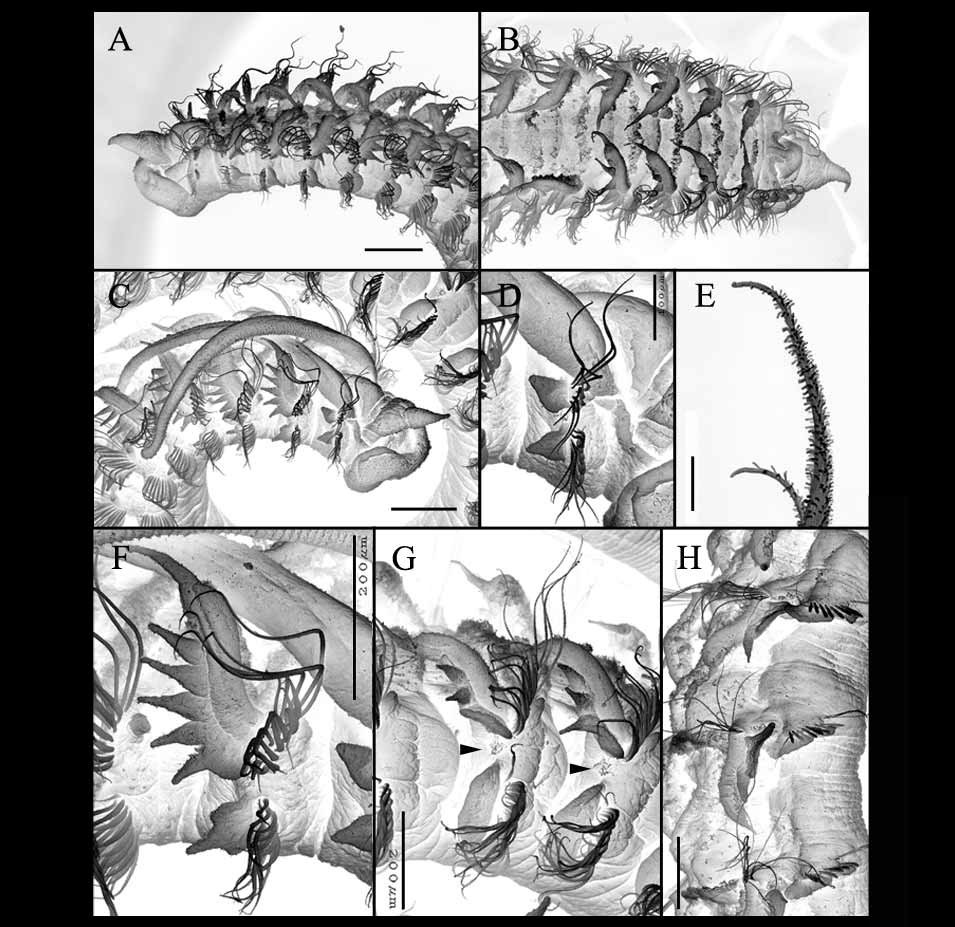
FIGURE 9. Scolelepis alisonae sp. n. A – H, paratypes (USNM 1096806) SEM micrographs. A, anterior end, oblique lateral view (palps missing); B, anterior end, dorsal view; C, anterior end, lateral view; D, setiger 1 and base of palp, lateral view; E, tips of neurosetae from setiger 3; F, setiger 2, lateral view; G, setiger 19 & 20 (left to right), lateral view (arrowheads indicate lateral organs); H, setigers 49 – 51 (top to bottom), lateral view. Scale bars: A – C = 250 µm; D = 100 µm; E – H = 200 µm.
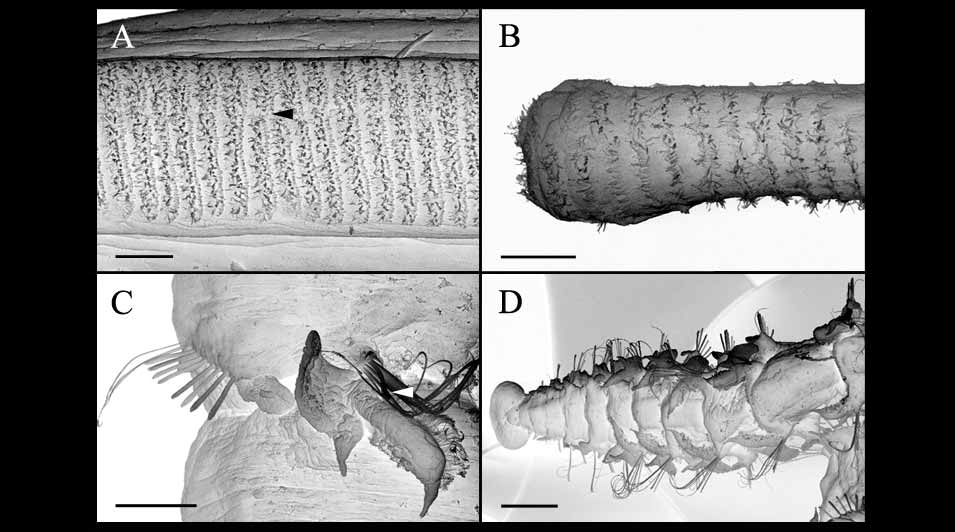
FIGURE 10. Scolelepis alisonae sp. n. A – D, paratypes (USNM 1096806) SEM micrographs. A, proximal region of palp, frontal view (arrowhead indicates faint division in transverse rows of cilia; short rows top, long rows bottom; note that at point of arrowhead there are two long rows below and only one short row above); B, distal tip of palp, frontal view; C, setiger 68, oblique dorsolateral view; D, posterior setigers and pygidium, dorsal view. Scale bars: A – B = 25 µm; C = 100 µm; D = 250 µm.
No known copyright restrictions apply. See Agosti, D., Egloff, W., 2009. Taxonomic information exchange and copyright: the Plazi approach. BMC Research Notes 2009, 2:53 for further explanation.
|
Kingdom |
|
|
Phylum |
|
|
Class |
|
|
Order |
|
|
Family |
|
|
Genus |
Scolelepis alisonae
| Williams, Jason D. 2007 |
S. laciniata
| Eibye-Jacobsen 1997 |
S. hutchingsae
| Dauer 1985 |
S. kudenovi Hartmann-Schröder, 1981
| Hartmann-Schroder 1981 |
S. blakei Hartmann-Schröder, 1980
| Hartmann-Schroder 1980 |
S. carunculata
| Blake & Kudenov 1978 |
S. chilensis ( Hartmann-Schröder, 1962 )
| Hartmann-Schroder 1962 |
1 (by plazi, 2016-04-05 05:04:00)
2 (by ImsDioSync, 2016-11-28 01:10:21)
3 (by ImsDioSync, 2016-11-28 01:58:10)
4 (by ImsDioSync, 2017-02-08 10:35:05)
5 (by ImsDioSync, 2018-06-29 18:57:47)
6 (by ExternalLinkService, 2019-09-26 22:05:22)
7 (by ExternalLinkService, 2022-01-30 21:00:26)
8 (by ExternalLinkService, 2022-02-23 10:58:01)
9 (by plazi, 2023-10-25 08:38:10)