
Luridiblatta fenestrata, Bohn, 2022
|
publication ID |
https://doi.org/10.11646/zootaxa.5215.1.1 |
|
publication LSID |
lsid:zoobank.org:pub:8882FE42-0615-4BBA-9E71-457B8639102A |
|
DOI |
https://doi.org/10.5281/zenodo.7403391 |
|
persistent identifier |
https://treatment.plazi.org/id/03AED24E-FFA3-DF2C-A5BE-F898CC4AFB73 |
|
treatment provided by |
Plazi (2022-12-06 08:03:17, last updated 2024-11-24 20:36:37) |
|
scientific name |
Luridiblatta fenestrata |
| status |
sp. nov. |
3. Luridiblatta fenestrata , sp. nov.
Figs. 5F View FIGURE 5 , 13A–N View FIGURE 13 , 14A–C View FIGURE 14 , 15A–I View FIGURE 15 , 35C View FIGURE 35 , 36I–K View FIGURE 36 , 37 View FIGURE 37 , 43 View FIGURE 43
Diagnosis. As in L. vesiculosa with very short T7 glandular pouches, otherwise well characterised by the unique structure of the windows of the anterior pit wall showing a lateral constriction brought about by a sharp transversal crest of the window frame protruding towards mesally (tc in Fig. 13F View FIGURE 13 ).
Etymology. The species name refers to the above mentioned structure resembling some windows (in Latin: fenestra, - ae) of ancient churches.
Material studied. Type material. Holotype, 1♂, MOROCCO, Forêt de la Mamora , 5 km NE Sidi-Allal-Bahraoui (30 km E Rabat), 150 m, 20.VIII.1984, leg. B. & H.Bohn (completely on two slides, Ma 16/19). (Coll. Bohn, ZSMC).
Additional material. MOROCCO. 29♂, 43♀, 36L, 6O, same data as holotype (slides: ♂, Ma 16/1,5-16,20–28; ♀, Ma 16/4,18,29–34; L, Ma 16/17,35,36). (Coll. Bohn, ZSMC) .
Description. Size. Male. Length of pronotum 1.47–1.60 (mean 1.53) mm, length of tegmina 2.1–2.45 (mean 2.34) mm. (N = 16/16). Female. Length of pronotum 1.85 mm, length of tegmina 2.04–2.28 (mean 2.19) mm. (N = 2/8).
Wings. Apex of tegmina in both sexes usually straight ( Fig. 13H,I View FIGURE 13 ), more seldom very weakly concave or convex; hindwings of variable shape, not broadening towards the apex ( Fig. 16J,K View FIGURE 16 ).
Male abdomen. Tergites: Fig. 16A–G. T View FIGURE 16 6 View FIGURE 6 as described under characters of the genus and the species group ( Fig. 5F View FIGURE 5 ). T7. Pit size: minor, largest with size 1–2 ( Figs. 14C View FIGURE 14 , 15B View FIGURE 15 ). Anterior pit wall (aw) with relatively large windows (w), which are bilobed by a sharp transversal crest (tc) protruding from the surrounding window frame (wf) towards mesally; stabilising bracelet (s) strongly broadening towards the bottom of the pit; posterior pit wall (pw) with a pair of well developed hemispherical bulges (bu), sometimes having a strongly sclerotised (internal?) surface ( Figs. 13F View FIGURE 13 , 14A,B View FIGURE 14 , 15C View FIGURE 15 ). In these figures, as well as in Fig. 15A View FIGURE 15 , the posterior wall of the pit has largely remained in an vertical position, the bulges thus appearing as “hanging drops”. Glandular pouches (gp) very short, scarcely surpassing the anterior border of the tergite ( Fig. 14A–C View FIGURE 14 ); tubules moderately long, virtually stretched fairly reaching the respective lateral border of the tergite (tu in Fig. 14B,C View FIGURE 14 ); both gland types seem to have their openings into the pit near the window embayment, the arrows in Fig. 15C,D View FIGURE 15 point to the endpieces of both systems visible through the windows. Genital hook with relatively normally shaped shaft, below the apical process not or only weakly bulged out ( Fig. 13L–N View FIGURE 13 ).
Colouration pattern in both sexes as typical for the first two species-groups, but dark markings often lighter coloured.
Distribution. So far only known from the type locality in the Forêt de la Mamora, near Rabat, Morocco.
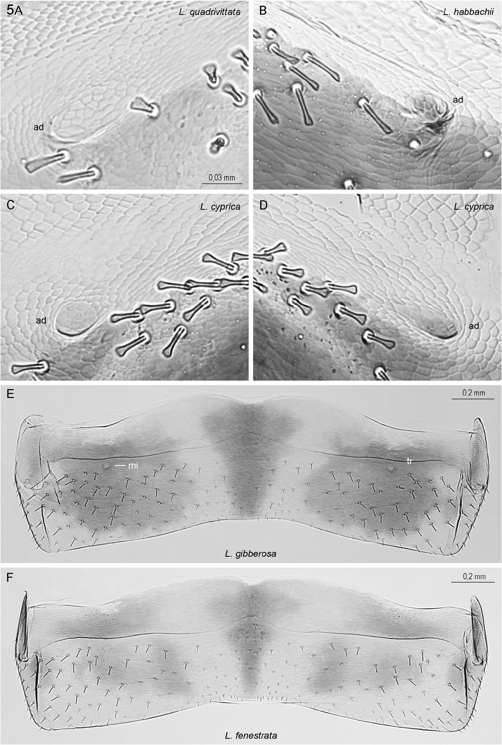
FIGURE 5A–F. Structures of male T6. A-D: Higher enlargement of anterior deepenings ad with their surroundings, (A,C) from the left side of T6, (B,D) from the right side. E,F: Continuation of overview pictures of male T6, species L. gibberosa (E) and L. fenestrata (F) (longitubulata-group).—Abbreviations: ad anterior deepening; mi insertion site of dorso-ventral muscles, homologon to the site where the anterior deepenings of the species of the trivittata-group develop; tr transversal ridge.— Enlargements: Same scale for A–D.—Identifications:A (Is 11/1), B (Al 36/7, holotype), C (Cy 25/1), D (Cy 23/1), E (Ma 14/10), F (Ma 16/19, holotype).

FIGURE 6A–D. Structures of male T6. Continuation of overview pictures of male T6, species: L. vesiculosa (longitubulata- group), with fully developed transversal ridge tr; L. cyprica, L. beybienkoi and L. graeca (beybienkoi-group), without transversal ridge, but with ad, pd anterior, posterior deepening; fo membraneous fold; arrowhead indicates w-shaped dark line.—Identifications: A (Ma 5/1), B (Cy 18/1, holotype), C (Ty 3/1), D (Kr 23/2, holotype).
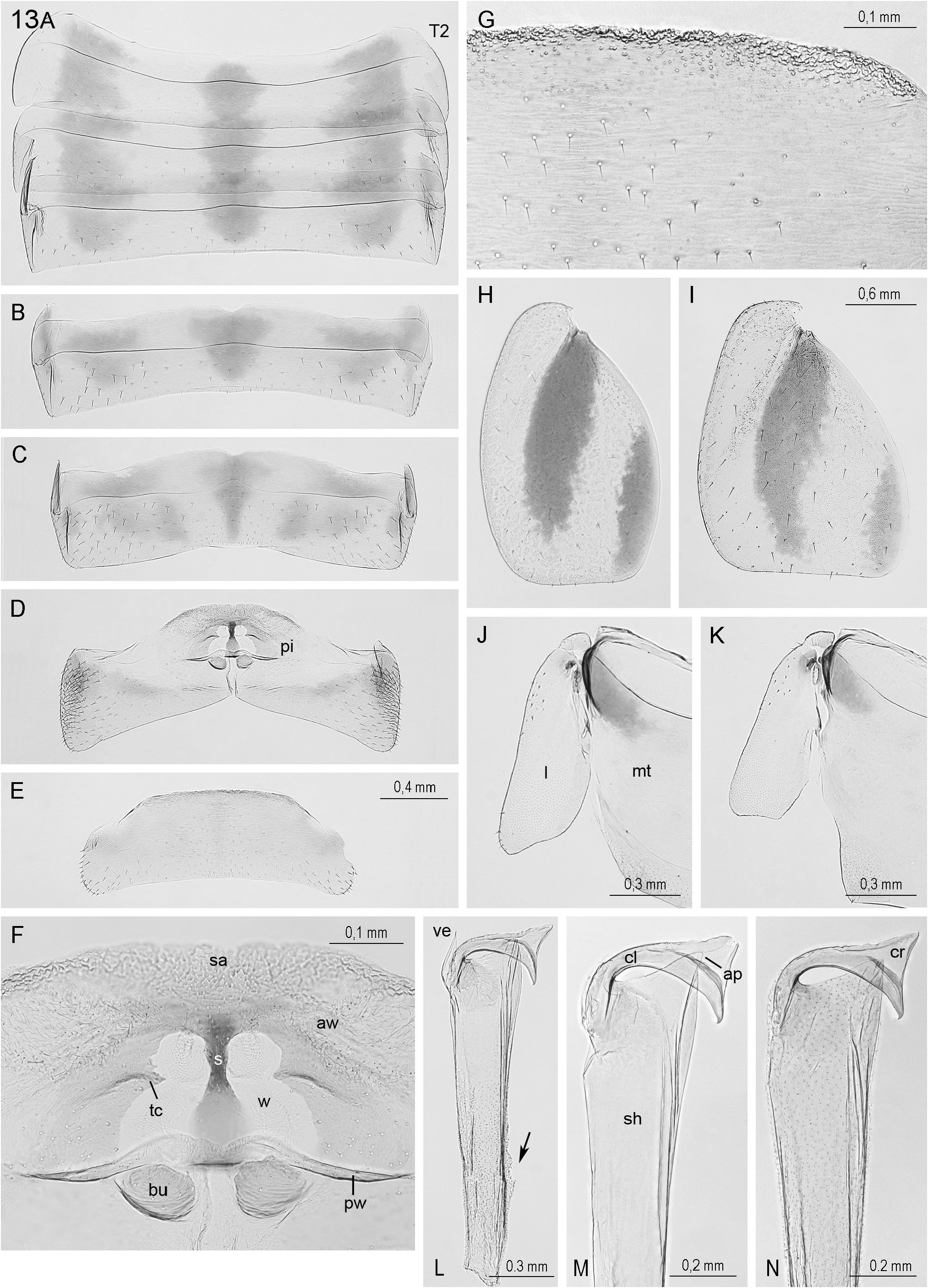
FIGURE 13A–N. Main structures of L. fenestrata.—A–G. Male tergites.A: T2–4; B: T5; C: T6; D,F: T7; (D) outline: glandular pit (pi); (F) more detailed: anterior pit wall aw with sa strongly sclerotised area; s stabilising bracelet, tc transversal crest, w transparent window; posterior pit wall pw with hemispherical bulge bu; E,G: T8, (G) anterior part in higher magnification, showing the rough cuticula along the anterior border.—H,I. Left tegmen of male (H) and female (I).—J, K. Hindwing lobe (l) with part of mt metanotum of male (J) and female (K).—L–N. Male hook with sh shaft and its apical process ap, ve velum, cl claw with triangular cr crest; the arrow points to remnants of the sheath into which the hook can be retracted, also still covering for most part hook in N.—Enlargements: Same scale for A–E, and for H,I.—Identifications: A–H (Ma 16/19, holotype), I (Ma 16/31), J (Ma 16/21), K (Ma 16/4), L (Ma 16/24), M (Ma 16/26), N (Ma 16/25).
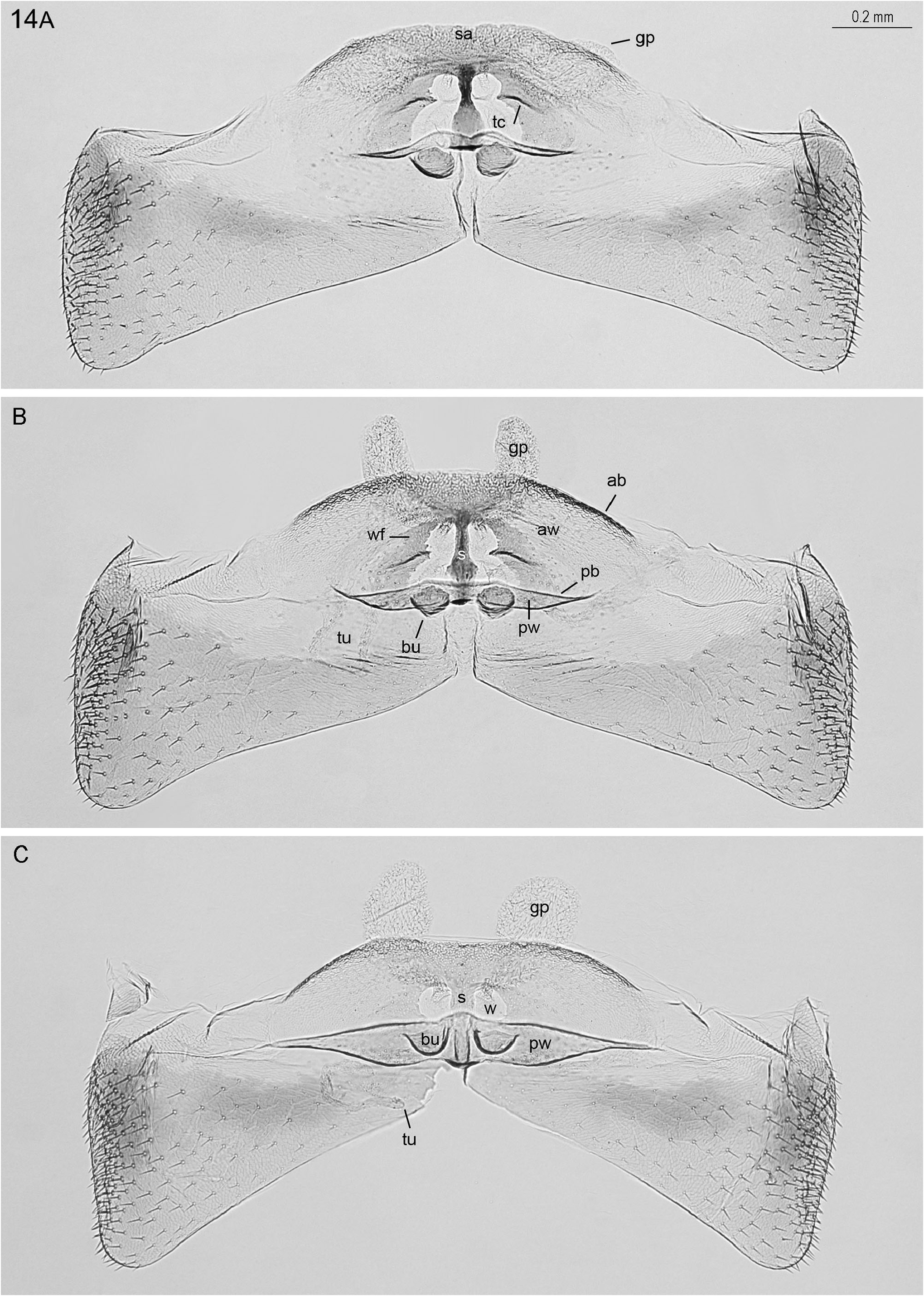
FIGURE 14A–C. Male T7 of L. fenestrata with differently developed glandular structures. Abbreviations: ab, pb anterior, posterior border of pit; aw anterior pit wall with s stabilising bracelet, sa strongly sclerotised area, tc transversal crest (in C covered by pw), w transparent window, and wf window frame; pw posterior pit wall with hemispherical bulges bu (in A,B strongly sclerotised); gp glandular pouches; tu tubules (in B on both sides of the label).—Enlargements: Same scale in A–C.— Identifications:A (Ma 16/19, holotype), B (Ma 16/20), C (Ma 16/8).
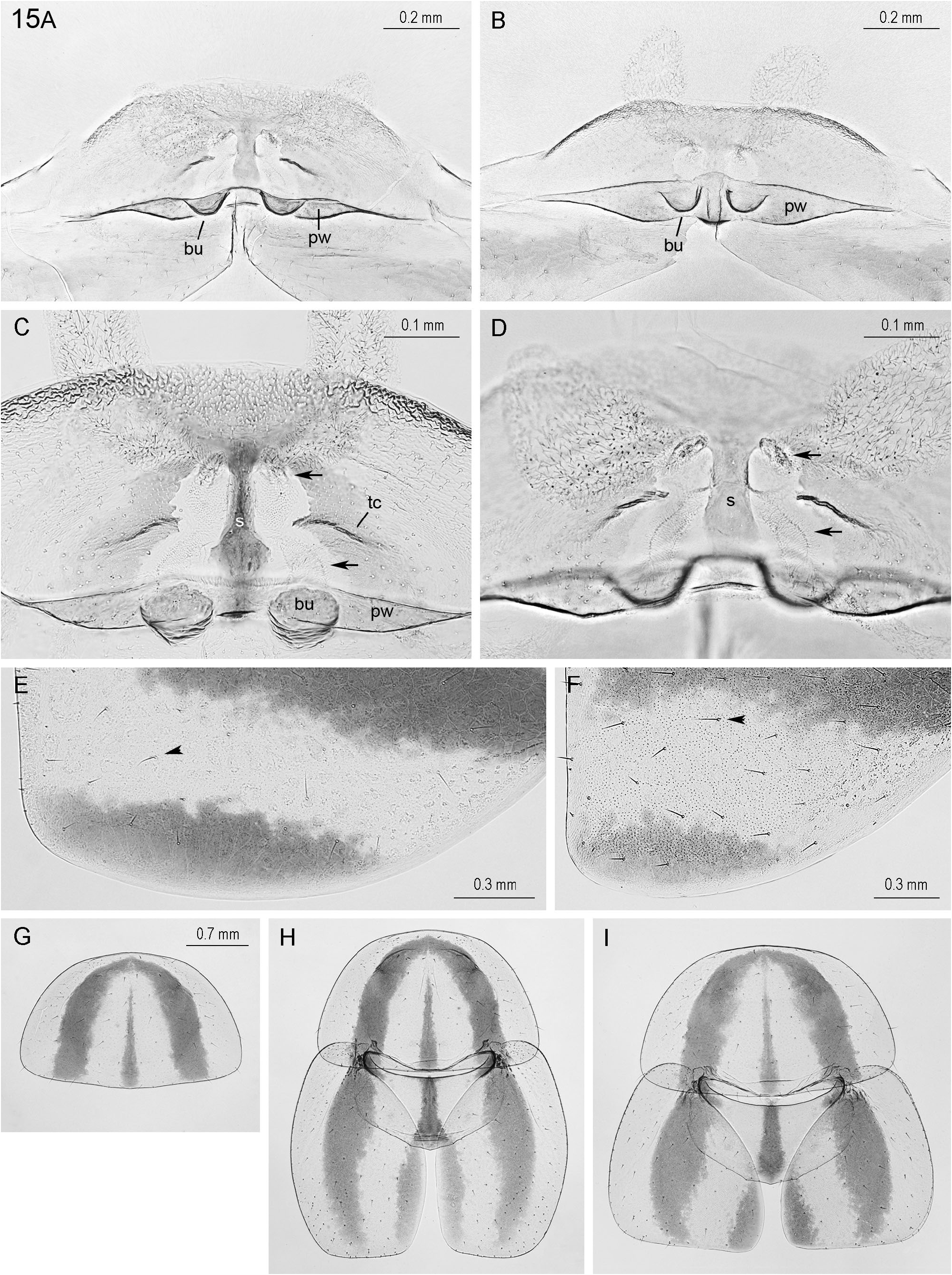
FIGURE 15A–I. Further structures of L. fenestrata.—A–D. Pit structures. A,B: The different appearance of the hemispherical bulges bu in the two specimens is certainly caused by the different orientation of the posterior pit wall pw, in (A) almost vertical, in (B) inclined. C,D: The arrows point to two tubulelike structures appearing in the transparent windows of each side: above the tailpiece of the respective glandular pouch, below the end piece of a tubule; both meat in the area between the mesal ends of the transversal crest tc and the stabilising bracelet s, where the two glands possibly have their openings into the pit hole.—E,F. Left male (E) and female (F) tegmen (anteriorly at the right, posterior border below), bristles of the lightly coloured area between the dark longitudinal stripes not inserted in the center of a dark spot (arrowheads, compare with Fig. 24F–I).—G–I. Thoracal nota and tegmina. Pronotum of holotype (G), pronotum, mesonotum and tegmina of a male (H) and a female (I).—Enlargements: Same scale for G–I.—Identifications: A,D (Ma 16/7), B (Ma 16/8), C (Ma 16/20), E,G (Ma 16/19, holotype), F (Ma 16/31), H (Ma 16/21), I (Ma 16/X).
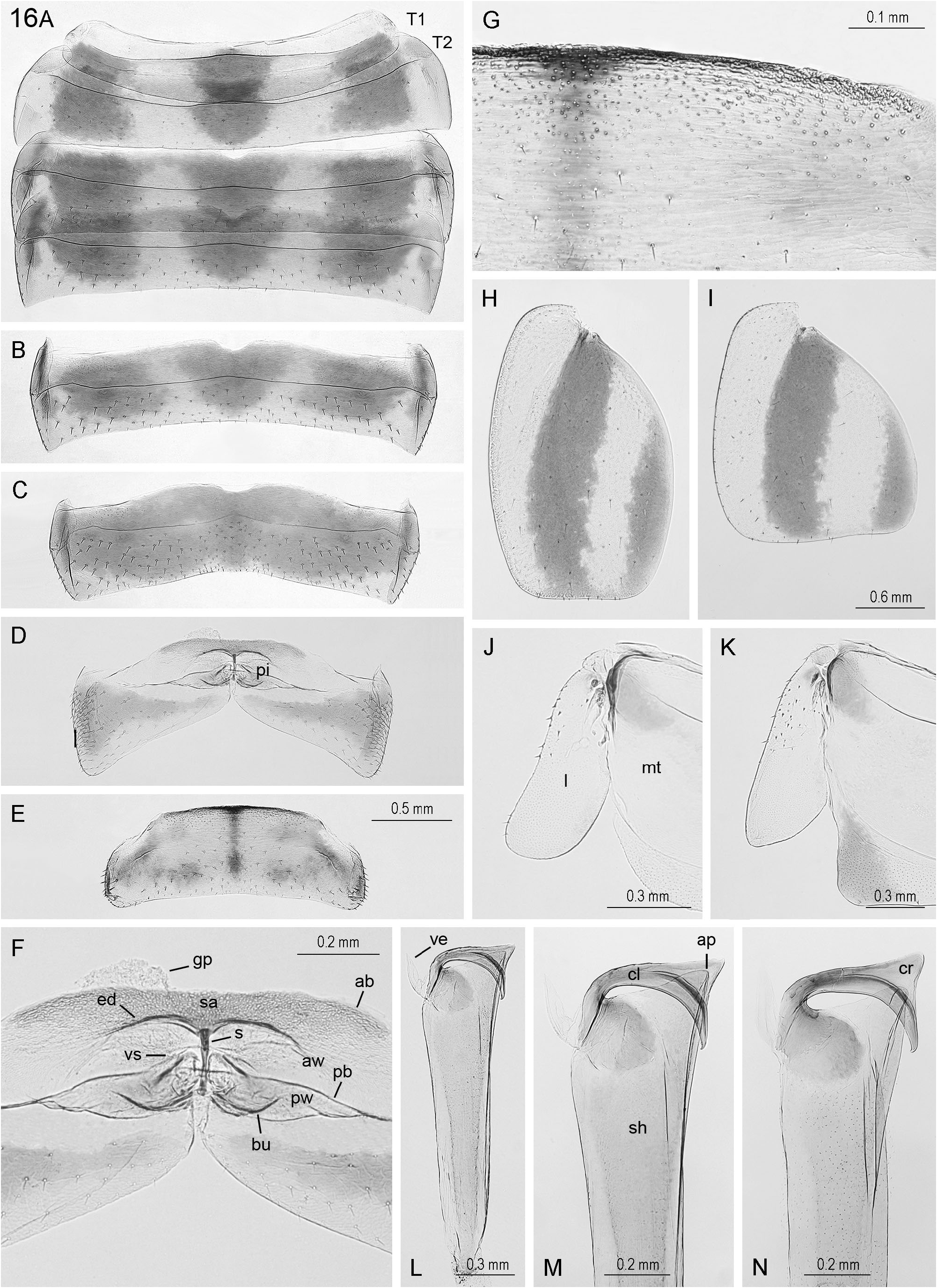
FIGURE 16A–N. Main structures of L. vesiculosa.—A–G. Male tergites.A: T1–4; B: T5; C: T6; D,F: T7; (D) outline: glandular pit pi; (F) more detailed: ab, pb anterior, posterior border of pit opening; aw anterior pit wall with sa strongly sclerotised area, ed edge of the excavation, and s stabilising bracelet; pw posterior pit wall with a pair of a relatively wide, but shallow posterior bulges bu; vs vesicular structures within the pit; gp glandular pouch. E,G: T8, (G) anterior part in more detail, showing the rough cuticle along the anterior border.—H,I. Left tegmen of male (H) and female (I).—J,K. Hindwing lobe l with part of metanotum mt of male (J) and female (K).—L–N. Male genital hook with shaft sh and its apical process ap; ve velum; cl claw and its triangular crest cr.—Enlargements: Same scale for A–E, and for H,I.—Identifications: A–D,F,H,J (Ma 5/8, holotype), E,G (Ma 37/1), I,K (Ma 5/7), L,M (Ma 38/5), N (Ma 38/3).

FIGURE 35 A–I. Colouration of female tergites in Luridiblatta species.—A–C. Longitubulata-group (comparable colouration also in L. gibberosa).—D. Trivittata-group (colouration of the female of L. habbachii not kown).—E–H. Beybienkoi-group (similar colouration also in L. beybienkoi).—Abbreviations of the dark patches: m mesal, sm submesal, l lateral, sl sublateral, ts transversal stripe; arrows point to the mesal patch of T1 showing characteristic differences between the first two and the third species group.—Identifications: A (Sp 47/15), B (Ma 3/20), C (Ma 16/Y), D (Sa 21/17), E (Is 13/4), F (Gr 1/8), G (Cy 25/3), H (Cy 30/3), I (Bo 1302).
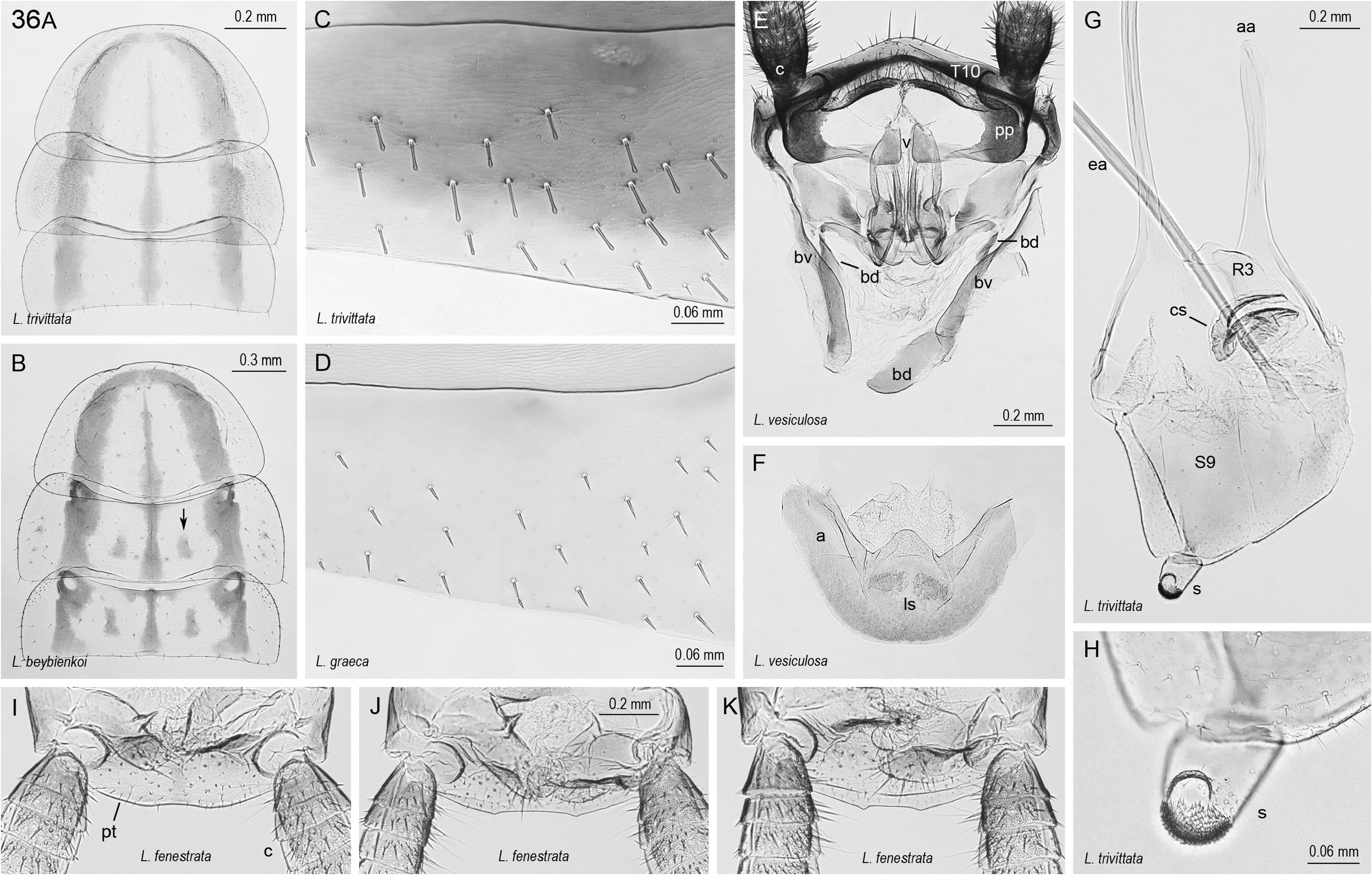
FIGURE 36 A–K. Characters of the genus Luridiblatta and its species groups.—A,B. Colouration of thoracal nota in halfgrown or older larvae of Luridiblatta. Meso- and metanotum in the species of the beybienkoi-group (B, L. beybienkoi) as compared to the other two species groups (A, L. trivittata) with two more dark patches (arrow).—C,D. Bristles on T5 in the species of the longitubulata- and trivittata- group (C, L. trivittata) mainly large and of spatular shape, with clavate tip, in the beybienkoi-group (D, L. graeca) small, with acute tip.—E,F. Female terminalia of L. vesiculosa, with genital sclerites, ventral view, posterior on top. Dorsal complex (E), with c cerci, pp paraprocts, v valves of ovipositor, bd, bv dorsal, ventral basivalvular sclerites; right (bd) much longer than left one, median part by (bv) covered. Ventral complex (F): laterosternal shelf ls, with relatively long arms a, right one apically damaged.—G,H. Male subgenital plate S9 (G), with genital sclerites (except the hook), dorsal view: anterior apodemes aa, endophallus apodeme ea, cleft sclerite cs, R3 sclerite, and stylus s; the latter in higher magnification (H), partly covered with tiny scales.—I–K. Male terminalia of L. fenestrata, with posterior border of the supraanal plate pt, cerci c.—Enlargements: Same scale for E,F and for I–K.—Identifications:A (It 34/7), B (Is 20/2), C (Sz 7/4), D (Kr 23/2, holotype), E,F (Ma 46/3), G,H (It 34/3), I,J,K (Ma 14/12,13,11).
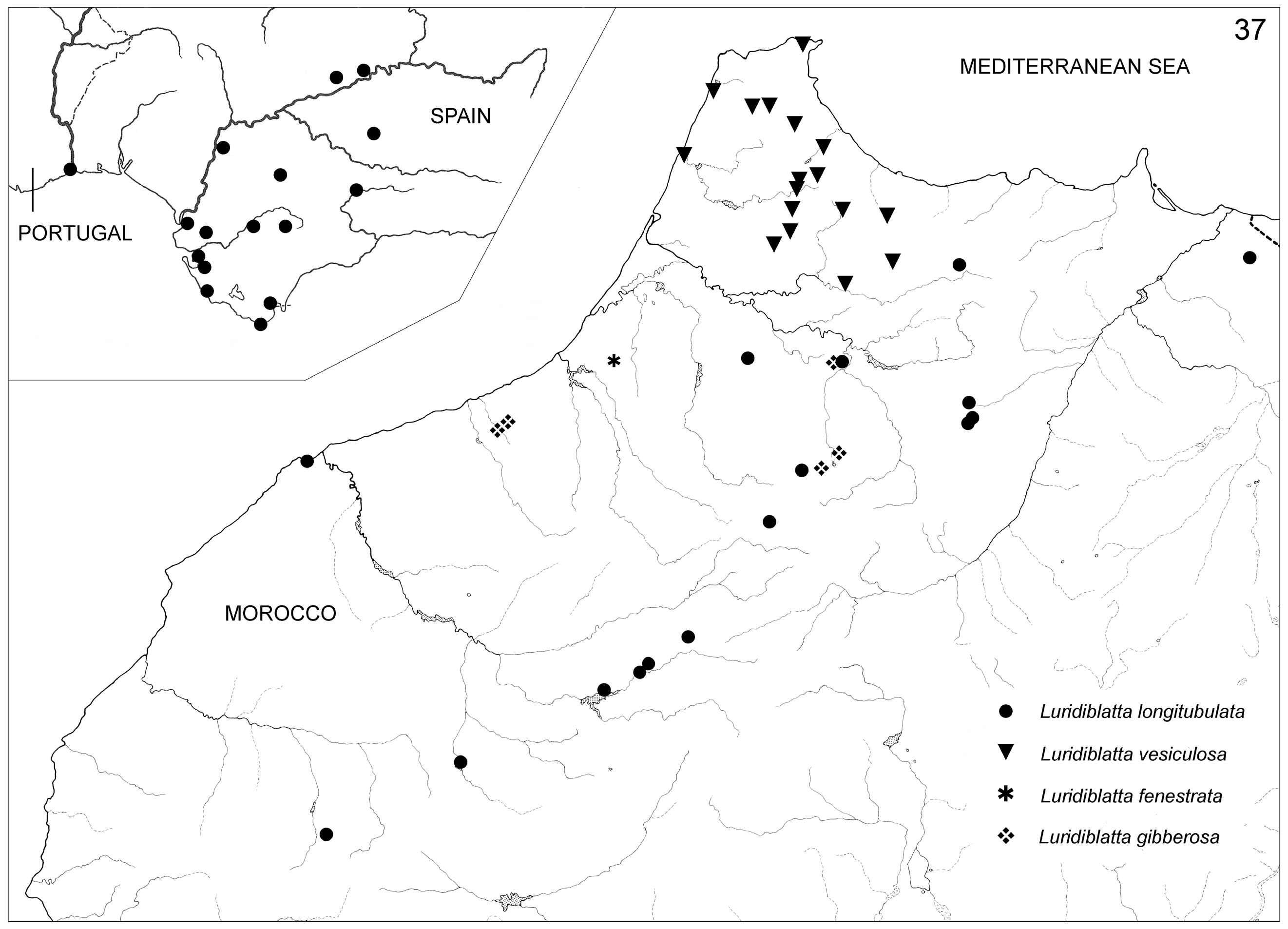
FIGURE 37. Distribution of the species of Luridiblatta occurring in Portugal, Southern Spain and Morocco. There are also one or two localities of L. longitubulata in Middle Algeria (Fig. 39).
No known copyright restrictions apply. See Agosti, D., Egloff, W., 2009. Taxonomic information exchange and copyright: the Plazi approach. BMC Research Notes 2009, 2:53 for further explanation.
|
Kingdom |
|
|
Phylum |
|
|
Class |
|
|
Order |
|
|
SuperFamily |
Blaberoidea |
|
Family |
|
|
SubFamily |
Ectobiinae |
|
Genus |
1 (by plazi, 2022-12-06 08:03:17)
2 (by ExternalLinkService, 2022-12-06 08:12:58)
3 (by juliana, 2022-12-06 20:12:42)
4 (by ExternalLinkService, 2022-12-07 16:11:23)
5 (by juliana, 2022-12-08 12:17:02)
6 (by ExternalLinkService, 2022-12-08 12:26:43)
7 (by plazi, 2023-11-07 23:51:08)
8 (by ExternalLinkService, 2023-11-08 03:12:04)