
Evoplosoma nizinskiae, Mah, 2020
|
publication ID |
https://doi.org/ 10.11646/zootaxa.4766.2.1 |
|
publication LSID |
urn:lsid:zoobank.org:pub:B47DC09C-181A-4DFE-B415-770AFFC11BD3 |
|
DOI |
https://doi.org/10.5281/zenodo.3803733 |
|
persistent identifier |
https://treatment.plazi.org/id/03AE8786-FFAE-D557-FF40-2793FC1259F5 |
|
treatment provided by |
Carolina |
|
scientific name |
Evoplosoma nizinskiae |
| status |
sp. nov. |
Evoplosoma nizinskiae View in CoL n. sp.
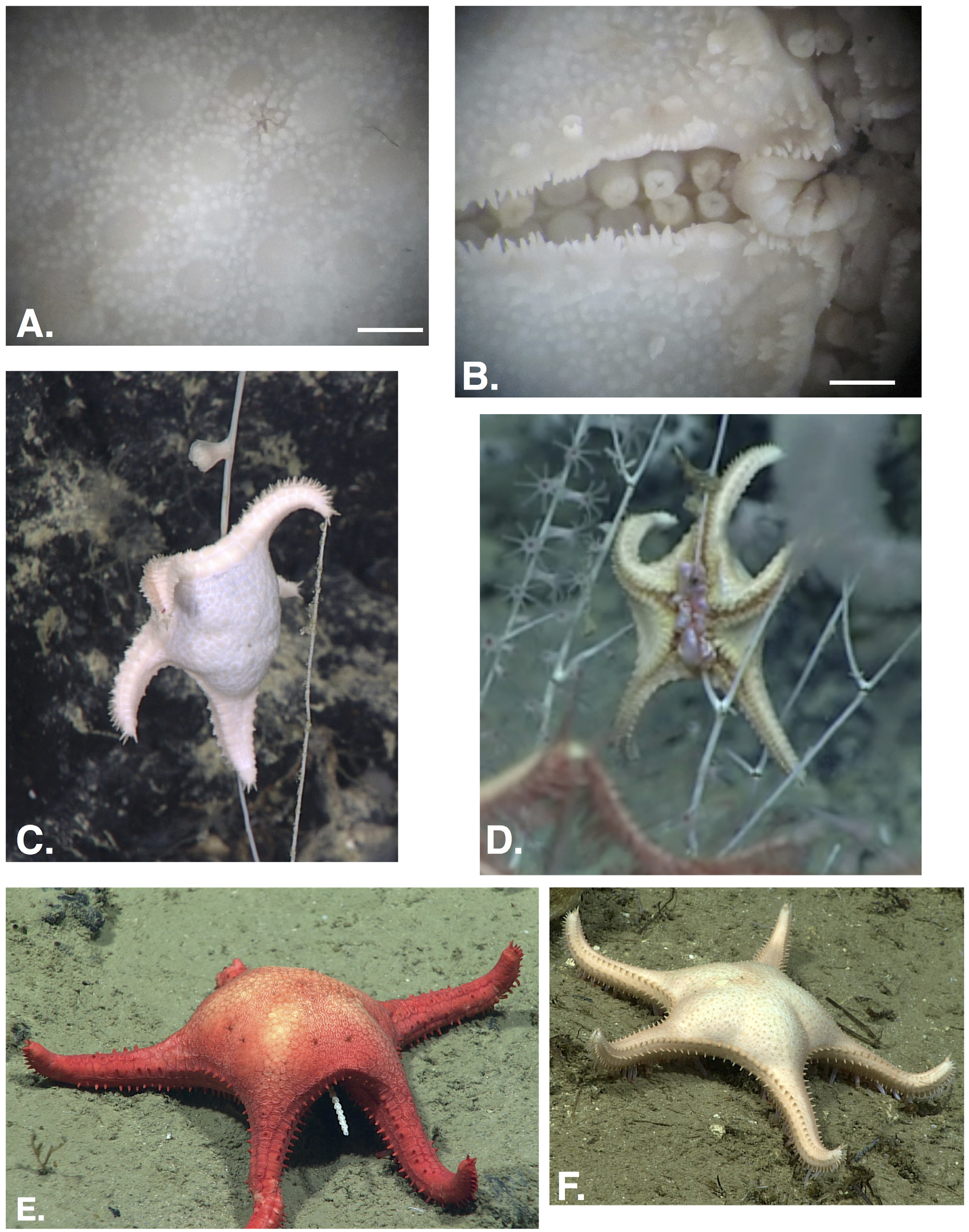
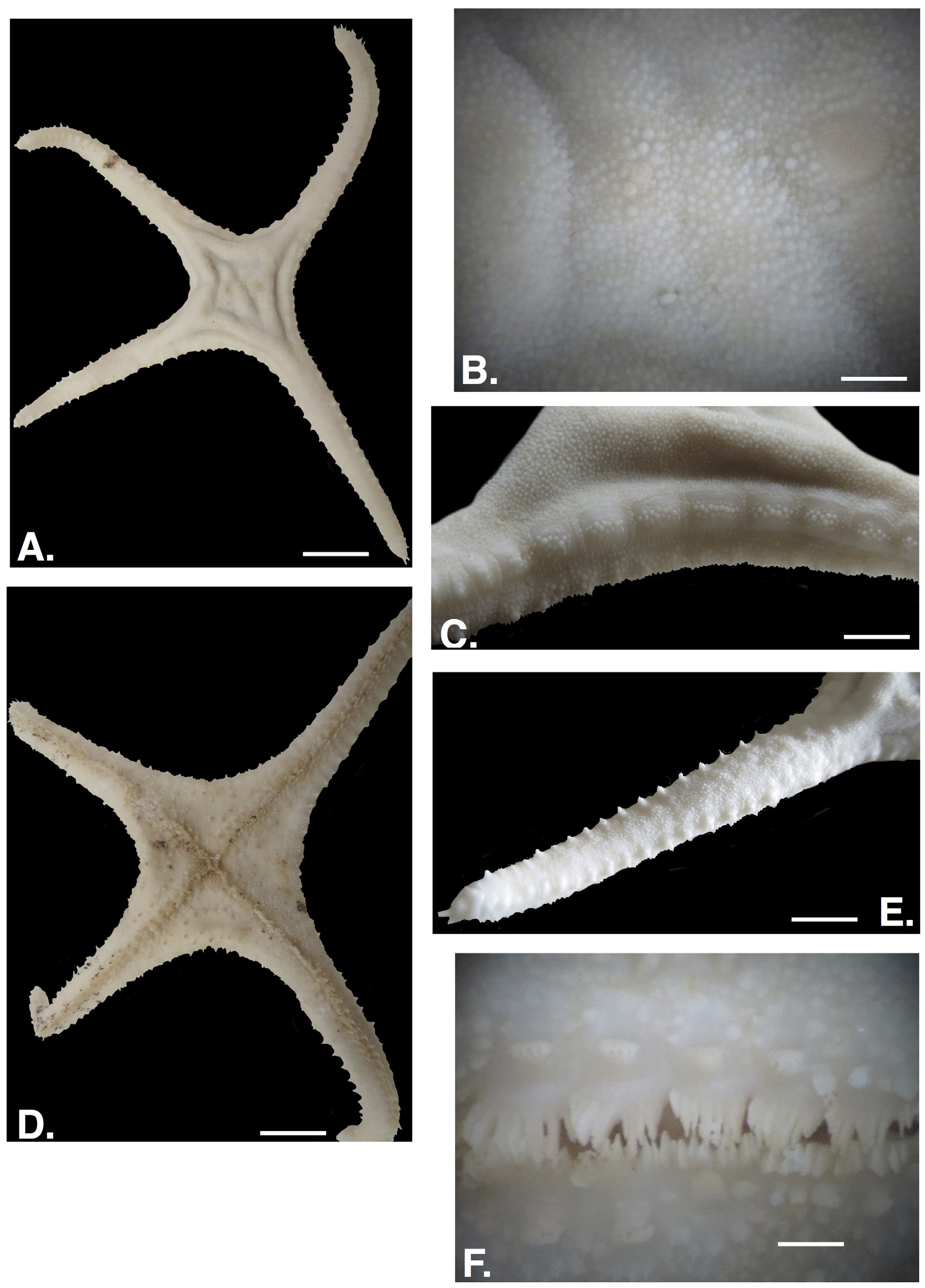
Figure 9 View FIGURE A–F, 10A–D
Etymology
This species is named for my colleague Dr. Martha Nizinski in honor of her tireless contributions to deep-sea coral ecology and NOAA’s Ocean Explorer program.
Diagnosis
This species is characterized by the lack of primary spines on the abactinal plates, the evenly distributed granules, the absence of primary spines from interradial superomarginal plates-but present on distal superomarginal
plates, mostly bare superomarginal plates save for coarse, widely spaced granules, prominent elongate subambulacral pedicellariae with three to four teeth per valve on each adambulacral plate and 7 to 10 leaflike furrow spines.
Occurrence: Gulf of Mexico, Caribbean Sea ( Puerto Rico), 2037–2549 m.
Description
Body strongly stellate (R/r=4.42), arms elongate with pointed arm tip, interradial arcs weakly curved to straight. Four arms (five on other observed specimens) ( Fig. 9A View FIGURE ).
Abactinal surface pulpy, shows strongly convex surface but with some folded and sagged regions, suggesting inflation when alive ( Fig 9A,B View FIGURE ). Abactinal plates extend from disk to arm tip. Abactinal plates round to irregular in shape, closely abutted continuous surface. Plate boundaries weakly defined. Surface covered by moderately coarse, widely spaced but evenly distributed granules ranging from round surface to pointed tips, approximately one to four, mostly one to three along a 1.0 mm line. Granules smaller distally becoming larger proximally on disk with some enlarged granules 0.5 mm width. Individual plate surfaces vary from completely 0 to 25 per plate. Plates lacking granules bare and smooth. Where granules are present, they form a periphery, approximately three to five per side with one to eight present centrally. The more granules present, the smaller they are. Pedicellariae present on disk, relatively few with approximately one per interradial region (four on this individual), approximately 1.0 mm in length, elongate, jaw-like with three to five teeth on each valve. Madreporite round, weakly pentagonal, weakly convex with a shallow channel present around it.
Superomarginals and inferomarginals 54–55 per interradius, with a lateral facing ( Fig. 9A, C View FIGURE ). All marginal plates with approximately 40–60 pointed granules forming the plate periphery, widely spaced, approximately 10–12 per side. Some superomarginals irregular in shape between fully formed superomarginal plates. Superomarginals larger and elongate interradially, (approximately ten plates) becoming wider (i.e. narrower) and more strongly convex distally along the arms. Interradial superomarginals, approximately ten to twelve plates lacking primary spines, each of these superomarginals with 20–30, mostly about 25 coarse, bullet-like, widely spaced granules. Central plate granules larger, smaller granules approximately 20% of the larger granules and present peripherally around the central granules. Superomarginals along the arms ( Fig. 9E View FIGURE ) mostly with a single prominent, bullet-shaped spine, (approximately 1–2.0 mm in length) remainder of superomarginal surface mostly bare, smooth, forming a continuous series along the arm ( Fig. 9C View FIGURE ). Most distalmost superomarginals with zero to eight small, round granules on plate surface around base of each spine. Inferomarginals with bald upper lateral plate surface, but with oral surface covered by 20 to 30 coarse, round to cylindrical granules with some appearing as incipient spines. Inferomarginal plates mostly with similar spines to those on superomarginals, with spines absent on more inferomarginals than superomarginals, especially distally. Inferomarginals present basal own arm. Where spines absent, coarse, widely spaced granules, 10–30 per inferomarginal plate surface. Shallow fasciolar grooves between superomarginal and inferomarginal plates. Both superomarginal and inferomarginal interradial plates with a relatively large (1.0–2.0 mm) elongate pedicellariae, similar to those on abactinal surface, on the lateral surface. Terminal plates triangular in shape, widely spaced granules present on terminal plate surface.
Actinal surface with only two to three series, mostly limited to disk and basal arm regions ( Fig. 9D View FIGURE ). Actinal plates with quadrate to irregularly shaped plates. Each plate surface with 2 to 20 pointed, evenly spaced granules. Most plates with 4 to 14 peripheral granules with 2 to 6 identical to large sized granules on each central actinal plate surface. Large, prominent pedicellariae, 6 to 9 per interradius, with some actinal plates bearing a single large pedicellariae with a bare smooth region around it. Pedicellariae identical to those on other surfaces. Peripheral granules around these “pedicellariae actinal plates” more widely spaced than other actinal plates.
Furrow spines 8 to 10 per plate arranged straight to weakly curved ( Fig. 9F View FIGURE ). Eight furrow spines proximally becoming ten distally. Furrow spines flattened, leaf-like, angular in cross section. Furrow spines interdigitate along the tube foot groove. Adambulacral plates on disk with large elongate pedicellariae, especially on proximal adambulacrals. Along arm and more distally adambulacral plates have both a prominent pedicellariae but also a single, blunt spine similar in size to the pedicellariae. Adambulacral spines becoming larger and more prominent than the pedicellariae on distalmost plates. Widely space pointed granules present on adambulacral plate adjacent to actinal plates side. Some adambulacral plates lacking spines and/or granules, surface covered by pointed to round granules present. Oral plate furrow spines, eight with central spine projecting into mouth, projecting into mouth. Spines narrow and leaf-like, triangular in cross section. Approximately seven or eight paired multi-spined granules present along either side of fossae on oral plate. Five to eight multi-spined granules present around periphery of each oral plate side.
In life, disk is white, arms a light orange to tan ( Figs 10 View FIGURE 10 C–F).
Comparisons with other Evoplosoma species
This species invites immediate comparison with Evoplosoma virgo Downey 1982 which also lacks primary spines on the abactinal and the interradial superomarginal plate surface. However Evoplosoma virgo has a dense covering of pointed granules covering the marginal plate surfaces, whereas E. nizinskiae n. sp. has a mostly bare and smooth superomarginal surface, save for widely spaced granules on the lateral edge. Then dense abactinal granulation in E. virgo occurs in discrete, round to polygonal patches versus the more evenly distributed granulation observed in E. nizinskiae . Furrow spines in E. virgo are three to five times as thick as those in E. nizinskia e n. sp. Evoplosoma nizinskiae is also more strongly stellate (R/r=4.42) with elongate arms versus E. virgo with a more weakly stellate form (R/r=3.5) and a broader disk.
However, the two species share similar pedicellariae morphology with both species displaying these pedicellariae on their adambulacral plates adjacent to the furrow spination. Both species also lack superomarginal spination interradially but possess a primary spine on more distalmost superomarginals along the arm.
Evoplosoma nizinskiae n. sp. is distinguished from the other Atlantic Evoplosoma spp. by the absence of numerous, thick primary spines present on the abactinal and actinal plates ( Fig. 9A, B View FIGURE ). Both Evoplosoma watlingi and Evoplosoma scorpio also possess a full series of prominent superomarginal spines from arm tip to arm tip, whereas Evoplosoma nizinskiae n. sp. lacks superomarginal spines interradially ( Fig. 9C View FIGURE ).
The unidentified Evoplosoma sp. 1 listed herein, also shows strong similarity with Evoplosoma nizinskiae n. sp., in showing a similar body shape, and color, but differs in having a full series of superomarginal spines.
Juvenile morphology
Body form stellate (R/r=2.25) but more weakly than the larger individual. Arms triangular but less elongate. Interradial arcs weakly curved to straight.
Abactinal surface composed of primarily round plates ( Fig. 10A View FIGURE 10 ), extending to arm tip. Plates becoming more irregular in shape distally, especially near arm tip. Each plate with four to 20 small round granules forming periphery of each plate. Central region devoid of granules, surface bare and smooth. Plates weakly convex becoming more flat distally. Anus surrounded by five triangular, blunt spines. Madreporite weakly convex, with four sides, rounded surrounded by five to six plates. Pedicellariae absent from abactinal surface.
At R=1.8, 19–21 superomarginal plates per interradius (arm tip to arm tip), 22–24 inferomarginal plates per interradius. Marginal plates blocky, with distinct angular lateral surface. Interradial superomarginals with no spines, but two proximal superomarginals on arm with distinct, blunt, bullet-shaped spines. on two arms. Remaining superomarginals without larger primary spine, most with 1 to 10 (mostly 6 to 8) widely spaced spiny tipped coarse granules present on lateral and abactinolateral surface. Central granules coarse and approximately twice as large as others on plate surface. Remainder of plate smooth and bare. Inferomarginal plate surface covered by 10–20 pointed granules, widely spaced in linear patterns. Round, bald region on ventral inferomarginal plate surface. Periphery of each marginal with 40–60 widely spaced, irregular to round granules with spiny tips, approximately 10–12 per side. Terminal plate triangular with one to three pointed but blunt spines projecting from each arm tip. Pedicellariae absent from marginal plate surface.
Actinal region with two series, limited to disk. Actinal plates quadrate to irregular in shape. Pedicellariae absent from actinal surface. Actinal plate surface covered by 2 to 16 pointed granules, widely spaced. A single elongate pedicellariae with three to four teeth per valve present on two inter radii. The remaining two actinal regions lacking pedicellariae.
Furrow spines six to seven ( Fig. 10B View FIGURE 10 ), flattened, with shortest spines farthest from center. Furrow spine in straight to weakly curved series. Proximalmost three or four adambulacral plates with prominent elongate single pedicellariae on surface. Distalmost remaining adambulacral plate surface with prominent, single blunt spines. Adambulacral plate surface with four to seven pointed granules. Oral plates with eight furrow spines identical to other furrow spines but with a single angular spine projecting into mouth. Oral plate surface each with five pairs triangular granules flanking each side of central fossae. Other short thorny granules irregularly present on oral plate surface.
Color in life almost white but with light orange arm color.
Comments on Juvenile form
The paratype, USNM 1550630 is smaller and shows several differences from the holotype, but characters strongly suggest similarity and it is regarded herein as a juvenile of Evoplosoma nizinskiae n. sp. If correct, this provides a rare opportunity to examine the growth in a deep-sea goniasterid species. It showed a significantly less stellate body from (R/r=2.25) versus the more stellate body form (R/r=4.42) at R=6.2 as well as triangular versus the elongate arms in the holotype. The abactinal plates are round to polygonal in outline ( Fig. 10A View FIGURE 10 ), and although many, if not most, of the plates are bare, several plates show single to few granules suggesting incipient development of the granulation that is seen in the larger specimen ( Fig. 9A View FIGURE ). Furrow spine number also shows some overlap with the paratype showing primarily six or seven furrow spines ( Fig. 10B View FIGURE 10 ) and the larger specimen displaying seven to ten, primarily eight ( Fig. 9B View FIGURE ).
Feeding Observations
Of the four in situ observations of this species ( Fig. 10 View FIGURE 10 C–E), three showed E. nizinskiae n. sp. feeding. In two instances, ( Fig. 10C, E View FIGURE 10 ) individuals had climbed up onto higher branches and extended their pyloric stomachs onto the polyps of their isidid octocoral (aka bamboo coral) prey.
Images Examined
Smooth Escarpment Ridge, Gulf of Mexico. 28.00404, -86.43931, 2054 m EX1711_IMG_20171205T154320Z_ROVHD.jpg
KC560 site, Gulf of Mexico, 26.42891, -92.35167, 1918 m
EX1711_IMG_20171213T194231Z_ROVHD.jpg
West Florida Escarpment, Gulf of Mexico. 24.91667, -84.49046, 2181 m. EX1803_VID_20180430T164500Z_ROVHD_Low.mp4.tiff
Material Examined
Holotype. USNM 1507289 View Materials DeSoto Canyon, Gulf of Mexico, 28.28, -87.22, 2549 m, Coll. D. Wagner, ROV Deep Discoverer from NOAA Ship Okeanos Explorer EX 1803 25 April 2018. 1 wet spec. R=6.2 r=1.4. EX1803_ IMG_20180425T163351Z_ROVHD.jpg
Paratype. USNM 1550630 Virgin Islands Trough, Puerto Rico, Caribbean Sea. 17.7732, -65.4269, 2037 m, Coll. D. Wagner with Deep Discoverer ROV, NOAA Ship Okeanos Explorer , 5 Nov. 2018. 1 wet spec. 1 wet spec. R =1.8 r=0.8. EX 1811_IMG_20181105 T 170423Z_ ROVHD.jpg (feeding)
No known copyright restrictions apply. See Agosti, D., Egloff, W., 2009. Taxonomic information exchange and copyright: the Plazi approach. BMC Research Notes 2009, 2:53 for further explanation.