
Otatea ximenae, Ruiz-Sanchez & Clark
|
publication ID |
https://doi.org/10.11646/phytotaxa.609.2.1 |
|
DOI |
https://doi.org/10.5281/zenodo.8274500 |
|
persistent identifier |
https://treatment.plazi.org/id/03AD87BF-FFFA-0C14-FF45-2ACBFEEFFC6B |
|
treatment provided by |
Plazi (2023-08-22 09:30:37, last updated 2024-11-26 08:52:37) |
|
scientific name |
Otatea ximenae |
| status |
|
Otatea ximenae View in CoL View at ENA ( Fig. 14 View FIGURE 14 )
Material examined: — MEXICO. Oaxaca: Asunción Nochixtlán, Km 190 of the Puebla-Oaxaca highway, 17°23’9.1”N, 97°8’8.9”W 1928 m, 2 February 2020, Ruiz-Sanchez & M GoogleMaps . A GoogleMaps . García-Martínez 645 ( IBUG!) .
Culm anatomy description: —Culm solid. Epidermis with a layer of axially elongated lignified cells, 12 µm length, with occasional rectangular silica bodies on top ( Fig. 16C View FIGURE 16 ). Cortex with one layer of thick-walled cells followed by 2 layers of wider, thinner-walled ovoidal cells (3 layers in total, 30 µm length; Fig. 16C View FIGURE 16 ). Vascular bundles in 5–6 alternating cycles ( Fig. 16A View FIGURE 16 ). Peripheral, transitional, and central vascular bundles surrounded by a sclerenchyma sheath defining their contour ( Figs. 16D–F View FIGURE 16 ). Ovate central vascular bundles, 309 ×304 μm; two round shaped metaxylem vessels, 75 × 83 μm; phloem with sieve tubes and companion cells located between two metaxylem vessels on the upper level, 78 × 98 µm; sieve tubes 19 × 20 µm ( Fig. 16F View FIGURE 16 ). Ground parenchyma with abundant starch grains. Frequent conical silica bodies across the ground tissue occupying intercellular spaces covering a mean area of 10 µm 2 ( Fig. 16B View FIGURE 16 ).
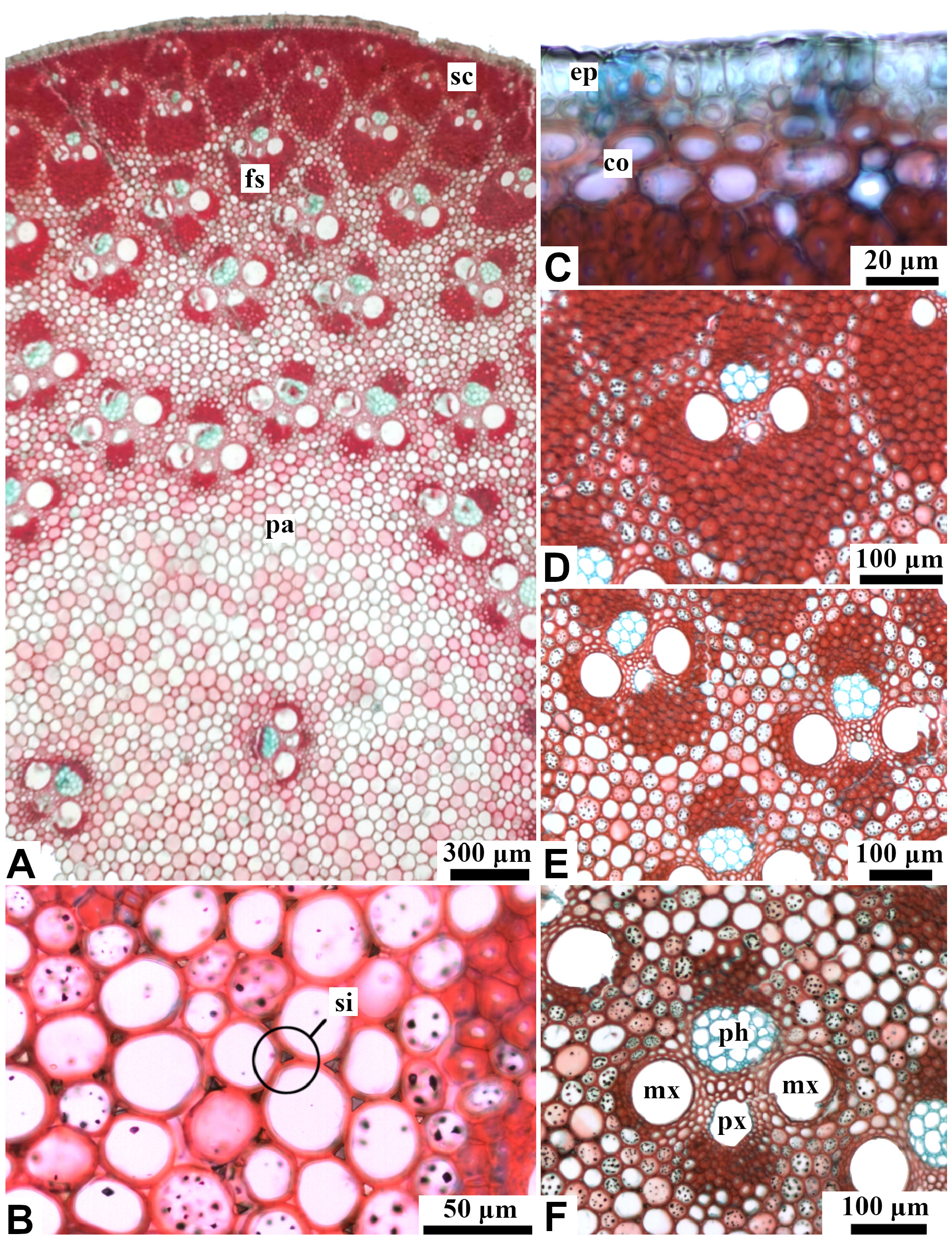
FIGURE 14. Culm cross section of Otatea ximenae. A. Cross section general view. B. Central area with vascular bundles following different orientations. C. Epidermis and hypodermis. D. Peripheral vascular bundle. E. Transitional vascular bundle. F. Central vascular bundle. Abbreviations: co, cortex; ep, epidermis; fs, fiber sheath; mx, metaxylem; pa, parenchyma; ph, phloem; px, protoxylem; sc, sclerenchyma; si, silica bodies.

FIGURE 16. Culm cross section of Otatea sp. nov. 2. A. Cross section general view. B. Epidermis and cortex. C. Peripheral vascular bundle. D. Transitional vascular bundle. E. Central vascular bundle. Abbreviations: co, cortex; ep, epidermis; fs, fiber sheath; mx, metaxylem; pa, parenchyma; ph, phloem; px, protoxylem; sc, sclerenchyma; si, silica bodies.
| M |
Botanische Staatssammlung München |
| A |
Harvard University - Arnold Arboretum |
| IBUG |
Universidad de Guadalajara |
No known copyright restrictions apply. See Agosti, D., Egloff, W., 2009. Taxonomic information exchange and copyright: the Plazi approach. BMC Research Notes 2009, 2:53 for further explanation.
|
Kingdom |
|
|
Phylum |
|
|
Class |
|
|
Order |
|
|
Family |
|
|
Genus |