
Theridiosomatidae
|
publication ID |
https://doi.org/10.3897/zookeys.11.160 |
|
publication LSID |
lsid:zoobank.org:pub:C631A347-306E-4773-84A4-E4712329186B |
|
persistent identifier |
https://treatment.plazi.org/id/03ACB915-FF94-7418-FF4E-402AFBA5FAD5 |
|
treatment provided by |
Plazi (2020-04-27 10:43:22, last updated 2024-11-26 01:02:42) |
|
scientific name |
Theridiosomatidae |
| status |
|
Key to Gaoligongshan Theridiosomatidae View in CoL View at ENA
1 Females ....................................................................................................... 2
Males.......................................................................................................... 9
2(1) Abdomen subtriangular, tapered posteriorly ( Fig. 8B View Figure 8 ). Spermathecae separated by nearly their diameter ( Fig. 11F View Figure 11 )... Coddingtonia euryopoides View in CoL sp. n.
– Abdomen subspherical, not distinctly tapered posteriorly. Spermathecae juxtaposed ( Fig. 3D View Figure 3 )........................................................................................ 3
3(2) Scape protrudes from beneath epigynal plate ( Figs 11C View Figure 11 , 14C)...................... ............................................................................. Wendilgarda muji View in CoL sp. n.
– Scape absent ( Fig. 11A View Figure 11 ) or if present ( Fig. 3E View Figure 3 ) then a simple extension of the epigynal plate.............................................................................................. 4
4(3) Posterior median eyes separated by at least their diameter ( Fig. 4C View Figure 4 ).............. ..................................................................... Ogulnius barbandrewsi View in CoL sp. n.
– Posterior median eyes separated by less than their diameter......................... 5
5(4) Epigynum subtriangular, pointed posteriorly with concave margins so medial part is more acute than lateral part ( Fig. 3E View Figure 3 )......... Baalzebub nemesis View in CoL sp. n.
– Epigynum otherwise, typically subrectangular ( Fig. 11A View Figure 11 )............................ 6
6(5) Epigynum with a deep atrium, height of opening about one third the width in posterior view ( Fig. 3B View Figure 3 ), without fleshy tissue at posterior margin of epigynum. Lateral pits absent....................................... Epeirotypus dalong View in CoL sp. n.
– Atrium absent ( Fig. 3F View Figure 3 ) or slit-like (Fig. 13A), height of opening (if visible) much less than one third the width in posterior view. Region between posterior margin of epigynum and abdomen often with fleshy tissue ( Fig. 3F View Figure 3 ). Lateral pits present ( Fig. 9B View Figure 9 ) ....................................................................... 7
7(6) Epigynum with pair of processes arising from posterolateral margin running towards each other ( Fig. 3H View Figure 3 ) ........................ Theridiosoma shuangbi View in CoL sp. n.
– Epigynum without pair of processes arising from posterolateral margin ...... 8
8(7) Posterior margin of epigynum with median longitudinal slit ( Figs 3F View Figure 3 , 9A View Figure 9 ). Abdomen tan with dark gray with silver patches ........................................... ........................................................................ Theridiosoma diwang View in CoL sp. n.
– Posterior margin of epigynum entire ( Fig. 11A View Figure 11 ). Abdomen dark gray with silver patches forming curved transverse stripe (Fig. l0A) .............................. ................................................................................... Zoma dibaiyin View in CoL sp. n.
9(1) With long, filliform embolic apophysis ( Figs 4G View Figure 4 , 14A)............................. 10
– Embolic apophysis absent ( Fig. 2C View Figure 2 )..................... Epeirotypus dalong View in CoL sp. n.
10(9) Posterior median eyes separated by about their diameter............................... ..................................................................... Ogulnius barbandrewsi View in CoL sp. n.
– Posterior median eyes separated by less than half their diameter................ 11
11(10) Palpal tibia with one trichobothrium. Median apophysis sclerotized with concave dorsal margin (Fig. 12E).......................... Wendilgarda muji View in CoL sp. n.
– Palpal tibia with two trichobothria. Median apophysis fleshy, with pointed dorsal apex (Fig. 10D) ................................................ Zoma dibaiyin View in CoL sp. n.

Figure 2. Epeirotypus dalong sp. n. holotype from Fugong Co., male palp. A, prolateral; B, retrolateral; C, ventral. C: conductor; MA: median apophysis; PC: paracymbium; ST: subtegulum; T: tegulum; arrow in C indicates field of tubercles on mesal lobe of tegulum.

Figure 3. A, B, Epeirotypus dalong sp. n. from Fugong Co.; C, D, Ogulnius barbandrewsi sp. n.; C from Guocai He at Fucai; D from No. 12 Bridge Camp area; E, Baalzebub nemesis sp. n. from No. 12 Bridge Camp area; F, G, Theridiosoma diwang sp. n. from Xiao Hei Shan Nature Reserve; H, I, Theridiosoma shuangbi sp. n. from Xiao Hei Shan Nature Reserve. A, C, E, F, H, epigynum, ventral view; B, epigynum, posterior view; D, G, I, cleared vulva, dorsal view. CD: copulatory duct; S: spermatheca; arrow in A indicates transverse submarginal groove; in G and I indicate lateral pits, in H indicates base of one of two opposing lateral apophyses.

Figure 4. Ogulnius barbandrewsi sp. n. from Guocai He at Fucai. A-D, habitus; E-G, male palp. A, male, lateral; B, female, lateral; C, female, dorsal; D, female, ventral; E, prolateral; F, retrolateral; G, ventral. C: conductor; EA: embolic apophysis; MA: median apophysis; T, tegulum; arrow in D indicates sternal pit.

Figure 8. A, Baalzebub nemesis sp. n. from No. 12 Bridge Camp area; B, Coddingtonia euryopoides sp. n. from Zaotang He at Baihualing village; C-E, Theridiosoma diwang sp. n. from Xiao Hei Shan Nature Reserve, female. A, B, D, female, dorsal; C, female, lateral; E, female, ventral.
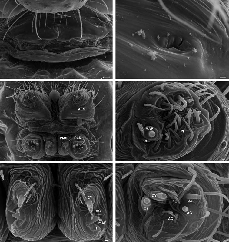
Figure 9. Theridiosoma diwang sp. n. from Xiao Hei Shan Nature Reserve, SEM of female. A, epigynum; B, epigynum, detail showing lateral pore; C, spinnerets; D, ALS; E, PMS; F, PLS. AC: aciniform gland spigot; AG: aggregate gland spigot; CY: cylindrical gland spigot; FL: flagelliform gland spigot; MAP: major ampullate gland spigot; mAP: minor ampullate gland spigot; n: nubbin; PI: piriform gland spigot, PLS: posterior lateral spinneret, PMS: posterior median spinneret, t: tartipore.
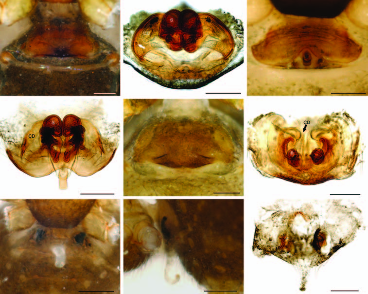
Figure 11. A, B, Zoma dibaiyin sp. n. from Pee He; C, D, Wendilgarda muji sp. n. from Xiao Hei Shan Nature Reserve; E, F, Coddingtonia euryopoides sp. n. from Zaotang He at Baihualing village; G-I, Mysmena changouzi sp. n. from from No. 12 Bridge Camp area. A, C, E, G, epigynum, ventral view; B, D, F, I, cleared vulva, dorsal view; H, epigynum, lateral view. Arrow in B indicates lateral pit.
No known copyright restrictions apply. See Agosti, D., Egloff, W., 2009. Taxonomic information exchange and copyright: the Plazi approach. BMC Research Notes 2009, 2:53 for further explanation.
|
Kingdom |
|
|
Phylum |
|
|
Class |
|
|
Order |
|
|
Family |