
Tupigea angelim Huber, 2011
|
publication ID |
https://doi.org/10.1080/00222933.2010.524319 |
|
persistent identifier |
https://treatment.plazi.org/id/03A887D8-FF8B-FFAE-BB1B-D83DFE35FB03 |
|
treatment provided by |
Carolina (2021-04-06 18:35:39, last updated by Plazi 2023-11-02 05:01:27) |
|
scientific name |
Tupigea angelim Huber |
| status |
sp. nov. |
Tupigea angelim Huber View in CoL sp. nov.
( Figures 4 View Figure 4 A–C, 5A–E, 6A–H)
Tupigea View in CoL sp. 1: Astrin et al. 2006: 445, 2007: 20.
Types
Male holotype and 2♂, 4♀ paratypes from Brazil, São Paulo, Ubatuba, Fazenda Angelim (23 ◦ 23.6 ′ S, 45 ◦ 03.8 ′ W), ∼ 50 m above sea level (a.s.l.), 16–18 December 2003 (B.A. Huber), holotype and 1♀ in IBSP , 2♂, 3♀ paratypes in ZFMK .
Etymology
The specific name is a noun in apposition, taken from the type locality.
Diagnosis
Easily distinguished from known congeners by armature on male chelicerae (pair of frontal apophyses, Figure 5C View Figure 5 ) and by dorsal worm-shaped projection of bulb ( Figures 5A View Figure 5 , 6C View Figure 6 ); also by male palp (modifications of femur, procursus shape, embolic division of bulb, Figures 5A,B View Figure 5 , 6C,D View Figure 6 ), and female genitalia ( Figures 5D,E View Figure 5 , 6G View Figure 6 ).
Male (holotype)
Total body length 1.6, carapace width 0.67. Leg 1: 14.9 (3.7 + 0.2 + 3.8 + 5.9 + 1.3), tibia 2: 2.0, tibia 3: 1.4, tibia 4: 1.9, tibia 1 L / d: 72. Habitus as in Figure 4A View Figure 4 ; carapace ochre-yellow, darker around thoracic furrow, ocular area and clypeus light brown, clypeus medially with dark brown band that is lined by two cuticular ridges ( Figures 6A,B View Figure 6 ), sternum and legs ochre-yellow, legs with darker rings subdistally on femora and tibiae, abdomen greenish-grey with large darker marks on dorsal half. Distance PME–PME 70 µm, diameter PME 70 µm, distance PME–ALE 35 µm; AME absent, only irregular pigment spots. Ocular area slightly elevated, thoracic furrow shallow but distinct, clypeus unmodified ( Figure 6A View Figure 6 ). Chelicerae as in Figure 5C View Figure 5 , with distinctive pair of frontal apophyses. Sternum wider than long (0.46 / 0.38), unmodified. Palps as in Figure 5A,B View Figure 5 , coxa with indistinct retrolateral apophysis, trochanter barely modified, femur with proximal projection, tiny cones on prolatero-ventral side, tiny retrolateral apophysis, and small ventral apophysis distally, patella relatively long ventrally, procursus short, distally with distinctive sclerotized and membranous elements, bulb large, with distinctive dorsal worm-shaped projection and partly membranous embolic division with terminal apophysis and transparent flap. Palpal tarsal organ exposed ( Figure 6E View Figure 6 ). Legs without spines and curved hairs, with some vertical hairs on tibiae dorsally; retrolateral trichobothrium on tibia 1 at 22%; tarsus 1 with 16 pseudosegments, quite distinct. Anterior lateral spinnerets with two spigots each; gonopore without epiandrous spigots ( Figure 6F View Figure 6 ).
Variation. Tibia 1 in four other males: 3.7, 3.9 (type locality), 3.0, 3.3 (Rio das Pedras); in males from Rio das Pedras the tiny cones ventrally on the femur are replaced by a single slightly larger apophysis; otherwise palps and chelicerae identical.
Female
In general similar to male but without darker median band and cuticular ridges on clypeus; carapace medially and ocular area darker brown; tibia 1 in eight females: 1.6–2.0 (mean 1.85). Epigynum ( Figures 4B View Figure 4 , 5D View Figure 5 , 6G View Figure 6 ) frontal plate darkened medially, posterior margin sclerotized, posterior plate with pair of frontal indentations; internal genitalia as in Figure 5E View Figure 5 . In females from Rio das Pedras the posterior margin of the frontal plate is barely sclerotized and less protruding, but the internal genitalia appear indistinguishable. Two females with genital plugs ( Figure 4C View Figure 4 , 6H View Figure 6 ).
Distribution
Known from two localities in São Paulo and Rio the Janeiro states ( Figure 2 View Figure 2 ).
Material examined
BRAZIL: São Paulo: Ubatuba, Fazenda Angelim : 3♂, 4♀ types above; same data, 5♀ in pure ethanol, in ZFMK. Rio de Janeiro: Reserva Ecológica Rio das Pedras (22 ◦ 59.5 ′ S, 44 ◦ 06.0 ′ W), 50 m a.s.l., night collecting, 25 September 2009 (B.A. Huber), 1♂, 3♀ in ZFMK; same locality but 22 ◦ 59.5 ′ S, 44 ◦ 06.0 ′ to 06.8 ′ W, 50–200 m a.s.l., day collecting, 26 September 2009 (B.A. Huber, A. Giupponi), 1♂, 3♀ in MNRJ (1♂, 2♀) and ZFMK (1♀) .
Astrin JJ, Huber BA, Misof B, Kluetsch CFC. 2006. Molecular taxonomy in pholcid spiders (Pholcidae, Araneae): evaluation of species identification methods using CO 1 and 16 S rRNA. Zool Scr. 35: 441 - 457.
Astrin JJ, Misof B, Huber BA. 2007. The pitfalls of exaggeration: molecular and morphological evidence suggests Kaliana is a synonym of Mesabolivar (Araneae: Pholcidae). Zootaxa 1646: 17 - 30.
Ribeiro MC, Metzger JP, Martensen AC, Ponzoni FJ, Hirota MM. 2009. The Brazilian Atlantic Forest: how much is left, and how is the remaining forest distributed? Implications for conservation. Biol Conserv. 142: 1141 - 1153.
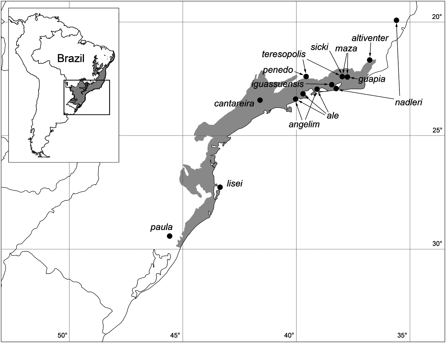
Figure 2. Known distribution of Tupigea. Grey shade in inset: extension of Atlantic Forest in Brazil (inset modified from Ribeiro et al. 2009). Grey shade in main map: extension of Serra do Mar biogeographic sub-region, from Silva and Casteleti (2003) in Ribeiro et al. (2009).
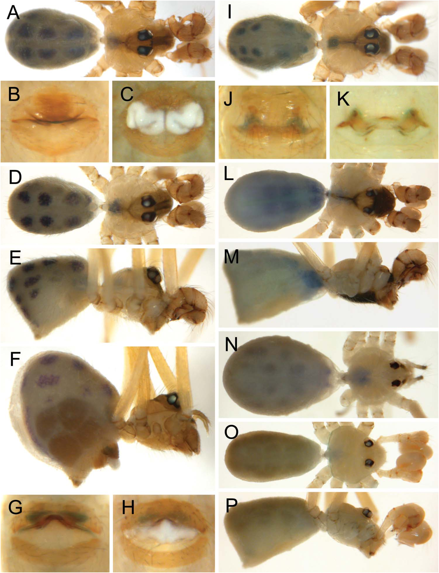
Figure 4. Preserved specimens of Tupigea species showing habitus, epigynum and genital plugs. (A–C) T. angelim, male (A), epigyna without (B) and with (C) genital plug. (D–H) T. penedo sp. nov., male (D, E), female (F), epigyna without (G) and with (H) genital plug. (I, J) T. maza, male (I) and epigynum (J). (K–N) T. teresopolis, epigynum (K), male (L, M), and female (N). (O, P) T. ale sp. nov., male.
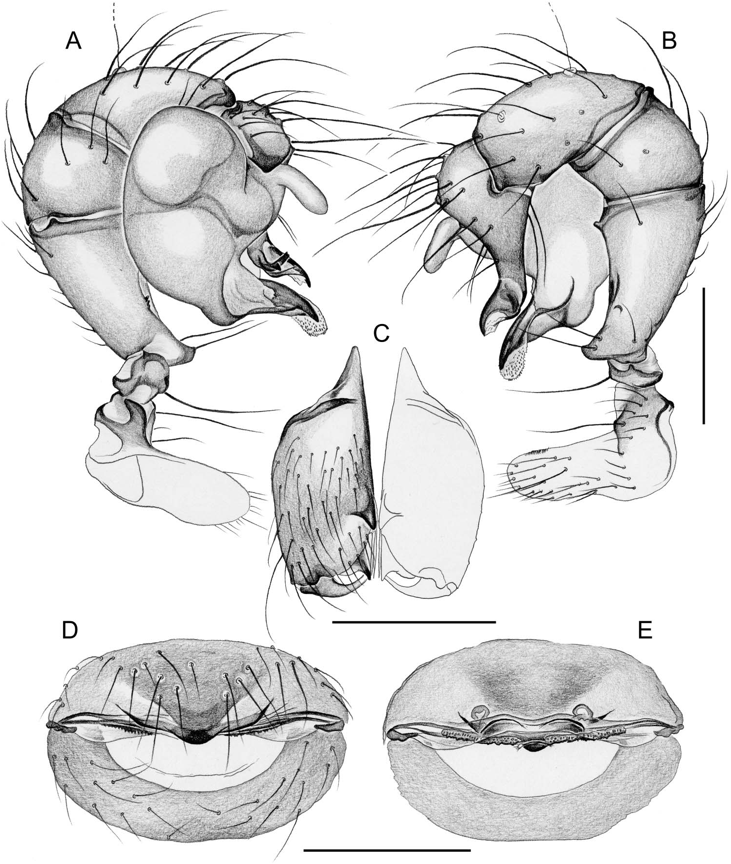
Figure 5. Tupigea angelim sp. nov. Male left palp in prolateral (A) and retrolateral (B) views, male chelicerae, frontal view (C), and cleared female genitalia in ventral (D) and dorsal (E) views. Scale bars: 0.2 mm.
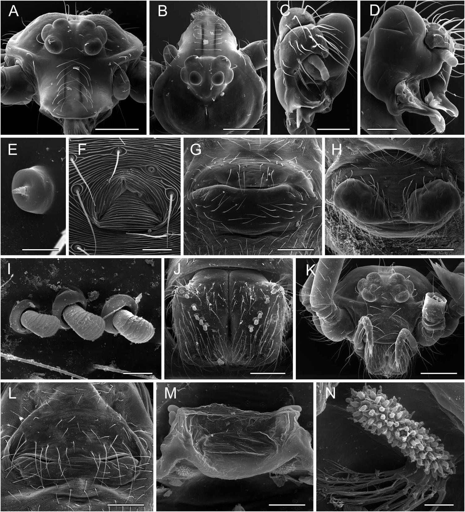
Figure 6. Scanning electron micrographs of Tupigea angelim sp. nov. (A–H) and T. maza (I–N). (A, B) Male prosoma, frontal and dorsal views, showing ridges on clypeus; (C) right palp in dorsal view; (D) left palp in prolatero-dorsal view; (E) male palpal tarsal organ; (F) male gonopore; (G, H) epigynum without (G) and with (H) genital plug. (I) Modified hairs on male chelicerae; (J) male chelicerae, frontal view; (K) female prosoma; (L) epigynum; (M) cleared epigynum, dorsal view; (N) pore plate. Scale bars: 5 µm (E), 10 µm (I, N), 20 µm (F), 50 µm (M), 80 µm (D, J), 100 µm (C, G, H, L), 200 µm (A, B, K).
No known copyright restrictions apply. See Agosti, D., Egloff, W., 2009. Taxonomic information exchange and copyright: the Plazi approach. BMC Research Notes 2009, 2:53 for further explanation.