
Tupigea Huber, 2000
|
publication ID |
https://doi.org/10.1080/00222933.2010.524319 |
|
persistent identifier |
https://treatment.plazi.org/id/03A887D8-FF86-FFB4-B8E5-DBB3FDB2FA1F |
|
treatment provided by |
Carolina (2021-04-06 18:35:39, last updated by Plazi 2023-11-02 05:01:27) |
|
scientific name |
Tupigea Huber, 2000 |
| status |
|
Tupigea Huber, 2000 View in CoL View at ENA
Tupigea Huber 2000: 314 View in CoL ; type species: T. lisei Huber, 2000 View in CoL .
Diagnosis
See Huber (2000). A character not previously mentioned that seems to be diagnostic for Tupigea is the paired receptaculum-like structures in the female internal genitalia. They have previously been illustrated for T. nadleri , T. paula and T. cantareira
(Continued)
Note: Grey-shaded entries are those species that were found at only one locality. Collection codes are provided for unnamed species to facilitate retrieval.
(figs. 1298 and 1307 in Huber 2000 and fig. 39 in Machado et al. 2007), and also occur in T. angelim , T. penedo , T. ale , T. guapia , T. maza and T. teresopolis ( Figures 5E View Figure 5 , 7E View Figure 7 , 8D,E View Figure 8 , 10B,E View Figure 10 ). They were not shown in the figure of the type species T. lisei (fig. 1271 in Huber 2000), but this may be because they are very poorly visible unless the cleared genitalia are stained, and this was not done in Huber 2000.
Description
See Huber (2000). Short vertical hairs on male legs may not only occur on tibiae but also on femora ( T. penedo , T. ale , T. guapia , T. teresopolis ; Figure 9A View Figure 9 ). Prolateral trichobothrium absent on tibia 1, present on other tibiae. Sexual dimorphism usually inconspicuous (except for longer male legs and larger female abdomen; Figure 4 View Figure 4 D–F), but distinct in T. teresopolis ( Figures 3D,E View Figure 3 , 4 View Figure 4 L–N). Females sometimes with conspicuous genital plugs ( Figures 4C,H View Figure 4 , 6H View Figure 6 ). Pore plate morphology extremely variable among species ( Figures 6N View Figure 6 , 9M View Figure 9 , 11M View Figure 11 ). Males and females with comb-hairs distally on tarsus 4 ( Figure 11C View Figure 11 ).
Composition and distribution
The genus now includes 13 species, but two of them continue to be known only from females and to be tentatively assigned ( T. altiventer , T. iguassuensis ; Huber 2000). The genus appears to be largely restricted to the Serra do Mar biogeographical sub-region of the Atlantic Forest ( Figure 2 View Figure 2 ), but at least in the north and in the south it extends into the neighbouring sub-regions.
Natural history
Tupigea includes both leaf-litter species and species that live in close association with the underside of green (alive) leaves ( Figure 3 View Figure 3 A–G). Leaf-litter species are rather dark, short-legged, have a globose abdomen and eye triads close together ( Figure 4A,D,I View Figure 4 ) while leaf-dwelling species are pale greenish (the green colour gets lost in ethanol), have longer legs, and triads are farther apart ( Figures 3 View Figure 3 A–E, 4L–P). The leaf-dwelling T. ale and T. teresopolis were observed to build very fine single-layered webs with a diameter of about 20–40 cm, extending from the underside of a leaf towards the nearby vegetation ( Figure 3F View Figure 3 ). During the day, these spiders are always found hanging from that part of the web that is attached to the underside of the leaf. Males and females of these two and of other species often share one web. When disturbed, they slowly move to another part of the sheet; they could never be induced to vibrate. In contrast, the litter-dwelling T. penedo runs quickly when disturbed; no web could be seen in this (or any other litter-dwelling) species.
Huber BA. 2000. New World pholcid spiders (Araneae: Pholcidae): a revision at generic level. Bull Am Mus Nat Hist. 254: 1 - 348.
Machado EO, Yamamoto FU, Brescovit AD, Huber BA. 2007. Three new ground living pholcid species (Araneae: Pholcidae) from Parque Estadual da Cantareira, Sao Paulo, Sao Paulo, Brazil. Zootaxa 1582: 27 - 37.
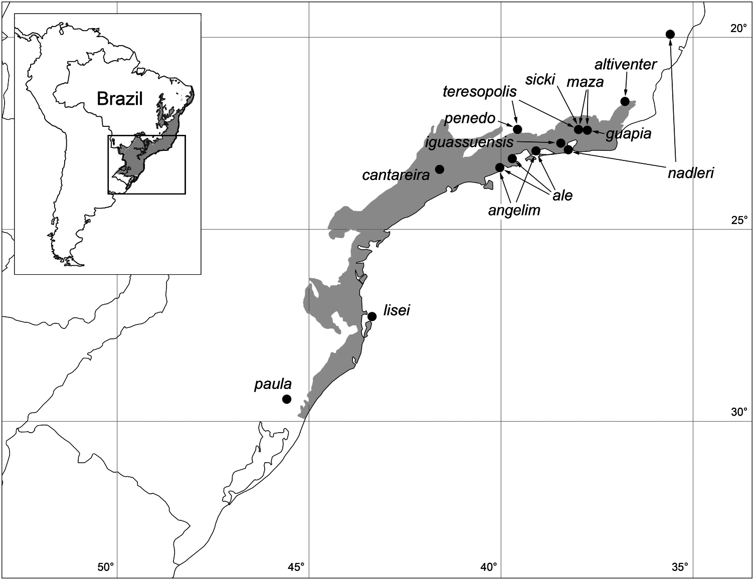
Figure 2. Known distribution of Tupigea. Grey shade in inset: extension of Atlantic Forest in Brazil (inset modified from Ribeiro et al. 2009). Grey shade in main map: extension of Serra do Mar biogeographic sub-region, from Silva and Casteleti (2003) in Ribeiro et al. (2009).
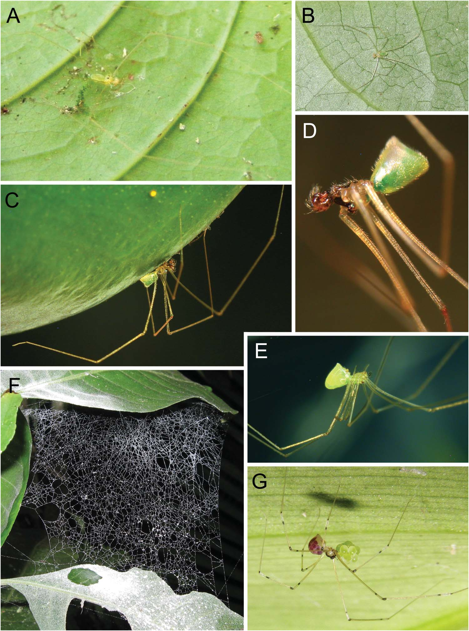
Figure 3. Leaf-dwelling Tupigea species and their web in the natural habitat. (A, B) T. ale from Rio das Pedras (A) and Paraty (B). (C–F) T. teresopolis from Penedo, males (C, D), female (E), and web dusted with starch to improve visibility (F). (G). Unidentified species from Guapiaçú (“Br09-32” in Table 2), female with egg sac.
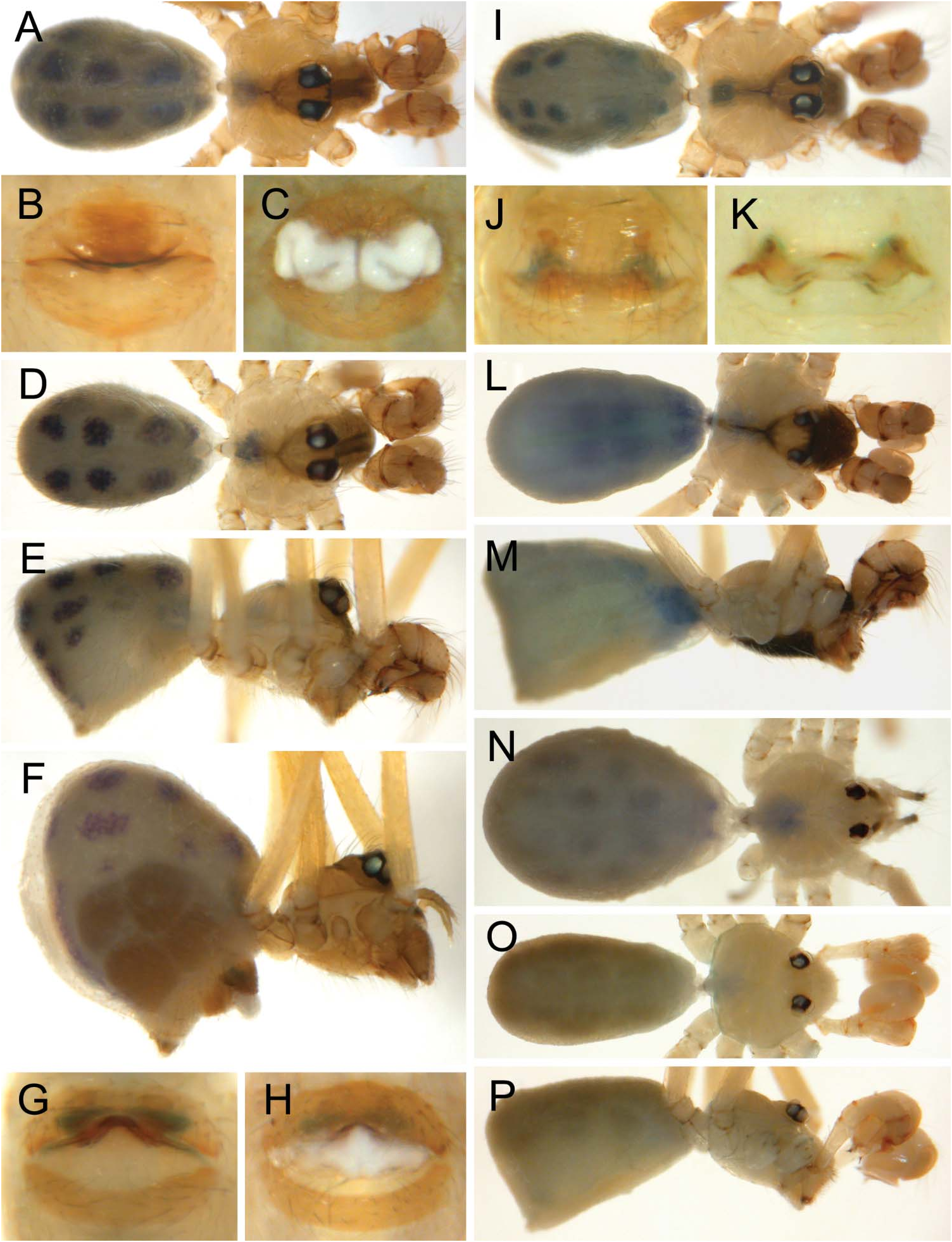
Figure 4. Preserved specimens of Tupigea species showing habitus, epigynum and genital plugs. (A–C) T. angelim, male (A), epigyna without (B) and with (C) genital plug. (D–H) T. penedo sp. nov., male (D, E), female (F), epigyna without (G) and with (H) genital plug. (I, J) T. maza, male (I) and epigynum (J). (K–N) T. teresopolis, epigynum (K), male (L, M), and female (N). (O, P) T. ale sp. nov., male.
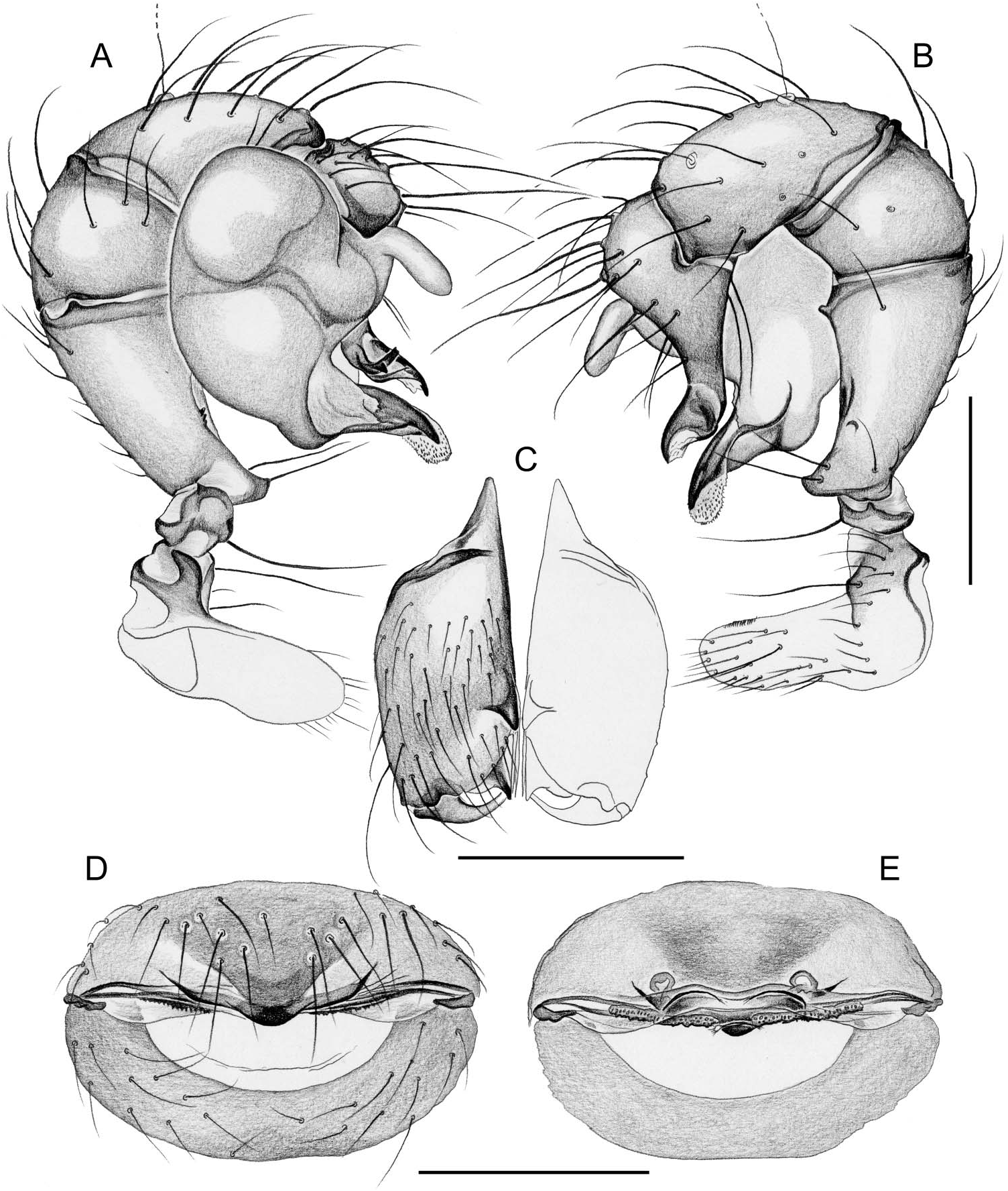
Figure 5. Tupigea angelim sp. nov. Male left palp in prolateral (A) and retrolateral (B) views, male chelicerae, frontal view (C), and cleared female genitalia in ventral (D) and dorsal (E) views. Scale bars: 0.2 mm.
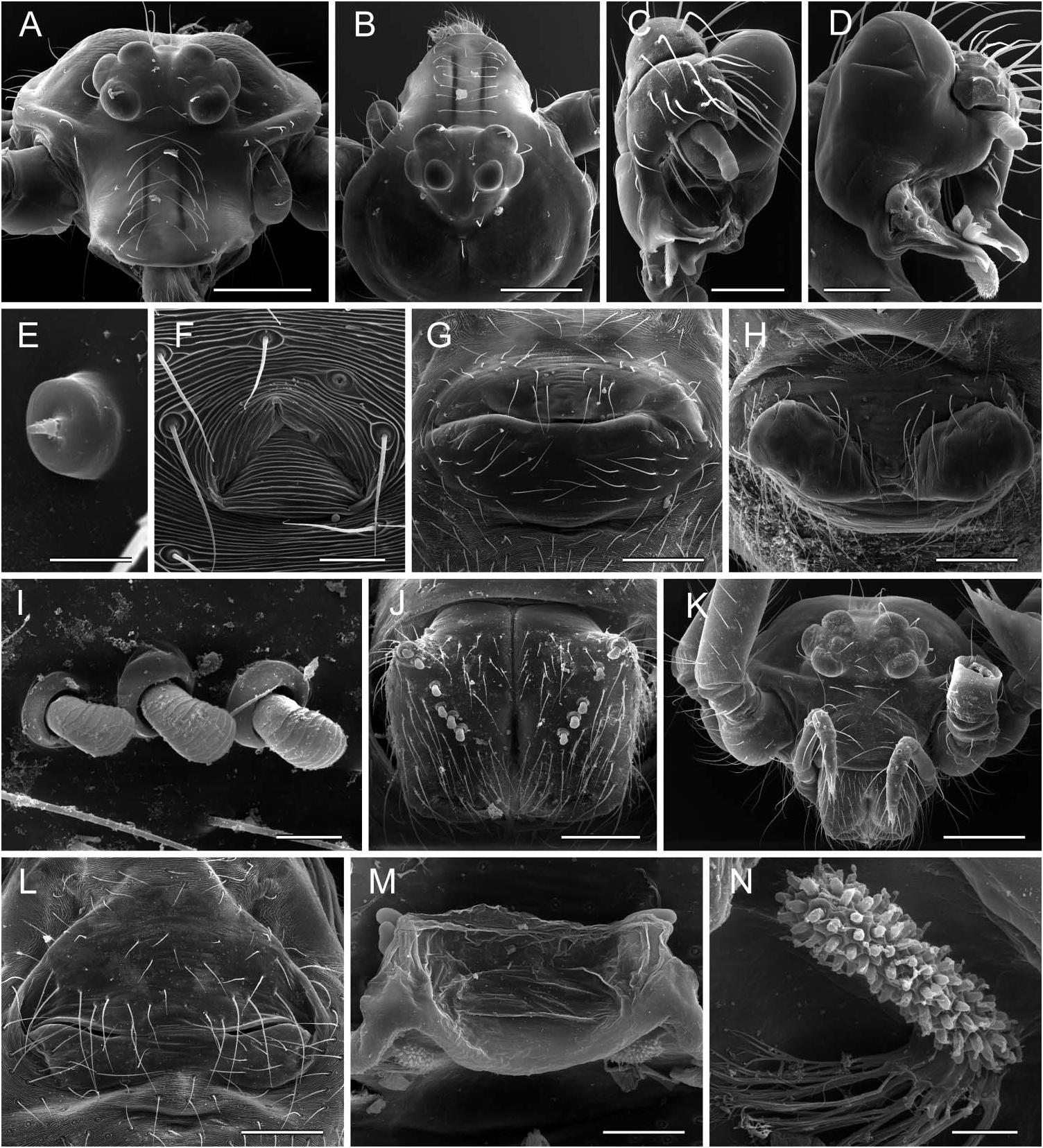
Figure 6. Scanning electron micrographs of Tupigea angelim sp. nov. (A–H) and T. maza (I–N). (A, B) Male prosoma, frontal and dorsal views, showing ridges on clypeus; (C) right palp in dorsal view; (D) left palp in prolatero-dorsal view; (E) male palpal tarsal organ; (F) male gonopore; (G, H) epigynum without (G) and with (H) genital plug. (I) Modified hairs on male chelicerae; (J) male chelicerae, frontal view; (K) female prosoma; (L) epigynum; (M) cleared epigynum, dorsal view; (N) pore plate. Scale bars: 5 µm (E), 10 µm (I, N), 20 µm (F), 50 µm (M), 80 µm (D, J), 100 µm (C, G, H, L), 200 µm (A, B, K).
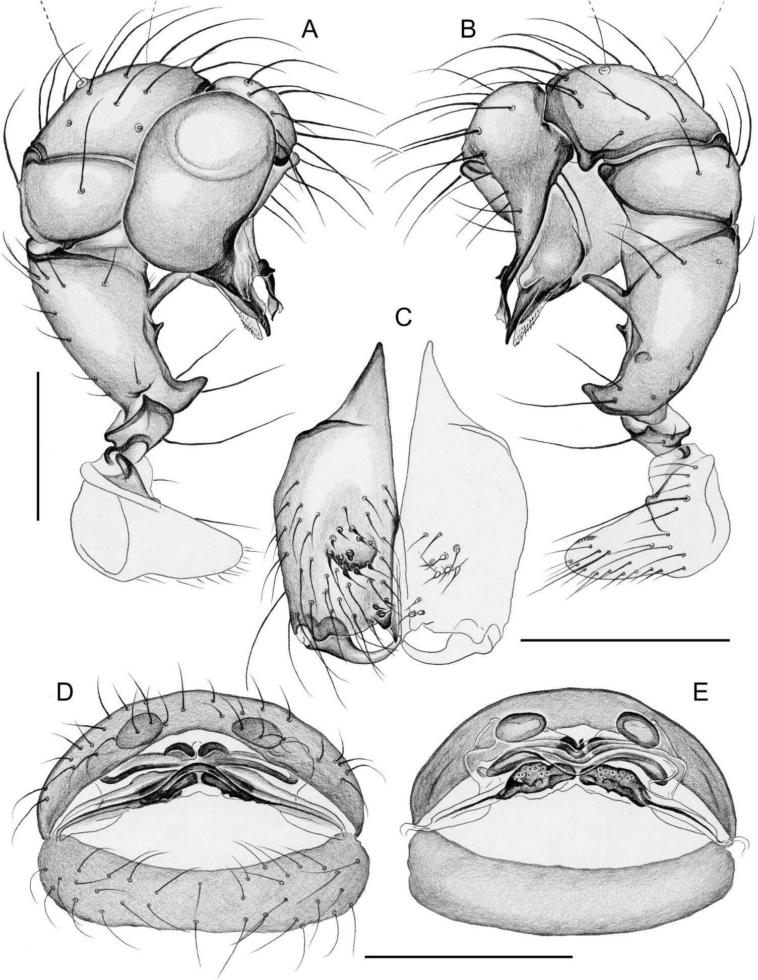
Figure 7. Tupigea penedo sp. nov. Male left palp in prolateral (A) and retrolateral (B) views, male chelicerae, frontal view (C), and cleared female genitalia in ventral (D) and dorsal (E) views. Scale bars: 0.2 mm.

Figure 8. Tupigea ale sp. nov. (A–D) and T. guapia sp. nov (E). Male left palp in prolateral (A) and retrolateral (B) views, male chelicerae, frontal view (C), and cleared female genitalia in dorsal views (D, E). Scale bars: 0.2 mm.

Figure 9. Scanning electron micrographs of Tupigea ale sp. nov. (A–E) and T. guapia sp. nov. (F–M). (A) Male femur 4, mid-section; (B) modified hair-bases on male chelicerae; (C) epigynum, female from Ubatuba; (D–F) male chelicerae, males from Rio das Pedras (D), Ubatuba (E) and Guapiaçú (F); (G, H) male prosoma in lateral and frontal views; (I) male spinnerets; (J) male gonopore; (K) epigynum; (L) cleared female genitalia, dorsal view; (M) detail of poreplate. Scale bars: 6 µm (M), 10 µm (B, I), 20 µm (J), 50 µm (A), 80 µm (C–F, K, L), 200 µm (G, H).
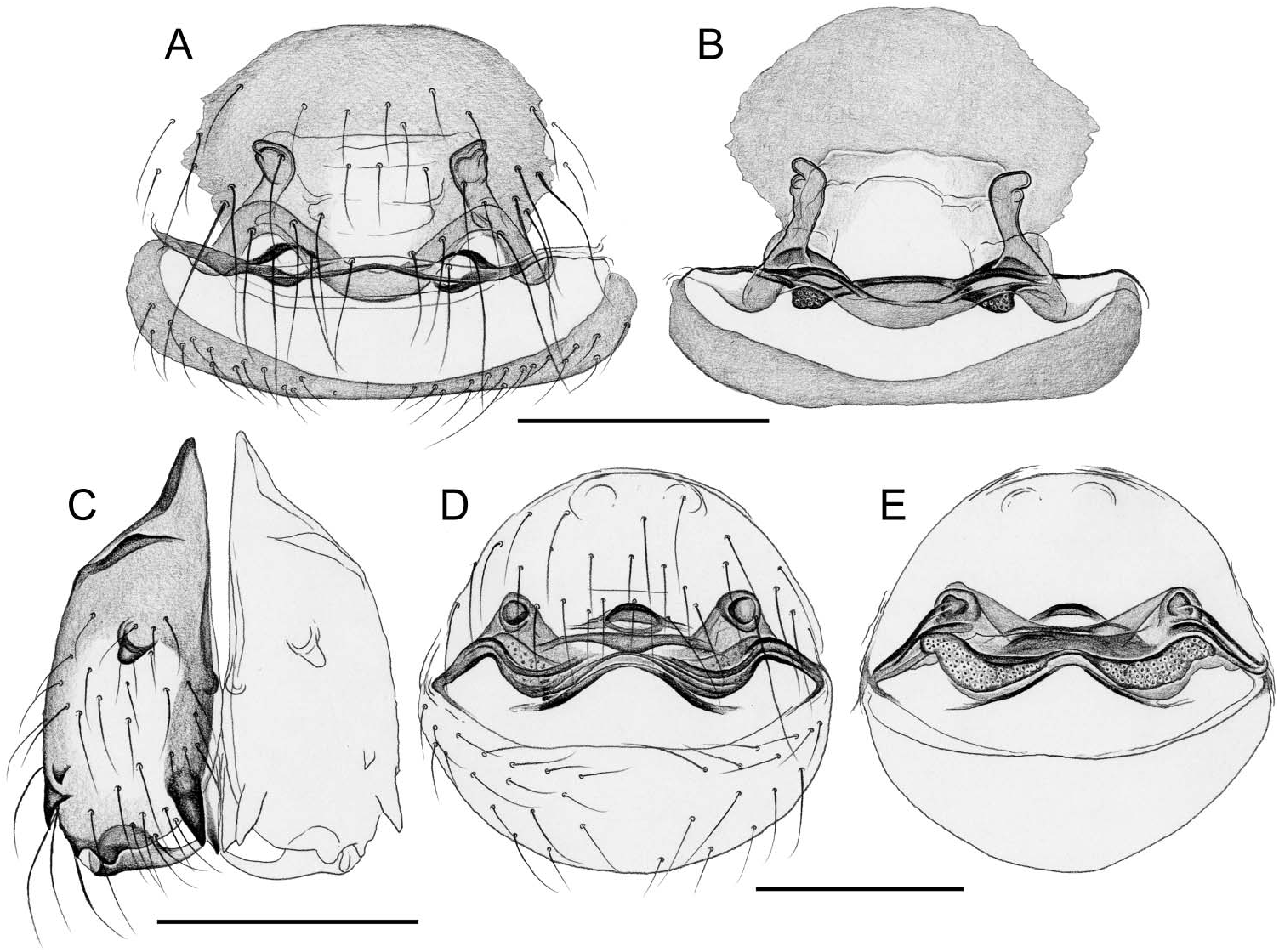
Figure 10. Tupigea maza (A, B) and T. teresopolis (C–E). Cleared female genitalia in ventral (A, D) and dorsal (B, E) views, and male chelicerae of male from Penedo (C). Scale bars: 0.2 mm.
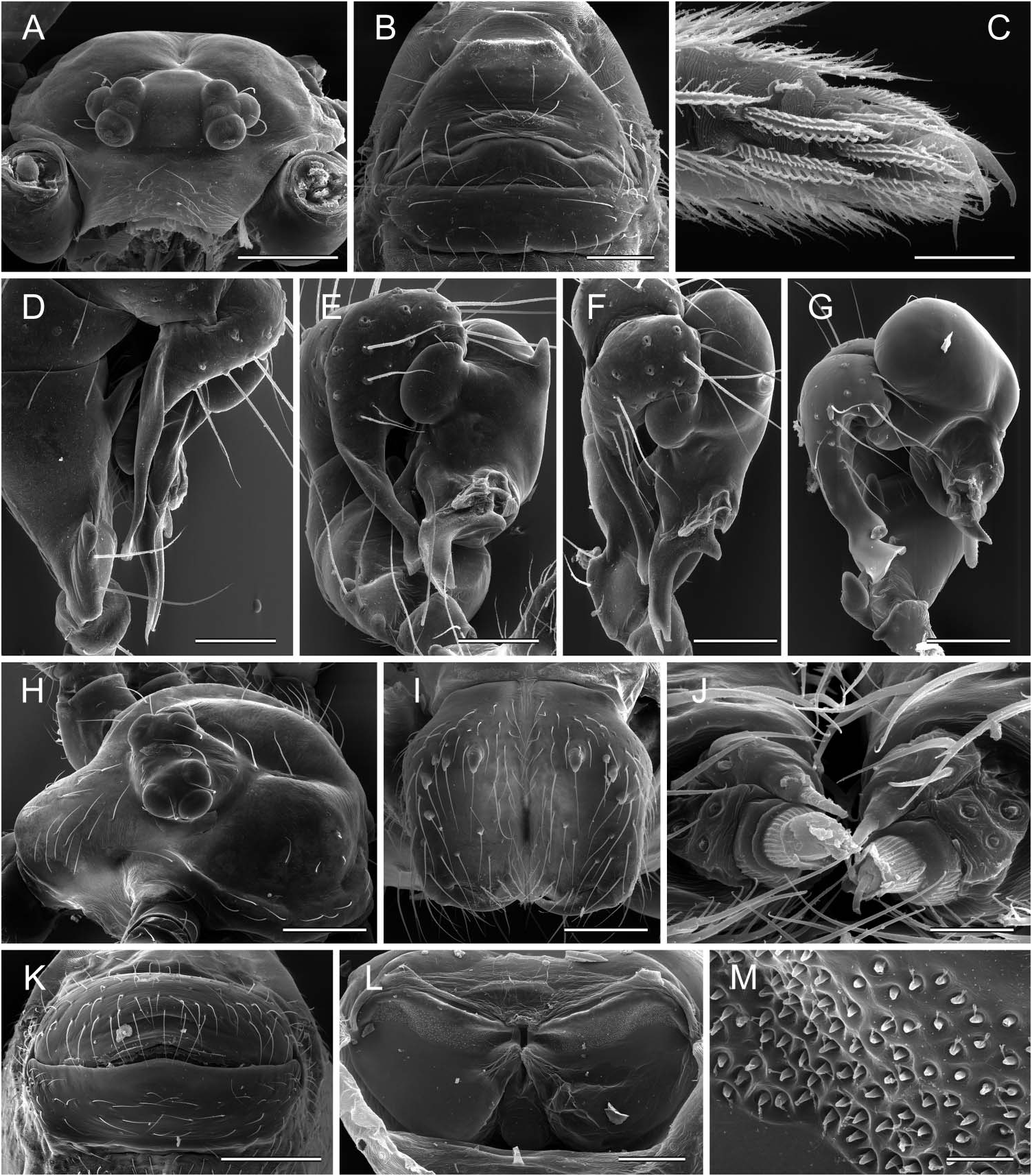
Figure 11. Scanning electron micrographs of Tupigea teresopolis (A–F), T. penedo sp. nov. (G), and T. cantareira (H–M). (A) Male prosoma, frontal view; (B) epigynum; (C) female tarsus 4 tip with comb-hairs; (D–G) male right palps in retrolateral (D), retrolatero-dorsal (E) and dorsal (F, G) views; (H) male prosoma, dorsolateral view; (I) male chelicerae; (J) female anterior lateral spinnerets; (K) epigynum; (L) cleared female genitalia, dorsal view; (M) detail of pore plate. Scale bars: 10 µm (J, M), 20 µm (C), 80 µm (D, E), 100 µm (B, F, G, I, L), 200 µm (A, H, K).
No known copyright restrictions apply. See Agosti, D., Egloff, W., 2009. Taxonomic information exchange and copyright: the Plazi approach. BMC Research Notes 2009, 2:53 for further explanation.
|
Kingdom |
|
|
Phylum |
|
|
Class |
|
|
Order |
|
|
Family |
Tupigea Huber, 2000
| Huber, Bernhard A. & Rheims, Cristina A. 2011 |
Tupigea
| Huber BA 2000: 314 |