
Macroplea Samouelle, 1819
|
publication ID |
https://doi.org/10.5281/zenodo.278456 |
|
DOI |
https://doi.org/10.5281/zenodo.6184801 |
|
persistent identifier |
https://treatment.plazi.org/id/03A28793-FFB3-FFE8-FF4E-519BAC16048E |
|
treatment provided by |
Plazi (2016-04-11 02:47:03, last updated 2024-11-29 13:31:37) |
|
scientific name |
Macroplea Samouelle, 1819 |
| status |
|
Genus Macroplea Samouelle, 1819 View in CoL
Macroplea Samouelle, 1819: 211 View in CoL (Type species Donacia zosterae Fabricius, 1801 ). Apelma Billberg, 1820: 57 (Type species Donacia zosterae Fabricius, 1801 ).
Haemonia Dejean, 1821: 114 (Type species Donacia zosterae Fabricius, 1801 ).
Diagnosis. Dorsum and legs yellow or brown ( Figs 1, 3 View FIGURES 1 – 4 , 24, 26 View FIGURES 24 – 27 , 50–53 View FIGURES 50 – 53 ); vertex with dense pubescence ( Figs 6 View FIGURES 5 – 20 , 29 View FIGURES 28 – 43 , 50–53 View FIGURES 50 – 53 ); antenna long, extending beyond middle of elytron ( Figs 1–4 View FIGURES 1 – 4 , 50, 52, 53 View FIGURES 50 – 53 ), or short, not extending beyond middle of elytron ( Figs 24–27 View FIGURES 24 – 27 ); pronotum with setae at anterior angles ( Figs 5 View FIGURES 5 – 20 , 28 View FIGURES 28 – 43 , 56–59 View FIGURES 54 – 59 ); pronotum with distinct anterior and posterior beads ( Figs 5 View FIGURES 5 – 20 , 28 View FIGURES 28 – 43 , 56–59 View FIGURES 54 – 59 ); punctures along elytral striae arranged more or less in paired rows ( Figs 1, 3 View FIGURES 1 – 4 , 50–53 View FIGURES 50 – 53 ), or in single rows ( Figs 24, 26 View FIGURES 24 – 27 ); elytron with or without spine at outer apical angle ( Figs 12 View FIGURES 5 – 20 , 35 View FIGURES 28 – 43 , 64–67 View FIGURES 60 – 67 ); metafemur slender, without tooth ( Figs 1–4 View FIGURES 1 – 4 , 24–27 View FIGURES 24 – 27 , 50–53 View FIGURES 50 – 53 ); profemur with a short linearly arranged brush of setae basally on posterior surface ( Figs 16 View FIGURES 5 – 20 , 36 View FIGURES 28 – 43 ); metatarsus with markedly reduced pubescence, fifth tarsomere elongate, at least as long as basal three combined ( Figs 10 View FIGURES 5 – 20 , 39 View FIGURES 28 – 43 , 68, 70, 71 View FIGURES 68 – 71 ); endophallus with two ELDs fused, enclosing MEG, without pELD ( Figs 21, 22 View FIGURES 21 – 23 , 47–49 View FIGURES 44 – 49 , 74 View FIGURES 72 – 75 , 75, 77 View FIGURES 76 – 78 , 78, 81–83 View FIGURES 79 – 84 ).
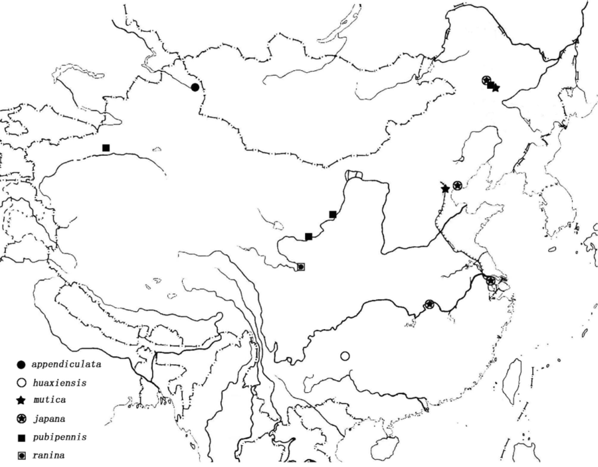
Distribution. Europe, northern Africa ( Algeria), Middle Asia, Siberia, Far East, China and Japan. Distribution in China: Heilongjiang, Hebei, Tianjin, Ningxia, Gansu, Xinjiang, Jiangsu, Hubei, Sichuan, Guizhou ( Fig. 85 View FIGURE 85 ).
Host plants. Ranunculus L. ( Ranunculaceae ), Carex L. ( Cyperaceae ), Brasenia Schreb. (Cabombaceae) , Potamogeton L. ( Potamogetonaceae ), Myriophyllum L. ( Haloragaceae ), Ruppia L. ( Zosteraceae ), Zostera L. ( Zosteraceae ), Sparganium L. ( Sparganiaceae ) ( Bieńkowski & Orlova-Bieńkowskaja, 2004), Vallisneria L. ( Hydrocharitaceae ), Ottelia Pers. (Hydrocharitaceae) and Hippuris L. ( Hippuridaceae ) (recorded in this paper).
Biology. Macroplea is a fully aquatic genus. They live in lentic or lotic water. Both M. appendiculata and M. mutica live in brackish as well as fresh water ( Borowiec, 1984; Kölsch et al., 2006; Saari, 2007; Mende et al., 2010). According to Mende et al. (2010: 101), immobility (cannot swim or fly) is believed to be a specific feature of M. mutica . However, three specimens of M. japana examined in this study were collected by light trap.
Remarks. Identification of species of Macroplea is difficult, despite the genus being less speciose. Many workers have used external morphology, male genitalia, ecological features, and molecular data to delimit species of this genus ( Freude et al., 1966; Daccordi & Ruffo, 1978; Mohr, 1985; Beenen & Winkelman, 1989; Hayashi & Shiyake, 2001; Kölsch et al., 2006). We have followed many of these workers to enhance the reliability of the key. Species of Macroplea can be separated from those of Neohaemonia Székessy in new world by the following characters: 1) vertex with dense pubescence (with a glabrous patch in Neohaemonia ); 2) pronotum with setae at anterior angles (such setae absent in Neohaemonia ); 3) pronotum with distinct anterior and posterior beads (such beads absent in Neohaemonia ); 4) profemur with a short linearly arranged brush of setae basally on posterior surface (such brush of setae absent in Neohaemonia ); 5) endophallus without pELD (pELD present in Neohaemonia ).
Beenen, R. & Winkelman, J. K. (1989) Aantekeningen over Chrysomelidae in Nederland (Coleoptera). Entomologische Berichten, Amsterdam, 49, 5, 69 - 71 (in Dutch).
Bienkowski, A. O. & Orlova-Bienkowskaja, M. J. (2004) Morphology, systematics and host plants of Palaearctic Donaciinae larvae. In: Jolivet, P., Santiago-Blay, J. A. & Schmitt, M. (Eds.), New Developments in the Biology of Chrysomelidae. SBP Academic Publishing, Netherlands, pp. 481 - 502.
Billberg, G. J. (1820) Enumeratio Insectorum in Museo Gust. Joh. Billberg. Typis Gadelianis, Stockholm, 138 pp. (in Latin)
Borowiec, F. (1984) Zoogeographical study on Donaciinae of the world (Coleoptera, Chrysomelidae). Polskie Pismo Entomologiczne, 53, 433 - 518.
Daccordi, M. & Ruffo, S. (1978) Sulla presenza del genere Macroplea Samouelle in Italia. Bollettino dell'Associazione Romana di Entomologia, 33, 56 - 65 (in Italian).
Dejean, P. F. M. A. (1821) Catalogue des colleopteres de la collection de M. Le Baron Dejean. Crevot, Paris, 136 pp. (in French).
Fabricius, J. C. (1801) Systema eleutheratorum secundum ordines, genera, species, adiectis synonymis, locis, observationibus, descriptionibus. Tomus II. Bibliopoli Academici Novi, Kiliae, 687 pp. (in Latin).
Freude, H., Harde, K. W. & Lohse, G. A. (1966) Unterfamilie: Donaciinae. In: Die Kafer Mitteleuropas, Vol 9. Phytophaga (Cerambycidae, Chrysomelidae). Goecke & Evers, Krefeld, pp. 100 - 109 (in German).
Hayashi, M. & Shiyake, S. (2001) The identity and distribution of Macroplea japana (Jacoby) (Coleoptera, Chrysomelidae, Donaciinae). Bulletin of the Osaka Museum of Natural History, 55, 15 - 22.
Kolsch, G., Bistrom, O. & Pedersen, B. V. (2006) Species delimitation in the leaf beetle genus Macroplea (Coleoptera, Chrysomelidae) based on mitochondrial DNA, and phylogeographic considerations. Insect Systematics & Evolutio n, 37, 467 - 479.
Mende, M., Bistrom, O., Meichssner, E. & Kolsch, G. (2010) The aquatic leaf beetle Macroplea mutica (Coleoptera: Chrysomelidae) in Europe: Population structure, postglacial colonization and the signature of passive dispersal. European Journal of Entomology, 107, 101 - 113.
Mohr, K. H. (1985) Beitrage zur Insektenfauna der DDR: Coleoptera-Chrysomelidae: Donaciinae, Orsodacninae, Criocerinae, Clythrinae. Beitrage zur Entomologie, 35, 219 - 262 (in German).
Saari, S. (2007) Meriuposkuoriaisen, Macroplea pubipennis (Coleoptera: Chrysomelidae), levinneisyys ja elinympaistovaatimukset Espoonlahdessa. University of Helsinki, Finland, 51 pp. (in Finnish).
Samouelle, G. (1819) The entomologist's useful compendium; or an introduction to the knowledge of British Insects. Thomas Boys, London, 496 pp.
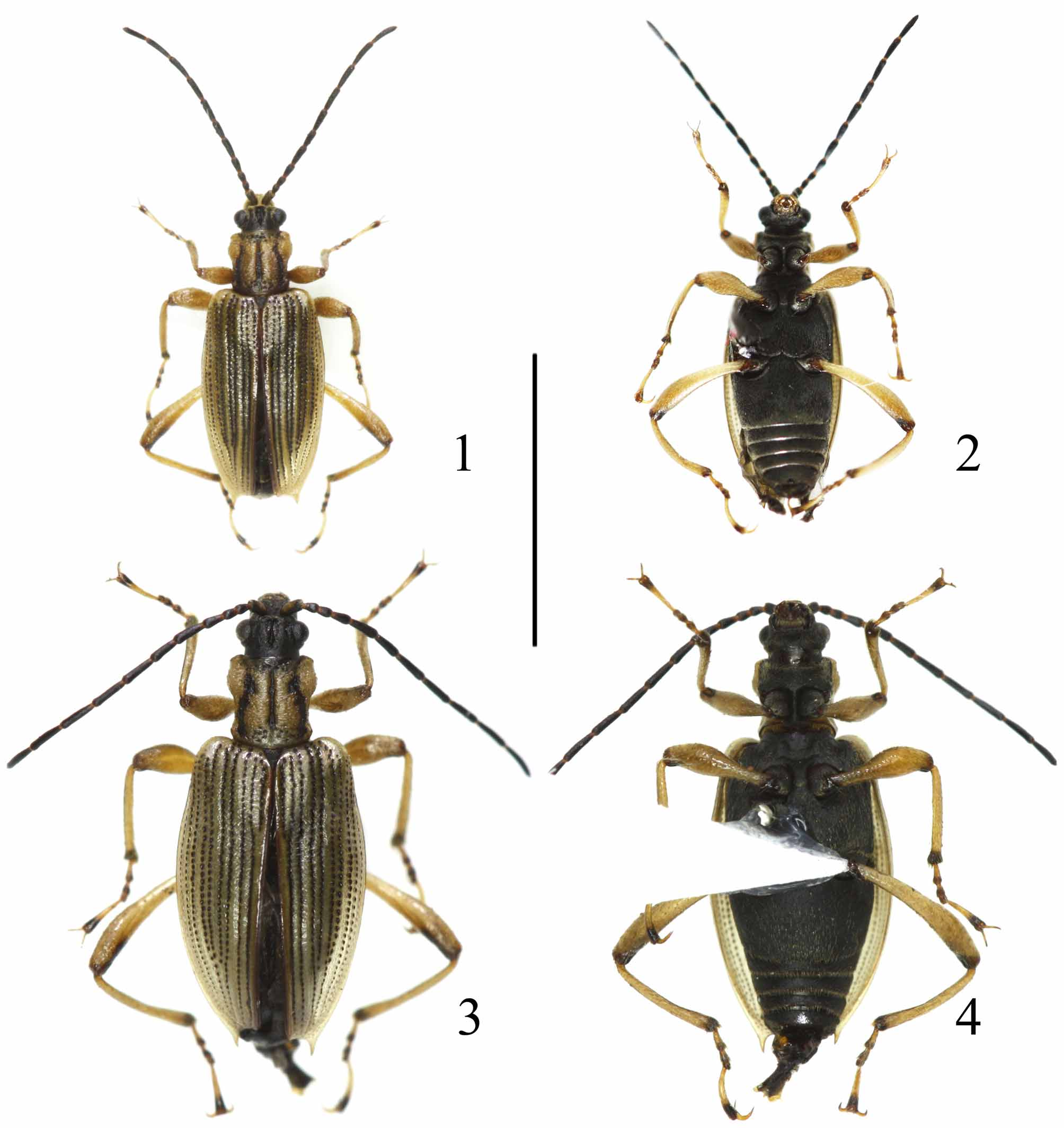
FIGURES 1 – 4. Macroplea huaxiensis sp. nov. 1. Holotype, male, dorsal view; 2. Paratype, male, ventral view; 3. Paratype, female, dorsal view; 4. Paratype, female, ventral view. Scale line = 5.0 mm.

FIGURES 24 – 27. Macroplea ranina sp. nov. 24. Paratype, male, dorsal view; 25. Paratype, male, ventral view; 26. Paratype, female, dorsal view; 27. Paratype, female, ventral view. Scale line = 5.0 mm.

FIGURES 50 – 53. Macraplea spp. 50. M. appendiculata from Xinjiang, male; 51. M. pubipennis from Ningxia, female; 52. M. japana from Jiangsu, female; 53. M. mutica from Tianjin, female. Scale lines = 5.0 mm.
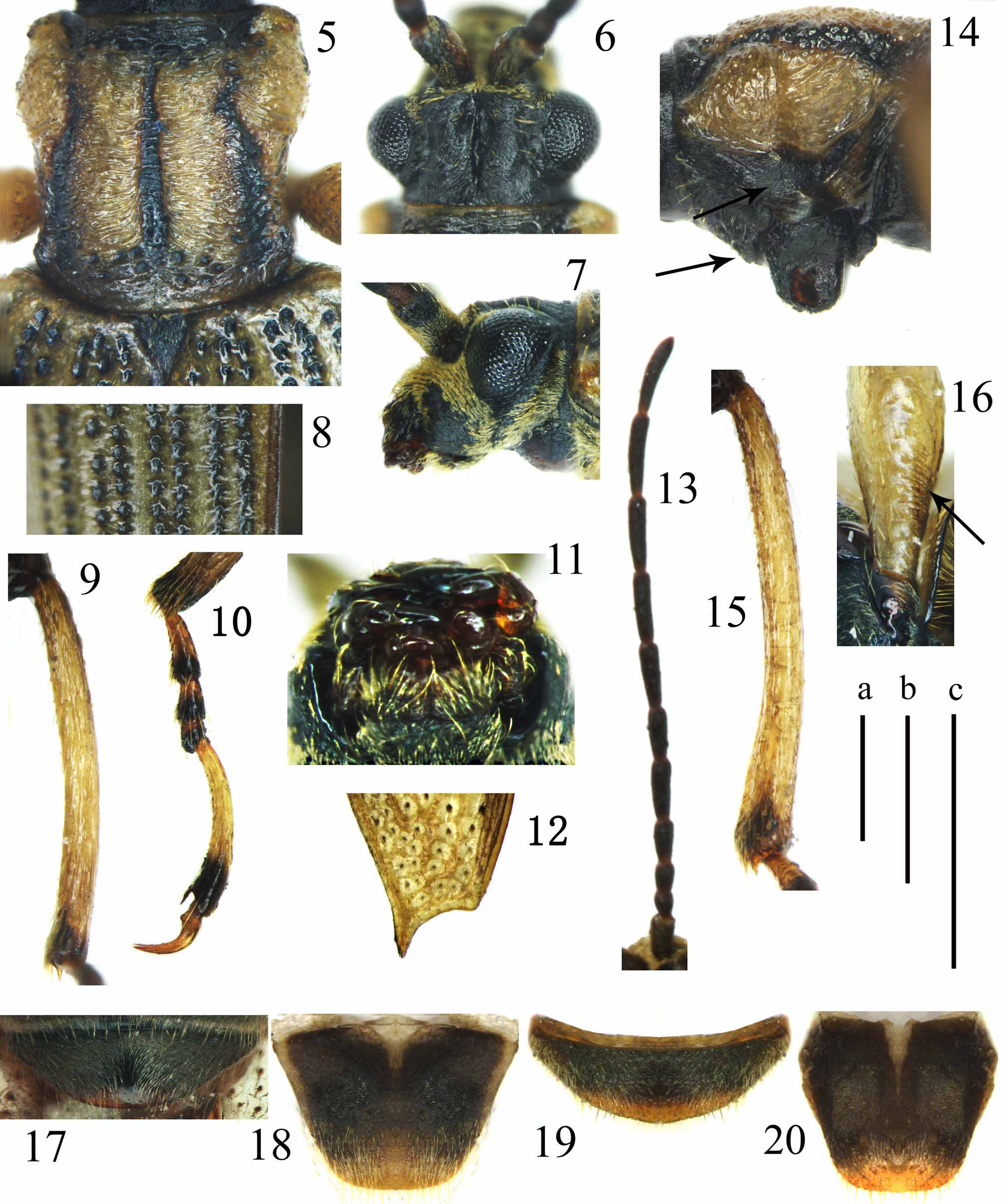
FIGURES 5 – 20. Macroplea huaxiensis sp. nov. 5 – 13, 17, 18. Male; 14 – 16, 19, 20. Female. 5. Pronotum, dorsal view; 6. Head, dorsal view; 7. Head, lateral view; 8. Left elytron, middle part; 9. Right metatibia, lateral view; 10. Right metatarsus, lateral view; 11. Mouth parts, ventral view; 12. Apex of left elytron; 13. Antenna; 14. Pronotum, left lateral view; 15. Right metatibia, lateral view; 16. Left profemur, ventral view; 17, 19. Apical abdominal sternum; 18, 20. Pygidium. Scale lines: a = 0.5 mm. (Figs 5 – 10, 12, 14 – 20); b = 1.0 mm. (Fig. 13); c = 0.5 mm. (Fig. 11).

FIGURES 28 – 43. Macroplea ranina sp. nov. 28 – 35, 38 – 41. Male; 36, 37, 42, 43. Female. 28. Pronotum, dorsal view; 29. Head, dorsal view; 30. Mouth parts, ventral view; 31. Left elytron, middle part; 32. Head and prothorax, left lateral view; 33. Antenna; 34. Part of abdominal sternum, showing long setae; 35. Apex of left elytron; 36. Left profemur, ventral view; 37, 38. Right metatibia, lateral view; 39. Right metatarsus, lateral view; 40, 42. Apical abdominal sternum; 41, 43. Pygidium. Scale lines: d = 0.5 mm. (Figs 28, 29, 31 – 33, 35 – 43); e = 0.5 mm. (Figs 30, 34).

FIGURES 54 – 59. Head, prothorax and pronotum of Macroplea spp. 54 – 56. M. japana from Jiangsu; 57. M. pubipennis from Ningxia; 58. M. appendiculata from Xinjiang; 59. M. mutica from Tianjin; 54. Head and prothorax, lateral view; 55. Vertex, dorsal view; 56 – 59. Pronotum, dorsal view. Scale lines = 0.5 mm.

FIGURES 60 – 67. Elytron of Macroplea spp. 60, 64. M. japana from Jiangsu; 61, 65. M. pubipennis from Ningxia; 62, 66. M. appendiculata from Xinjiang; 63, 67. M. mutica from Tianjin. 60 – 63. Left elytron, middle part; 64, 66, 67. Apex of left elytron; 65. Apexes of both elytra. Scale lines = 0.2 mm.
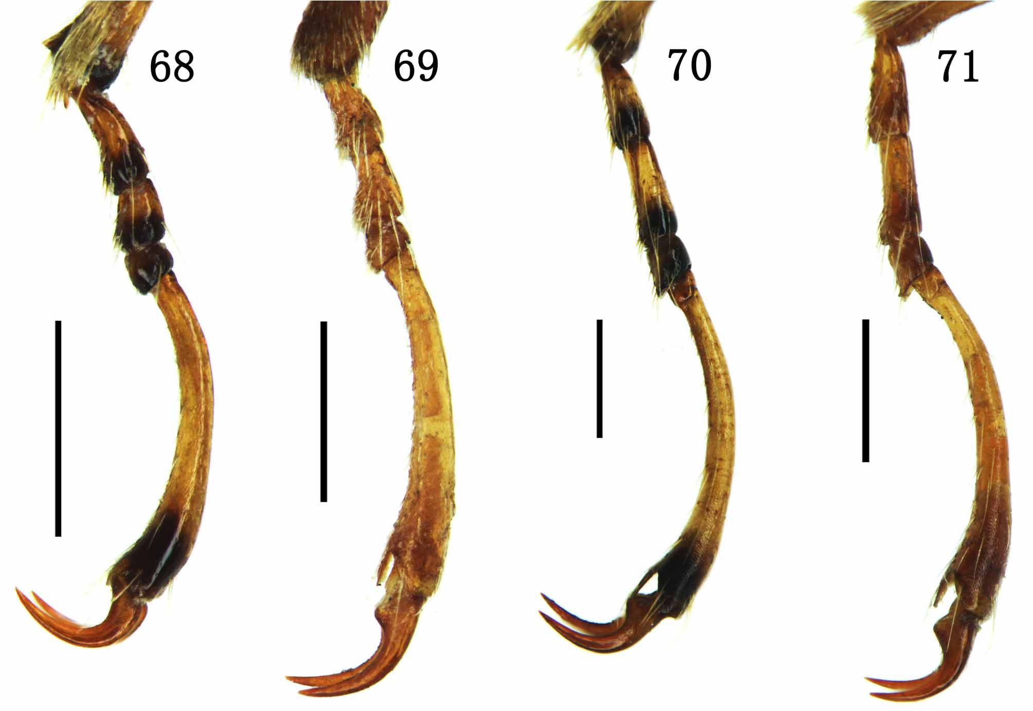
FIGURES 68 – 71. Tarsus of Macroplea spp. 68. M. japana from Jiangsu; 69. M. pubipennis from Ningxia; 70. M. appendiculata from Xinjiang; 71. M. mutica from Tianjin. 68, 70, 71. Right metatarsus; 69. Left protarsus. Scale lines = 0.5 mm
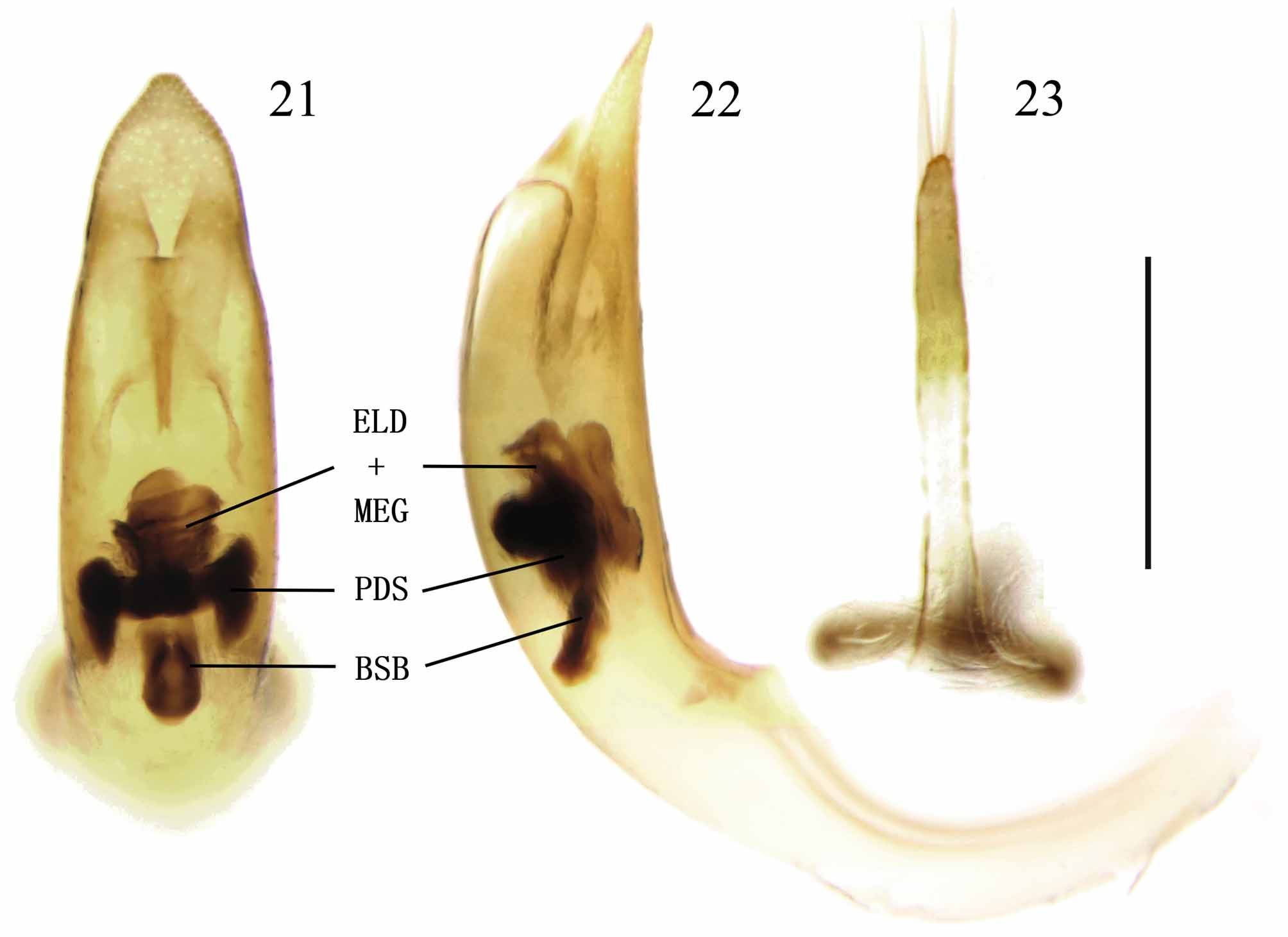
FIGURES 21 – 23. Macroplea huaxiensis sp. nov. Paratype. 21 – 22. Medial lobe with endophallus in it (21. Dorsal view, 22. Lateral view); 23. Tegmen, dorsal view. Scale line = 0.5 mm.
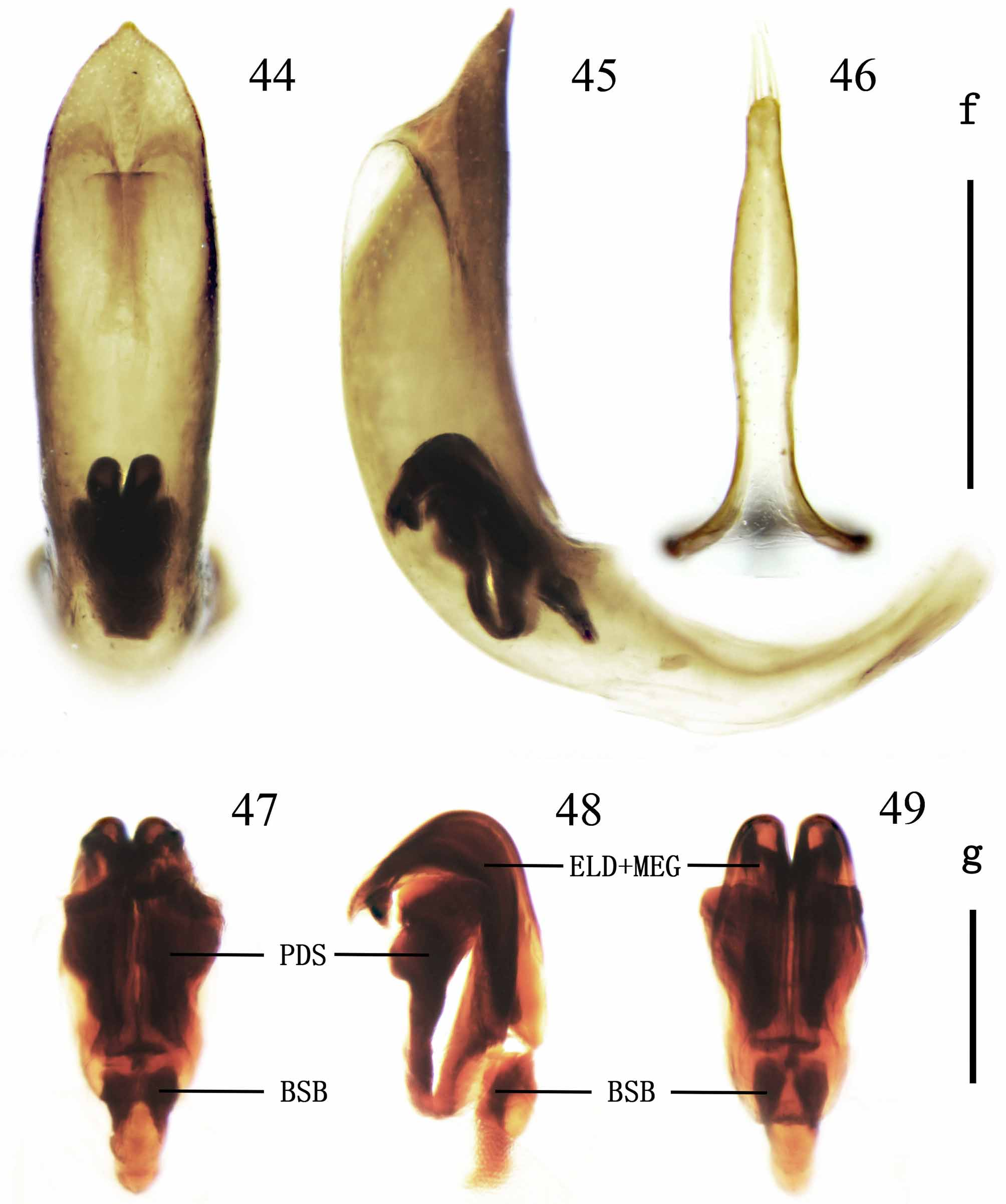
FIGURES 44 – 49. Macroplea ranina sp. nov. 44, 45. Median lobe with endophallus in it (44. Dorsal view; 45. Lateral view.); 46. Tegmen, dorsal view; 47 – 49. Endophallus (47. Dorsal view; 48. Lateral view; 49. Ventral view). Scale lines: f = 0.5 mm. (Figs 44 – 46); g = 0.2 mm. (Figs 47 – 49).
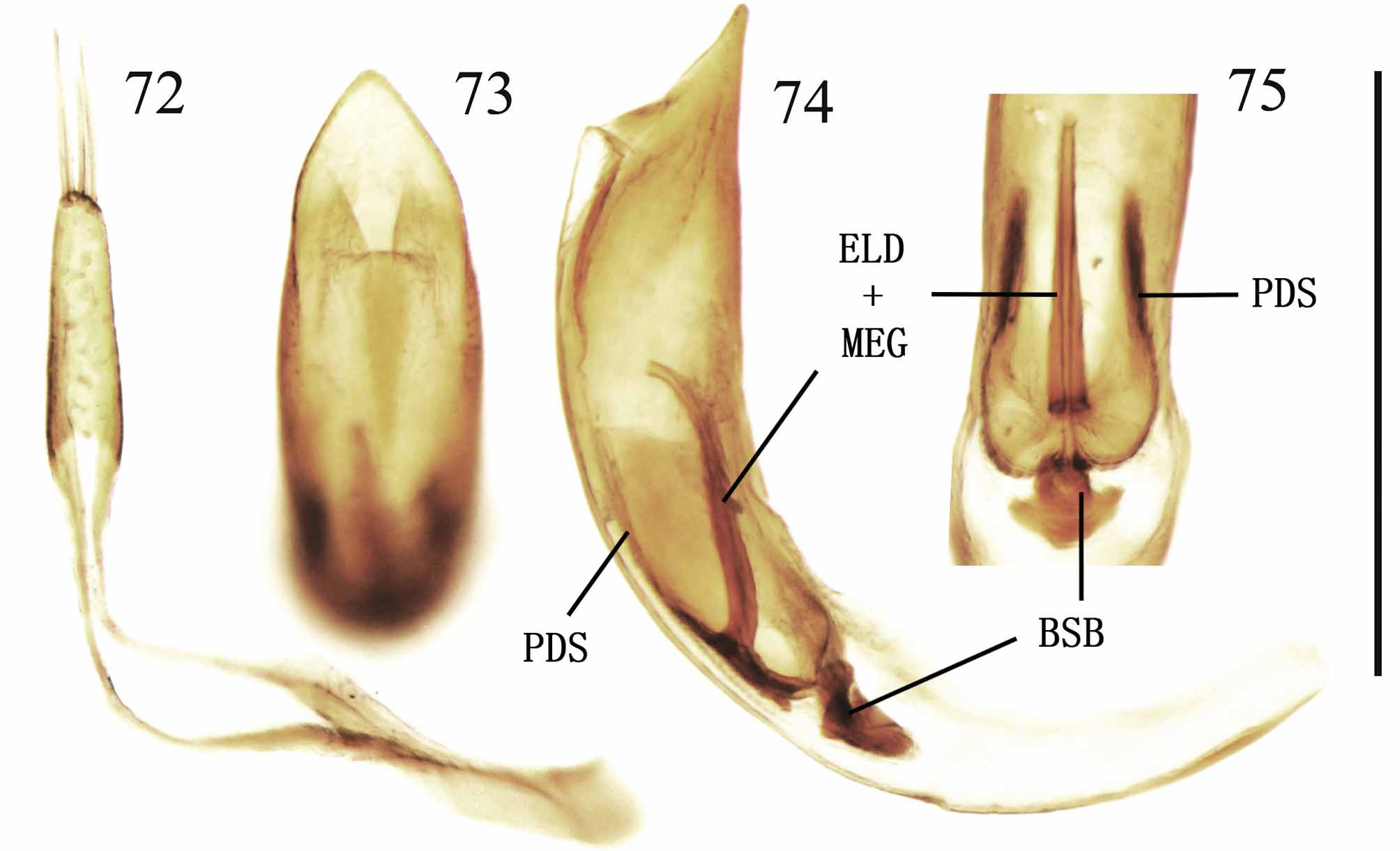
FIGURES 72 – 75. Macroplea japana from Jiangsu. 72. Tegmen, dorsal view of cap; 73, 74. Median lobe with endophallus in it (73. Dorsal view; 74. Lateral view); 75. Endophallus in median lobe, dorsal view. Scale line = 0.5 mm.
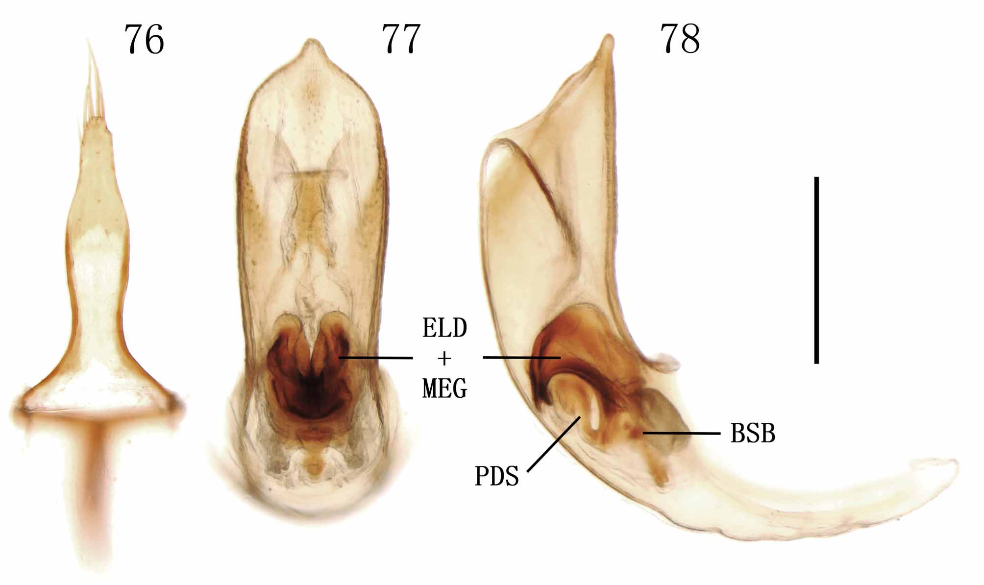
FIGURES 76 – 78. Macroplea appendiculata from Xinjiang. 76. Tegmen, dorsal view; 77, 78. Median lobe with endophallus in it (77. Dorsal view; 78. Lateral view). Scale line = 0.5 mm.

No known copyright restrictions apply. See Agosti, D., Egloff, W., 2009. Taxonomic information exchange and copyright: the Plazi approach. BMC Research Notes 2009, 2:53 for further explanation.
|
Kingdom |
|
|
Phylum |
|
|
Class |
|
|
Order |
|
|
Family |
|
|
SubFamily |
Donaciinae |
Macroplea Samouelle, 1819
| Lou, Qiaozhe, Yu, Peiyu & Liang, Hongbin 2011 |
Haemonia
| Dejean 1821: 114 |
Macroplea
| Billberg 1820: 57 |
| Samouelle 1819: 211 |
1 (by plazi, 2016-04-11 02:47:03)
2 (by ImsDioSync, 2017-02-08 23:14:23)
3 (by ImsDioSync, 2017-06-16 22:20:17)
4 (by ImsDioSync, 2017-06-16 23:35:20)
5 (by ExternalLinkService, 2019-09-26 20:35:17)
6 (by ExternalLinkService, 2022-01-30 13:57:15)
7 (by ExternalLinkService, 2022-02-20 12:26:52)
8 (by plazi, 2023-10-25 19:17:20)