
Macroplea
|
publication ID |
https://doi.org/10.5281/zenodo.278456 |
|
DOI |
https://doi.org/10.5281/zenodo.6184803 |
|
persistent identifier |
https://treatment.plazi.org/id/03A28793-FFB0-FFEB-FF4E-53F7ACEB0726 |
|
treatment provided by |
Plazi (2016-04-11 02:47:03, last updated 2024-11-29 13:31:37) |
|
scientific name |
Macroplea |
| status |
|
Key to species of Macroplea View in CoL
1 Punctures along elytral striae coarse, not arranged in paired rows ( Figs 24, 26 View FIGURES 24 – 27 , 31 View FIGURES 28 – 43 ); antenna relatively short, not extending beyond middle of elytron ( Figs 24–27 View FIGURES 24 – 27 ); metatarsus with apical tarsomere as long as basal three combined ( Fig. 39 View FIGURES 28 – 43 ); hypomeron with an oblong supracoxal pubescent patch ( Fig. 32 View FIGURES 28 – 43 ); BL = 5.4–8.5 mm. China (Sichuan).............. M. ranina sp. nov.
- Punctures along elytral striae fine, arranged more or less in paired rows ( Figs 1, 3 View FIGURES 1 – 4 , 50–53 View FIGURES 50 – 53 ); antenna relatively long, extending beyond middle of elytron ( Figs 1–3 View FIGURES 1 – 4 , 50, 52, 53 View FIGURES 50 – 53 ); metatarsus with apical tarsomere more than 1.2 times as long as basal three combined ( Figs 10 View FIGURES 5 – 20 , 68, 70, 71 View FIGURES 68 – 71 ); hypomeron with at most a small triangular supracoxal pubescent patch ( Figs 14 View FIGURES 5 – 20 , 54 View FIGURES 54 – 59 )....... 2
2 Pronotum and elytron pubescent, with conspicuous dense long hairs ( Figs 57 View FIGURES 54 – 59 , 61 View FIGURES 60 – 67 ); elytron without spine at outer apical angle ( Fig. 65 View FIGURES 60 – 67 ); pronotum without black spots or stripes on disc ( Fig. 57 View FIGURES 54 – 59 ); BL: 5.0–8.6 mm. China (Heilongjiang, Ningxia, Gansu, Xinjiang (Aksu)); Finland........................................................ M. pubipennis ( Reuter, 1875)
- Pronotum and elytron glabrous or with very sparse short hairs ( Figs 5, 8 View FIGURES 5 – 20 , 56, 58, 59 View FIGURES 54 – 59 , 60, 62, 63 View FIGURES 60 – 67 ); elytron with spine at outer apical angle ( Figs 12 View FIGURES 5 – 20 , 64, 66, 67 View FIGURES 60 – 67 ); pronotum with black spots or stripes on disc ( Figs 5 View FIGURES 5 – 20 , 56, 58, 59 View FIGURES 54 – 59 )........................ 3
3 Pronotum without coarse black punctures near apical and basal margin, medial longitudinal groove shallow, not black ( Figs 58, 59 View FIGURES 54 – 59 ); hypomeron without supracoxal pubescent patch; second metatarsomere longer than or equal to first ( Figs 70, 71 View FIGURES 68 – 71 )...... 4
- Pronotum with coarse black punctures near apical and basal margin, medial longitudinal groove deep, black ( Figs 5 View FIGURES 5 – 20 , 56 View FIGURES 54 – 59 ); hypomeron with a small triangular supracoxal pubescent patch ( Figs 14 View FIGURES 5 – 20 , 54 View FIGURES 54 – 59 ); first metatarsomere much longer than second ( Figs 10 View FIGURES 5 – 20 , 68 View FIGURES 68 – 71 )......................................................................................... 5
4 Median lobe of aedeagus relatively robust, apex with a denticle ( Figs 77, 78 View FIGURES 76 – 78 ); elytral outer apical spine generally long, with base narrow ( Fig. 66 View FIGURES 60 – 67 ); seond metatarsomere longer than first ( Fig. 70 View FIGURES 68 – 71 ); body relatively large, BL: 5.9–7.7 mm. China (Xinjiang (Altay)); Siberia; Middle Asia; Europe; Algeria..................................... M. appendiculata ( Panzer, 1794)
- Median lobe of aedeagus relatively slender, apex gradually narrowed ( Figs 79, 80 View FIGURES 79 – 84 ); elytral outer apical spine generally short, with base broad ( Fig. 67 View FIGURES 60 – 67 ); second metatarsomere equal to or slightly longer than first ( Fig. 71 View FIGURES 68 – 71 ); body relatively small, BL: 5.3–6.6 mm. China (Heilongjiang, Tianjin); Japan; Siberia; Mongolia; Middle Asia; Europe.... M. mutica ( Fabricius, 1792)
5 Pronotum and elytron smooth, without wrinkles ( Figs 56 View FIGURES 54 – 59 , 60 View FIGURES 60 – 67 ); lateral outline of elytra nearly parallel ( Fig. 52 View FIGURES 50 – 53 ); vertical tubercles distinctly convex ( Figs 54, 55 View FIGURES 54 – 59 ); black stripes on pronotal disc short ( Fig. 56 View FIGURES 54 – 59 ); body relatively small, BL: 3.5–4.5 mm. China (Heilongjiang, Hebei, Jiangsu, Hubei); Japan; East Siberia............................ M. japana ( Jacoby, 1885)
- Pronotum and elytron with wrinkles ( Figs 5, 8 View FIGURES 5 – 20 ); lateral outline of elytra not parallel, widest near middle ( Figs 1, 3 View FIGURES 1 – 4 ); vertical tubercles less convex ( Figs 6, 7 View FIGURES 5 – 20 ); black stripes on pronotal disc long ( Fig. 5 View FIGURES 5 – 20 ); body relatively large, BL: 5.1–6.4 mm. China (Guizhou).......................................................................... M. huaxiensis sp. nov.
Fabricius, J. C. (1792) Entomologia systematica emendata et aucta, secundum classes, ordines, genera, species, adjectis, synonimis, locis, observationibus, descriptionibus. Tomus I. Pars II. Proft, C. G., Hafniae, 538 pp. (in Latin).
Jacoby, M. (1885) Descriptions of the phytophagous Coleoptera of Japan, obtained by Mr. George Lewis during his second journey, from February 1880 to September 1881. Part I. Proceedings of the Scientific Meetings of the Zoological Society of London, 1885, 190 - 211.
Panzer, G. W. F. (1794) Fauna Insectorum Germanicae initia oder Deutschlands Insecten. Heft 24. Felsecker, Norimbergae, 24 pp. (in German).
Reuter, O. M. (1875) En ny Haemonia - art. Notiser ur Sallskapets pro Fauna et Flora Fennica Forhandlingar, 14, 326 - 327 (in German).

FIGURES 24 – 27. Macroplea ranina sp. nov. 24. Paratype, male, dorsal view; 25. Paratype, male, ventral view; 26. Paratype, female, dorsal view; 27. Paratype, female, ventral view. Scale line = 5.0 mm.

FIGURES 28 – 43. Macroplea ranina sp. nov. 28 – 35, 38 – 41. Male; 36, 37, 42, 43. Female. 28. Pronotum, dorsal view; 29. Head, dorsal view; 30. Mouth parts, ventral view; 31. Left elytron, middle part; 32. Head and prothorax, left lateral view; 33. Antenna; 34. Part of abdominal sternum, showing long setae; 35. Apex of left elytron; 36. Left profemur, ventral view; 37, 38. Right metatibia, lateral view; 39. Right metatarsus, lateral view; 40, 42. Apical abdominal sternum; 41, 43. Pygidium. Scale lines: d = 0.5 mm. (Figs 28, 29, 31 – 33, 35 – 43); e = 0.5 mm. (Figs 30, 34).
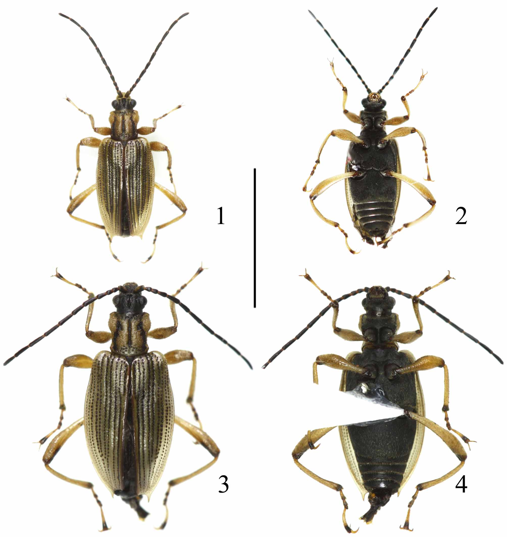
FIGURES 1 – 4. Macroplea huaxiensis sp. nov. 1. Holotype, male, dorsal view; 2. Paratype, male, ventral view; 3. Paratype, female, dorsal view; 4. Paratype, female, ventral view. Scale line = 5.0 mm.

FIGURES 50 – 53. Macraplea spp. 50. M. appendiculata from Xinjiang, male; 51. M. pubipennis from Ningxia, female; 52. M. japana from Jiangsu, female; 53. M. mutica from Tianjin, female. Scale lines = 5.0 mm.
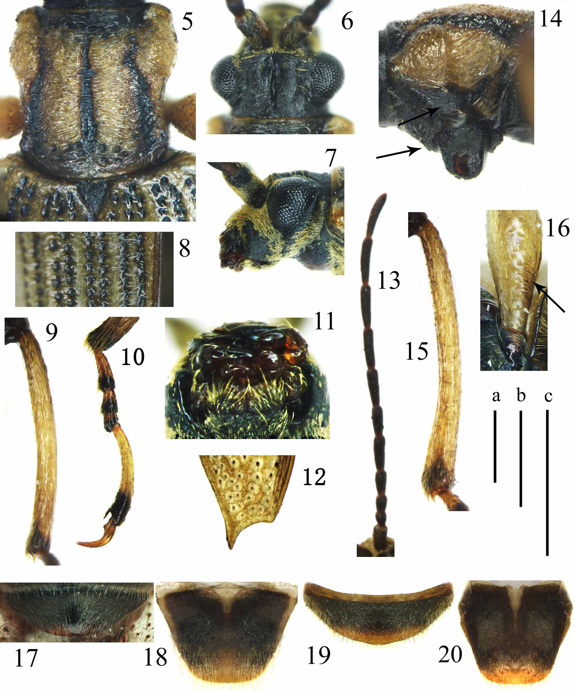
FIGURES 5 – 20. Macroplea huaxiensis sp. nov. 5 – 13, 17, 18. Male; 14 – 16, 19, 20. Female. 5. Pronotum, dorsal view; 6. Head, dorsal view; 7. Head, lateral view; 8. Left elytron, middle part; 9. Right metatibia, lateral view; 10. Right metatarsus, lateral view; 11. Mouth parts, ventral view; 12. Apex of left elytron; 13. Antenna; 14. Pronotum, left lateral view; 15. Right metatibia, lateral view; 16. Left profemur, ventral view; 17, 19. Apical abdominal sternum; 18, 20. Pygidium. Scale lines: a = 0.5 mm. (Figs 5 – 10, 12, 14 – 20); b = 1.0 mm. (Fig. 13); c = 0.5 mm. (Fig. 11).
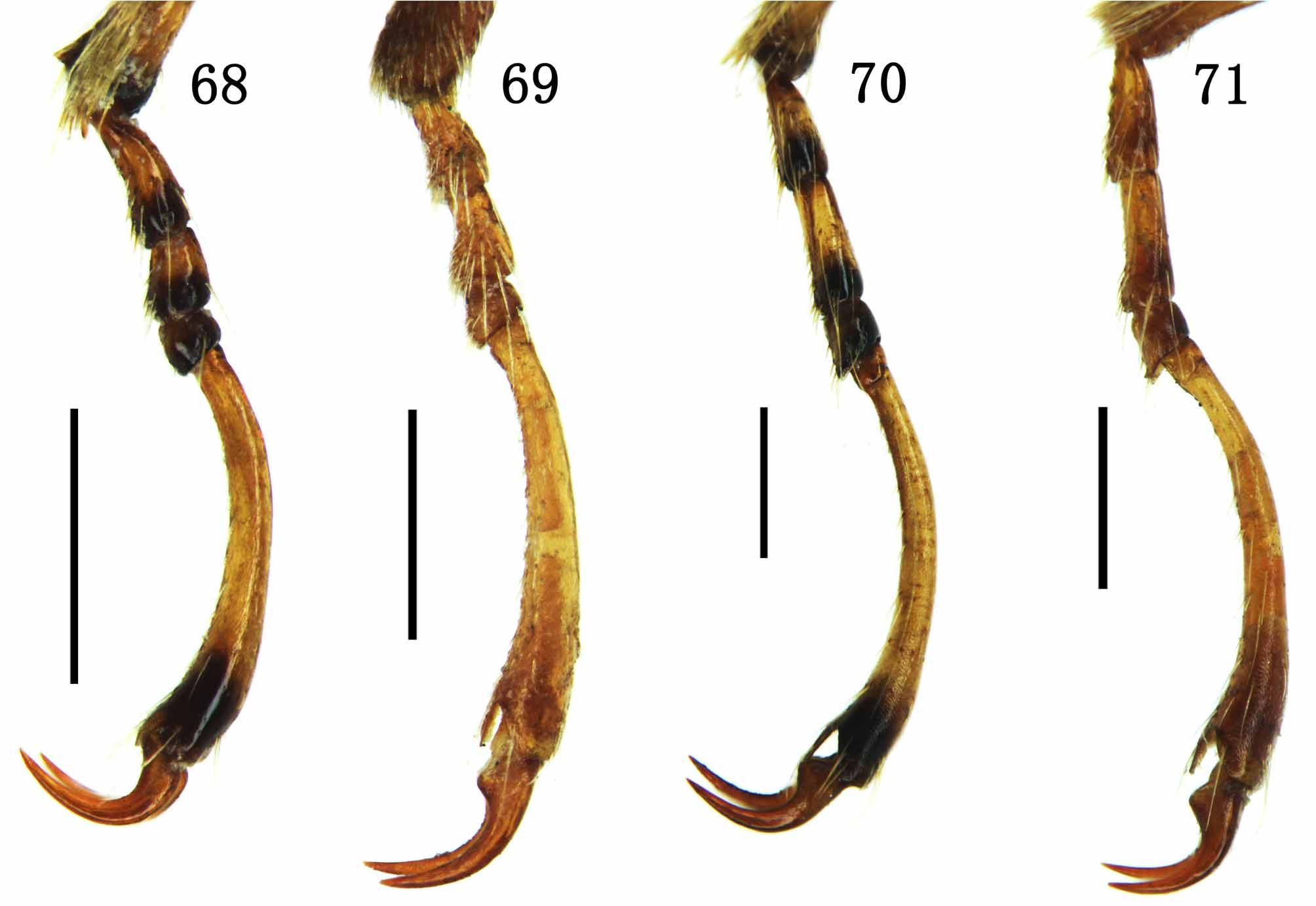
FIGURES 68 – 71. Tarsus of Macroplea spp. 68. M. japana from Jiangsu; 69. M. pubipennis from Ningxia; 70. M. appendiculata from Xinjiang; 71. M. mutica from Tianjin. 68, 70, 71. Right metatarsus; 69. Left protarsus. Scale lines = 0.5 mm

FIGURES 54 – 59. Head, prothorax and pronotum of Macroplea spp. 54 – 56. M. japana from Jiangsu; 57. M. pubipennis from Ningxia; 58. M. appendiculata from Xinjiang; 59. M. mutica from Tianjin; 54. Head and prothorax, lateral view; 55. Vertex, dorsal view; 56 – 59. Pronotum, dorsal view. Scale lines = 0.5 mm.

FIGURES 60 – 67. Elytron of Macroplea spp. 60, 64. M. japana from Jiangsu; 61, 65. M. pubipennis from Ningxia; 62, 66. M. appendiculata from Xinjiang; 63, 67. M. mutica from Tianjin. 60 – 63. Left elytron, middle part; 64, 66, 67. Apex of left elytron; 65. Apexes of both elytra. Scale lines = 0.2 mm.
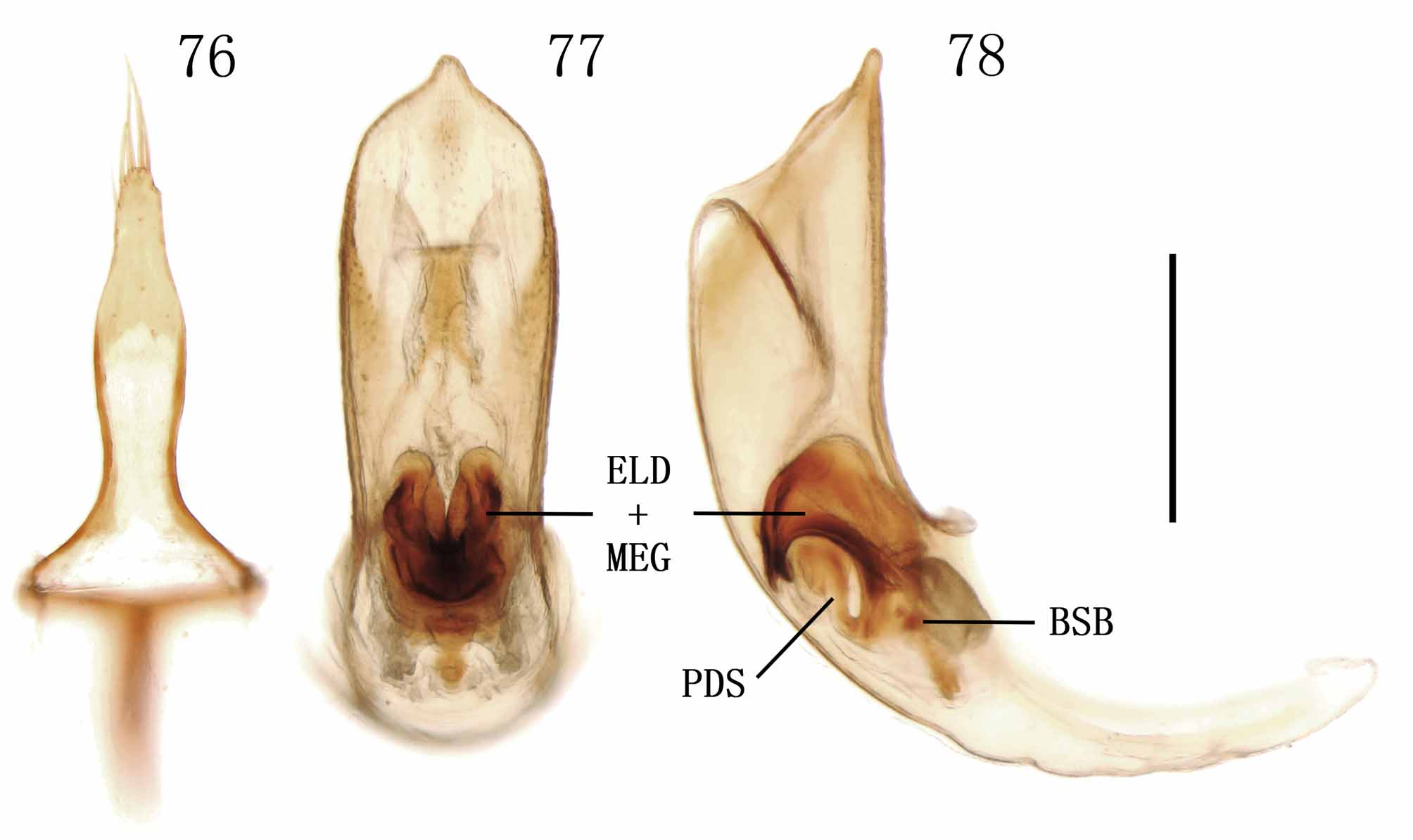
FIGURES 76 – 78. Macroplea appendiculata from Xinjiang. 76. Tegmen, dorsal view; 77, 78. Median lobe with endophallus in it (77. Dorsal view; 78. Lateral view). Scale line = 0.5 mm.

No known copyright restrictions apply. See Agosti, D., Egloff, W., 2009. Taxonomic information exchange and copyright: the Plazi approach. BMC Research Notes 2009, 2:53 for further explanation.
|
Kingdom |
|
|
Phylum |
|
|
Class |
|
|
Order |
|
|
Family |
|
|
SubFamily |
Donaciinae |
1 (by plazi, 2016-04-11 02:47:03)
2 (by ImsDioSync, 2017-02-08 23:14:23)
3 (by ImsDioSync, 2017-06-16 22:20:17)
4 (by ImsDioSync, 2017-06-16 23:35:20)
5 (by ExternalLinkService, 2019-09-26 20:35:17)
6 (by ExternalLinkService, 2022-01-30 13:57:15)
7 (by ExternalLinkService, 2022-02-20 12:26:52)
8 (by plazi, 2023-10-25 19:17:20)