
Meri yaciba, Rheims & Jäger, 2022
|
publication ID |
https://doi.org/10.11646/zootaxa.5135.1.1 |
|
publication LSID |
lsid:zoobank.org:pub:0CC0D586-E099-4593-9032-EA1885F00F3B |
|
DOI |
https://doi.org/10.5281/zenodo.6550553 |
|
persistent identifier |
https://treatment.plazi.org/id/039787EF-FFE9-C947-FF32-FBE9FCE4FF3A |
|
treatment provided by |
Plazi (2022-05-15 09:12:24, last updated by Valdenar 2025-02-13 17:59:30) |
|
scientific name |
Meri yaciba |
| status |
sp. nov. |
Meri yaciba View in CoL sp. nov.
Figs 297–308 View FIGURES 297–303 View FIGURES 304–308 , 317 View FIGURES 315–318
Type material. Holotype: VENEZUELA: Amazonas: ♂, Rio Yaciba [1.2333, ‑66.7333], 3 December 1953 ( AMNH) GoogleMaps . Paratype: VENEZUELA: Amazonas: 1♀, same locality as holotype, Igarapé, 7 December 1953, W.M. Beebe leg. ( AMNH) GoogleMaps .
Etymology. The specific name refers to the type locality; noun in apposition.
Diagnosis. Males of M. yaciba spec. nov. resemble those of M. pictitarsis ( Figs 221–224 View FIGURES 221–224 ) by the palp with RTA with a ventral branch ( Fig. 298 View FIGURES 297–303 ). They are distinguished from the latter species by RTA distally pointed with ventral branch roughly three times longer than wide ( Fig. 288–289 View FIGURES 285–291 ) and embolus with subdistal projection hook-shaped ( Fig. 300 View FIGURES 297–303 ) (RTA distally blunt with ventral branch triangular, slightly longer than wide and embolus with subdistal projection long, mediad and laminar in M. pictitarsis ). Females resemble those of M. pictitarsis comb. nov. ( Figs 225–227 View FIGURES 225–230 ) by the epigyne with MS widest anteriorly, partly covering LL and EP triangular. They are distinguished by the MS with posterior margin anterior to that of LL and EP more than 1.5 times longer than wide (MS with posterior margins in line with posterior margin of LL and EP slightly longer than wide in M. pictitarsis comb. nov.).
Description. Male (holotype): Total length 11.7. Prosoma 5.2 long, 4.5 wide. Opisthosoma 6.0 long, 3.0 wide. Eyes: diameters: 0.46, 0.40, 0.30, 0.35; interdistances: 0.20, 0.05, 0.45, 0.45, 0.27, 0.18. Legs: I: 29.9 (8.0, 2.9, 8.4, 8.4, 2.2); II: 32.3 (9.0, 2.9, 9.2, 8.9, 2.3); III: 21.5 (6.2, 2.2, 5.9, 5.5, 1.7); IV: 7.7, 2.1, 6.8, 6.9, 2.0). Palp: PTA trapezoid, roughly 2 times wider than long; RTA roughly 1.5 times longer than wide; cymbium with small retroproximal projection; subtegulum visible betwee 9–11:30 o’clock in ventral view; tegulum depressed close to conductor; conductor roughly the same width throughout; embolus curving subdistally ( Figs 297–300 View FIGURES 297–303 , 304–306 View FIGURES 304–308 ).
Female (paratype): Prosoma 6.0 long, 5.2 wide. Opisthosoma destroyed. Eyes: diameters: 0.50, 0.40, 0.30, 0.38; interdistances: 0.30, 0.30, 0.70, 0.61, 0.30, 0.25. Legs: I: 26.1 (7.2, 3.0, 7.0, 6.9, 2.0); II: 29.0 (8.0, 3.1, 8.0, 7.9, 2.0); III: 20.3 (6.0, 2.6, 5.0, 5.1, 1.6); IV: absent. Epigyne (broken anteriorly): MS as wide as long with lateral margins diverging anteriorly; HP triangular, opening at posterior margin of MS ( Figs 301 View FIGURES 297–303 , 307 View FIGURES 304–308 ). Vulva: internal ducts with FW C-shaped, mediad; GP longer than wide, antero mediad; SP rounded; FD antero mediad ( Figs 302–303 View FIGURES 297–303 , 308 View FIGURES 304–308 ).
Distribution. Only known from the type locality ( Fig. 317 View FIGURES 315–318 ).
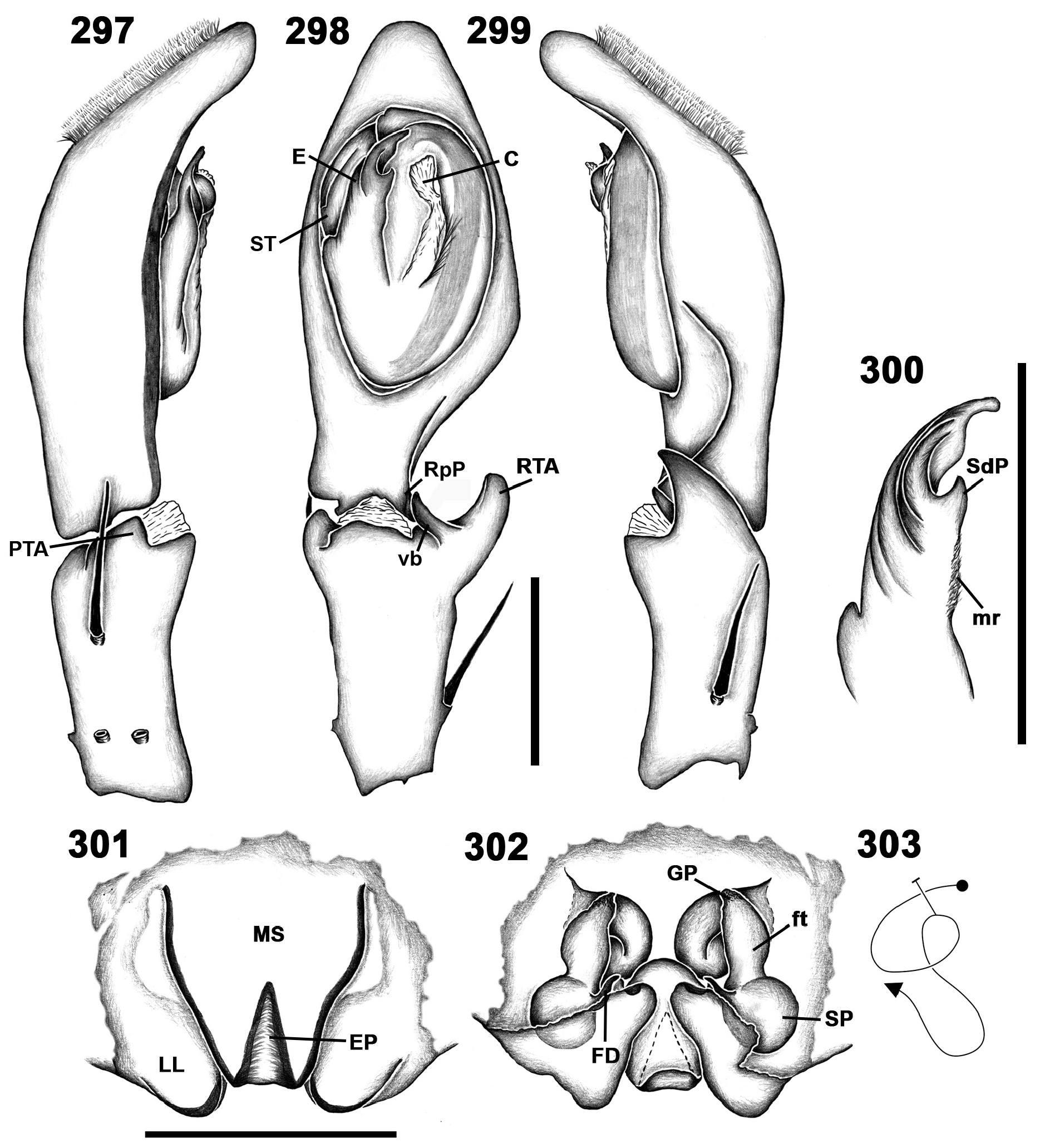
FIGURES 297–303. Meri yaciba spec. nov. 297–300 male (AMNH), left palp (297 prolateral, 298 ventral, 299 retrolateral, 300 detail of embolus); 301–303 female (AMNH) (301 epigyne, 302 vulva, 303 schematic course of internal duct system). C—conductor; E—embolus; EP—epigynal pocket; FD—fertilization duct; ft—first turn of duct system; GP—glandular projection; LL—lateral lobe; mr—membranous region; MS—median septum; PTA—prolateral tibial apophysis; RpP—retro proximal projection; RTA—retrolateral tibial projection; SdP—subdistal projection; SP—spermathecae; ST—subtegulum. Scale lines: 1 mm.

FIGURES 304–308. Meri yaciba spec. nov. 304–306 male (AMNH, holotype), left palp (304 prolateral, 305 ventral, 306 retrolateral); 307–308 female (AMNH, paratype) (307 epigyne, 308 vulva). Scale lines: 1 mm.
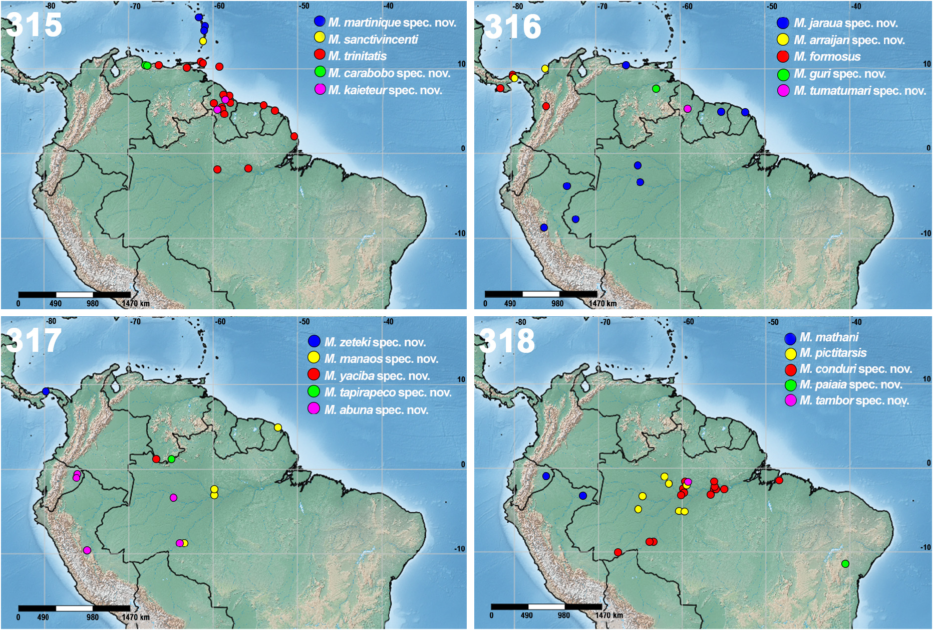
FIGURES 315–318. Distribution maps for species of Meri gen. nov. 315 (M. martinique spec. nov., M. sanctivincenti (Simon) comb. nov., M. trinitatis (Strand) comb. nov., M. carabobo spec. nov., M. kaieteur spec. nov.); 316 (M. jaraua spec. nov., M. arraijan spec. nov., M. formosus (Banks) comb. nov., M. guri spec. nov., M. tumatumari spec. nov.); 317 (M. zeteki spec. nov., M. manaos spec. nov., M. yaciba spec. nov., M. tapirapeco spec. nov., M. abuna spec. nov.); 318 (M. mathani (Simon) comb. nov., M. pictitarsis (Simon) comb. nov., M. conduri spec. nov., M. paiaia spec. nov., M. tambor spec. nov.);

FIGURES 221–224. Meri pictitarsis (Simon) comb. nov., male (IBSP 20795), left palp (221 prolateral, 222 ventral, 223 retrolateral, 224 detail of embolus). C—conductor; E—embolus; mr—membranous region; PTA—prolateral tibial apophysis; RTA— retrolateral tibial apophysis; SdP—subdistal projection; tp—tegular protrusion; vb—ventral branch of RTA. Scale lines: 1 mm.

FIGURES 285–291. Meri vanini spec. nov. 285–288 male (MPEG 30859, paratype), left palp (285 prolateral, 286 ventral, 287 retrolateral, 288 detail of embolus, ventral); 289–291 female (MZSP 37241, paratype) (289 epigyne, 290 vulva, 291 schematic course of internal duct system). C—conductor; E—embolus; EP—epigynal pocket; FD—fertilization duct; ft—first turn of duct system; GP—glandular projection; LL—lateral lobe; MS—median septum; PTA—prolateral tibial apophysis; RpP—retro proximal projection; RTA—retrolateral tibial projection; SdP—subdistal projection; SP—spermathecae; ST—subtegulum; tp—tegular protrusion. Scale lines: 1 mm.

FIGURES 225–230. Meri pictitarsis (Simon) comb. nov. 225–227 female (MNHN 1140, syntype) (225 epigyne, 226 vulva, 227 schematic course of internal duct system); 228–230 female (MNHN 1140, syntype) (228 epigyne, 229 vulva, 230 schematic course of internal duct system). EP—epigynal pocket; FD—fertilization duct; ft—first turn of duct system; GP—glandular projection; MS—median septum; SP—spermathecae. Scale lines: 1 mm.
| AMNH |
American Museum of Natural History |
No known copyright restrictions apply. See Agosti, D., Egloff, W., 2009. Taxonomic information exchange and copyright: the Plazi approach. BMC Research Notes 2009, 2:53 for further explanation.
|
Kingdom |
|
|
Phylum |
|
|
Class |
|
|
Order |
|
|
Family |
|
|
Genus |
1 (by plazi, 2022-05-15 09:12:24)
2 (by ExternalLinkService, 2022-05-15 09:48:01)
3 (by ExternalLinkService, 2022-05-15 09:58:04)
4 (by ExternalLinkService, 2022-05-15 10:13:17)
5 (by ExternalLinkService, 2022-05-15 10:22:23)
6 (by ExternalLinkService, 2022-07-12 03:45:20)
7 (by ExternalLinkService, 2022-07-12 05:11:50)
8 (by plazi, 2023-11-07 03:35:19)
9 (by ExternalLinkService, 2023-11-07 04:11:35)
10 (by felipe, 2024-11-18 14:19:44)