
Cylapinus minusculus Carvalho, 1986
|
publication ID |
https://doi.org/10.11646/zootaxa.5074.1.1 |
|
publication LSID |
lsid:zoobank.org:pub:7B3C6765-F0D2-4846-BB95-200258ECC0E1 |
|
DOI |
https://doi.org/10.5281/zenodo.5784476 |
|
persistent identifier |
https://treatment.plazi.org/id/039587FB-AE5E-FFB0-FF51-126F4339FC7D |
|
treatment provided by |
Plazi (2021-12-06 08:45:40, last updated 2024-11-28 20:55:07) |
|
scientific name |
Cylapinus minusculus Carvalho, 1986 |
| status |
|
Cylapinus minusculus Carvalho, 1986 View in CoL
( Figs 3a View FIGURE 3 , 7a View FIGURE 7 , 8a View FIGURE 8 , 10a View FIGURE 10 , 11a–d View FIGURE 11 , 12a–c View FIGURE 12 , 15d–f View FIGURE 15 , 20b View FIGURE 20 , 21b View FIGURE 21 )
Cylapinus minusculus Carvalho, 1986: 592 View in CoL , figs 10–13 (new species).
Cylapinus minusculus: Carvalho & Froeschner 1990: 319 View in CoL (list); Schuh 1995: 21 (catalog); Gorczyca 2006a: 13 (catalog)
Diagnosis. Recognized by the following set of features: scutellum from entirely pale to dark castaneous with pale apex ( Fig. 3a View FIGURE 3 ); corium with medial pale patch broad, bordering embolium and clavus ( Fig. 3a View FIGURE 3 ); bursa copulatrix broader than wide, anterior margin weakly removed from first gonapophyses ( Fig. 15f View FIGURE 15 ), sclerotized rings strongly curved, occupying almost entire lateral portion of bursa copulatrix ( Fig. 15e, f View FIGURE 15 ); vlp without any sclerotization ( Fig. 15d View FIGURE 15 ).
Redescription (composite description based on Carvalho 1986 and 18 additional specimens). Male. COLORATION ( Fig. 3a View FIGURE 3 , 7a View FIGURE 7 ). Head. Entirely dark castaneous to black, sometimes with pale patch on vertex; antennal segment I yellow, sometimes tinged with red; segment II varying from yellow with dark castaneous or dark brown apical two-thirds to entirely dark castaneous or dark brown; segments III and IV dark castaneous, basal one-fourth of segment III yellow; labium yellow, segment IV largely fuscous. Thorax. Pronotum. Dark castaneous to black. Mesoscutum and scutellum. Varying from entirely pale to dark castaneous with pale apex. Thoracic pleura. Varying from red or dark castaneous with yellow areas to entirely black; metathoracic scent gland evaporative area yellow. Hemelytron. Brown to dark brown with large, yellow areas; clavus brown to dark brown, sometimes with thin, yellow stripe along entire length; corium brown to dark brown with broad, yellow patch medially from clavus to embolium and cuneus varying from uniformly yellow to yellowish brown; membrane pale fuscous. Legs. Coxae yellow, sometimes weakly tinged with red; tibiae and femora yellow broadly tinged with red; tarsi varying from yellow to yellowish brown. Abdomen. Varying from yellow tinged with brown to entirely dark brown. TEXTURE AND VESTITURE. Vestiture as in generic description. Thorax. Thoracic pleura. Ventral surface of mesepimeron, anterior margin of metepisternum, posterior and ventral surface of metepisternum with characteristically perforated surface. STRUCTURE ( Figs 3a View FIGURE 3 , 7a View FIGURE 7 , 11a–d View FIGURE 11 ). Thorax. Pronotum. Collar narrow, narrower than antennal segment I diameter; calli moderately developed, narrower than eye diameter. Abdomen. Anterior margin of abdominal segment I narrowly covered with perforated surface. Genitalia. Endosoma with well–developed, straight medial sclerite ( Fig. 12a View FIGURE 12 ); secondary gonopore well differentiated ( Fig. 12a View FIGURE 12 ); left paramere C-shaped, apical process when viewed laterally weakly curved, almost straight, somewhat widened subapically, narrowly tapering toward apex; paramere body in lateral view thin weakly tapering toward apex; sensory lobe well-developed, rounded ( Fig. 12b View FIGURE 12 ); right paramere with apical process short, weakly narrowed toward apex when viewed laterally; sensory lobe prominent, rounded ( Fig. 12c View FIGURE 12 ).
Female. Like male in coloration, structure, texture, and vestiture. Genitalia. Bursa copulatrix extending laterally beyond first gonapophysis, crescent-like ( Fig. 15f View FIGURE 15 ); sclerotized rings paired, narrow, strongly arcuate ( Figs 15f, e View FIGURE 15 ); spermathecal gland originating from area situated between lateral oviducts ( Fig. 15f View FIGURE 15 ); ventral labiate plate without any sclerotization its surface weakly squamate ( Fig. 15d View FIGURE 15 ); vestibulum without any sclerite ( Fig. 20b View FIGURE 20 ); first valvula with ventral margin more or less arcuate, dorsal margin sinuate, strongly convex subapically ( Fig. 21b View FIGURE 21 ); second valvula with ventral margin arcuate, dorsal margin weakly sinuate, strongly serrate; posterior wall of bursa copulatrix membranous.
Measurements. (♂ / ♀, *: holotype measurements): Body. Length: 2.8*–3.3/3.40–3.45, width 1.20*–1.40/1.40– 1.55. Head. Length: 0.30*–0.35/0.30–0.36, width: 0.70*–0.83/0.85, interocular distance 0.25–0.30*/0.28. Antenna. Length of segment I: 0.28–0.50, 0.30*/0.50, II: 0.80*–0.90/0.88–0.90, III (mutilated in holotype): 0.90–0.92/0.95, IV mutilated in holotype): 1.45/1.70. Labium. Length of segment I: 0.33/0.35–0.40, II: 0.38/0.35–0.43, III: 0.35/0.28– 0.30, IV: 0.28/0.28–0.30. Pronotum. Length 0.52–0.60*/0.55–0.60, width of anterior margin:0.60–0.65/0.68–0.70, length of lateral margin: 0.53–0.58/0.58–0.60, width of posterior margin: 1.10*–1.2/1.22.
Biology. Unknown.
Distribution. Brazil (Rondônia), Ecuador (Orellana), Guyana (Cuyuni–Mazaruni).
Type material (not examined). Holotype (♂): Brazil, Rondônia, Ji–Paraná , II.1983, equipe J. R. Arias (Museu Nacional, Rio de Janeiro, Brazil).
Additional material examined. 1 ♀: Ecuador, Orellana Prov. [labeled Napo Prov. In error], Tiputini Biodiversity Stn., Tower 1, 0º 37’55” S, 76º08’39 W, 216 m, 4 July 1998, T.L. Erwin et al., collectors; insecticidal fogging of mostly bare green leaves, some with covering of lichenous or bryophytic plants, Lot # 1863, Transect # T–7; GoogleMaps 1 ♂: the same data except: 24 October 1994, Lot#1935, Transect #T–74; GoogleMaps 3 ♂, 1 ♀: Ecuador, Orellana Prov. [labeled Napo Prov. In error], Res. Ethnica Waorani, 1 km. S, Onkone Gare Camp, Trans. Ent. , 4 Feb. 1996, 220m, 00º39’10”S, 76 º26’00”W, T.L. Erwin, et al., insecticidal fogging of mostly bare green leaves, some with covering of lichenous or bryophytic plants in terre firme forest Lot 1407, Trans. T–1; GoogleMaps 4 ♂, 1 f #: the same data except: GoogleMaps 1 ♂: Lot 1468; 1 f #: 8 Feb. 1996, Lot 1476, Trans. T–8; GoogleMaps 1 ♂: 24 October, Transect T–4; GoogleMaps 1 ♂: 22 October 1998, Lot#1974, Transect#T–8; GoogleMaps 1 ♂: 3 July 1994, 73m mark Proj. MAXUS Lot 777; GoogleMaps 1 ♂: 20 Jan 1994, Project MAXUS, Ar–x–trans 7.93m, Lot 619; GoogleMaps 1 ♀: Guayana, Mazaruni, Potaro District Takutu Mountains , 6º15’ N, 59º5’ W, 14 December 1983; Malaise trap near stream in montane rainforest, EARTHWATCH Research Expedition, W. E. Steiner P. J. & Spangler collectors ( USNM) GoogleMaps .
Carvalho, J. C. M. (1986) Mirideos neotropicais, CCLVI: Dois generos e seis especies novos da tribo Cylapini (Hemiptera). Acta Amazonica, 16 / 17, 589 - 598. https: // doi. org / 10.1590 / 1809 - 43921987171598
Carvalho, J. C. M. & Froeschner, R. C. (1990) Taxonomic names proposed in the insect order Heteroptera by Jose Candido de Melo Carvalho from January 1985 to January 1989, with type depositories. Journal of the New York Entomological Society, 98, 310 - 346.
Gorczyca, J. (2006 a) The catalogue of the subfamily Cylapinae Kirkaldy, 1903 of the World (Hemiptera, Heteroptera, Miridae). Monographs of the Upper Silesian Museum. Vol. 5. Department of Natural History, Upper Silesian Museum, Bytom, 100 pp.
Schuh, R. T. (1995) Plant bugs of the World (Insecta: Heteroptera: Miridae). New York Entomological Society, New York, New York, 1329 pp.
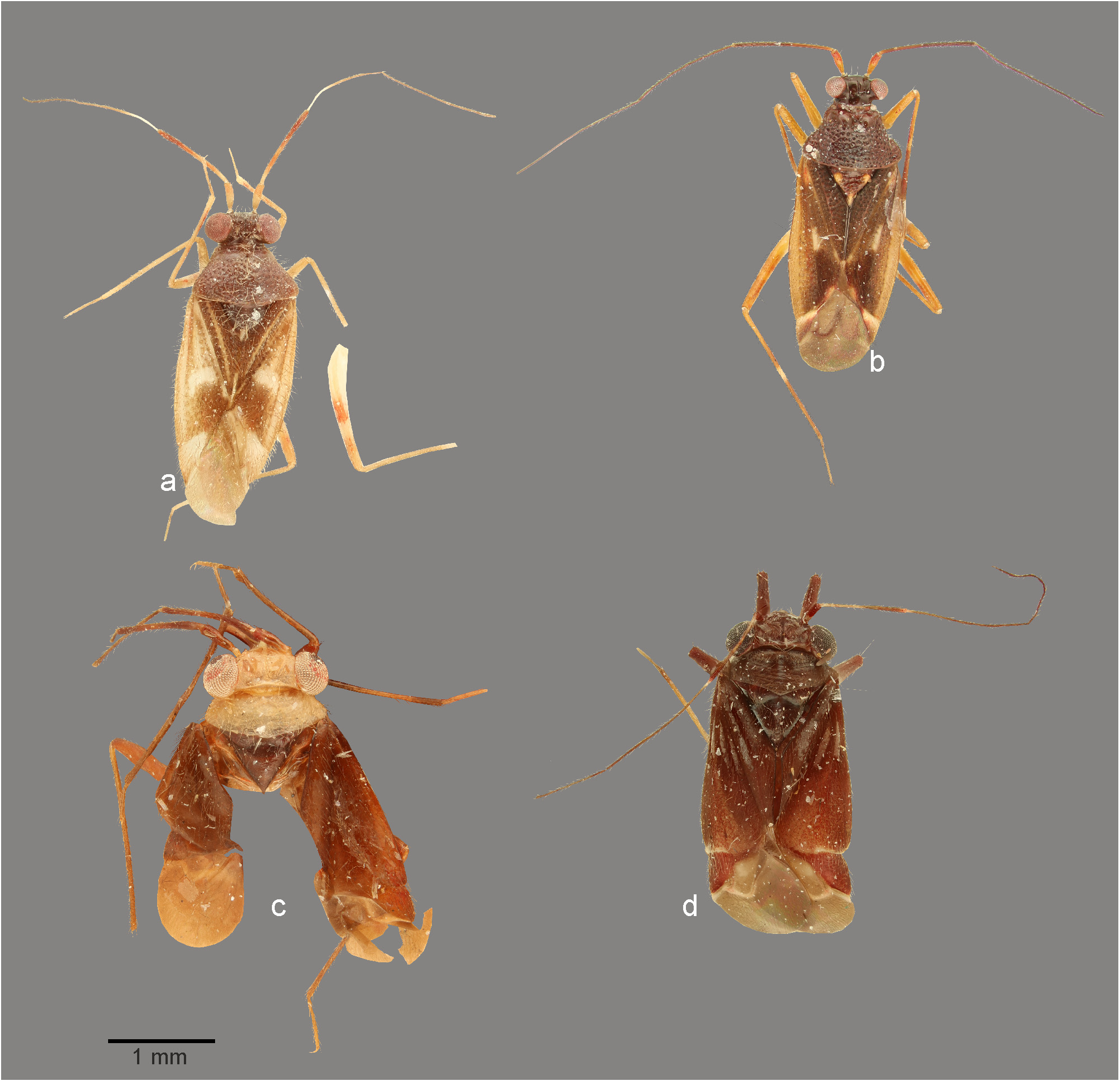
FIGURE 3. Dorsal habitus photographs. a. Cylapinus minusculus (♂, Ecuador); b. Cylapinus yasunagai (holotype); c. Cylapoides bicolor (holotype); d. Cylapoides unicolor (♂, Ecuador).
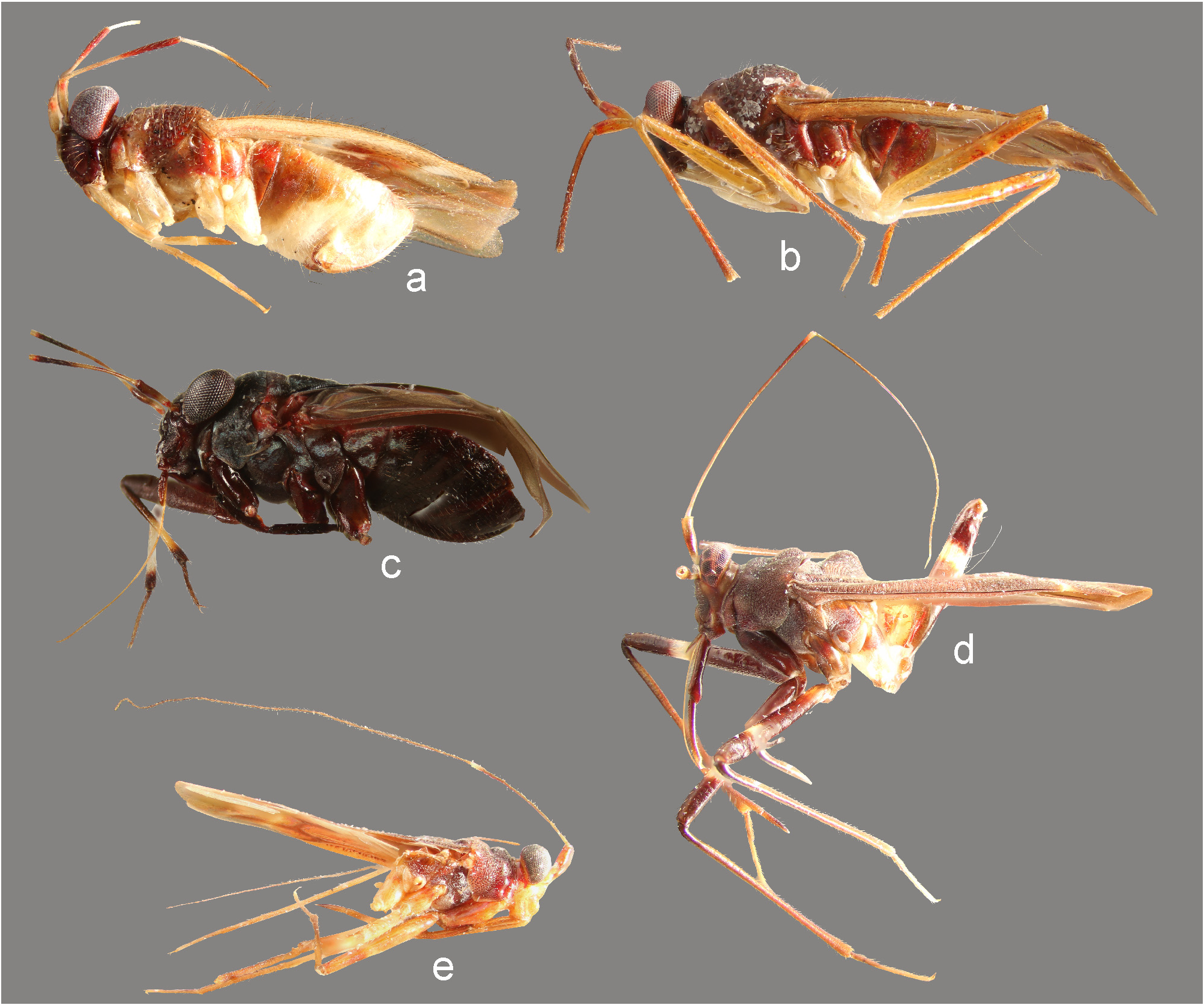
FIGURE 7. Lateral view. a. Cylapinus minusculus (♀); b. Cylapinus yasunagai (paratype); c. Cylapoides unicolor (♀); d. Peltidocylapus calyciformis (paratype); e. Peltidocylapus cerbereus (lectotype).

FIGURE 8. Head and pronotum, anterior (a–j, l, m) and lateral (k) views. a. Cylapinus yasunagai (paratype); b. Cylapoides unicolor (♀); c. Cylapus ruficeps Bergroth (♂); d. Cylapus tenuicornis (♂); e. Peltidocylapus calyciformis (paratype); f. Peltidocylapus caudatus (paratype); g. Peltidocylapus rugosus (holotype); h. Peltidocylapus simplex (paratype); i. Cylapomorpha sp. (♂); j, k. Vanniusoides clypeatus (paratype); l. Fulvius pallens (♂); m. Rhinocylapus vittatus (♀).

FIGURE 10. Scanning electron micrographs. a. Pronotum (left lateral view); b–h. Thoracic pleura; i–m. Metatarsus. n, o. Pretarsal claw. a. Valdasus schoechnerri; b. Amapacylapus amapariensis; c, i. Cylapus tenuicornis; d. Peltidocylapus scutellaris; e, j. Valdasus henryi; f. Bothriomiris dissimulans; g. Cylapocoris sp.; h. Psallops sp.; k, o. Vannius sp.; l. Fulvius sp.; m. Cylapocoris sp.; n. Cylapinus minusculus.

FIGURE 11. Scanning electron micrographs. a, e, g, m. Dorsal habitus; b, n. Lateral habitus; h, l. Scutellum; c, f, i. Thoracic pleura; d, j, o. Tarsus. k. Pretarsal claw. a–d. Cylapinus minusculus; e, f. Cylapoides unicolor; g–j. Peltidocylapus caudatus; k. Peltidocylapus calyciformis; l. Peltidocylapus ecuadorensis; m–o. Peltidocylapus scutellaris.

FIGURE 12. Male genitalia. a, d, g, j, p. Endosoma (dorsal view); b, e, h, m, s. Left paramere (right lateral view). c, f, i, o, u. Right paramere (left lateral view); n, t. Left paramere (dorsal view). k, q. Transparent portion of distal sclerotized portion of ductus seminis inside endosoma; l, r. Endosoma (ventral view). a–c. Cylapinus minusculus; d–f. Cylapinus yasunagai; g–i. Cylapoides unicolor; j–o. Peltidocylapus calyciformis; p–u. Peltidocylapus caudatus. ap = apical process; bp = basal process; dss = distal part of ductus seminis inside endosoma; pb = paramere body; sl = sensory lobe. Scale bars 0.2 mm.
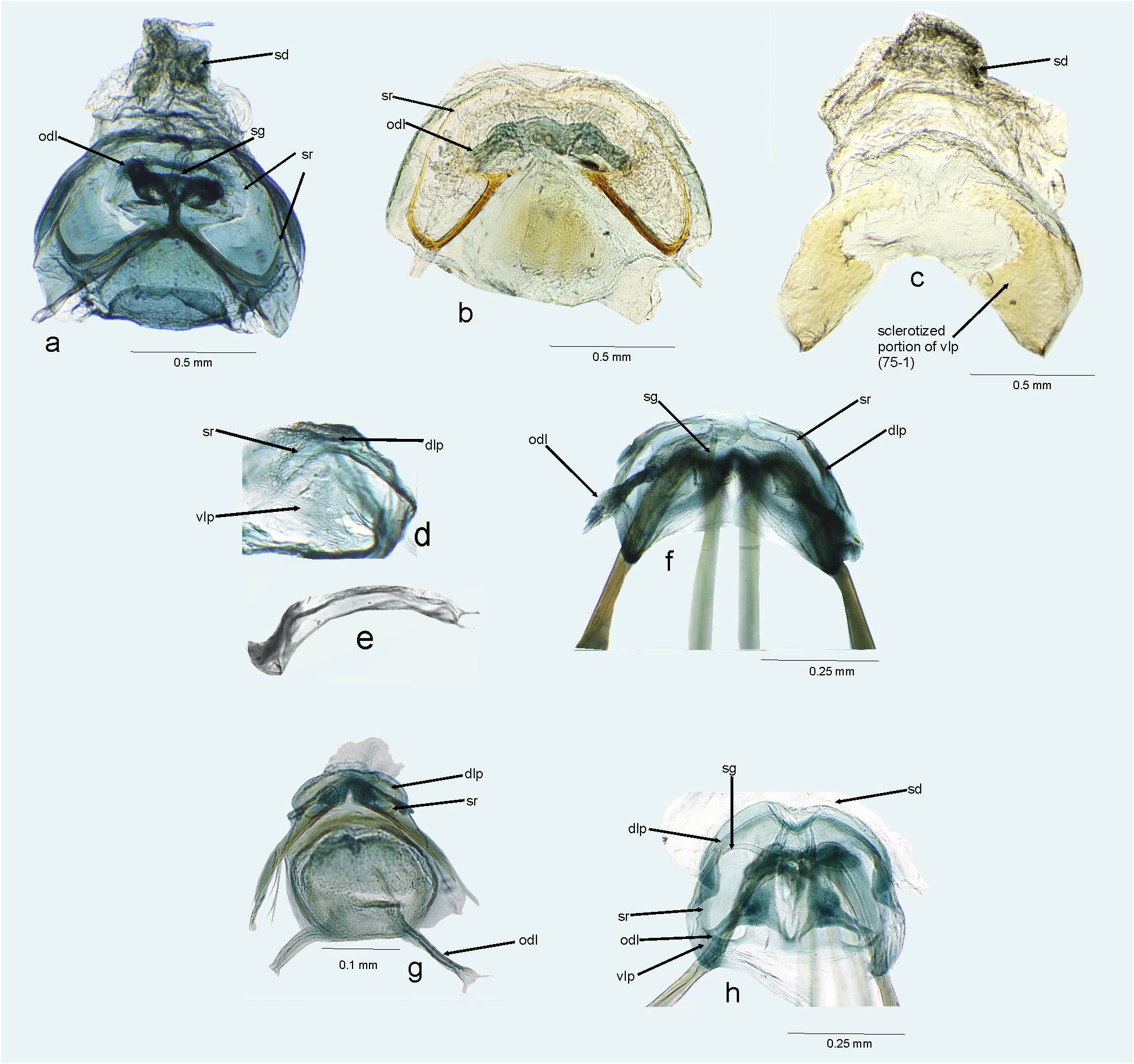
FIGURE 15. Female genitalia. a, b, f, g, h. Bursa copulatrix (dorsal view); c, d. Ventral labiate plate; e. Sclerotized rings of dorsal labiate plate. a. Amapacylapus amapariensis; b, c. Amapacylapus unicolor; d–f. Cylapinus minusculus; g. Cylapoides unicolor; h. Cylapomorpha sp. dlp = dorsal labiate plate; odl = lateral oviduct; sd = seminal depository; sg = spermathecal gland; sr = sclerotized ring; vlp = ventral labiate plate.

FIGURE 20. Female genitalia. Vestibular area. a. Amapacylapus amapariensis; b. Cylapinus minusculus; c. Cylapoides unicol- or; d. Cylapus antennatus; e. Cylapus citus; f. Cylapus tenuicornis; g. Peltidocylapus parallelus; h. Valdasus flavinotum.

FIGURE 21. Female genitalia, ovipositor. a–i. Apex of first gonapophysis; j–q. Apex of second gonapophysis. a, j. Amapacylapus amapariensis; b. Cylapinus minusculus; c, k. Cylapoides unicolor; d, l. Cylapomorpha sp.; e, m. Cylapus amazonicus; f, n. Cylapus citus; g, o. Cylapus ruficeps; h, p. Peltidocylapus ecuadorensis; i, q. Peltidocylapus parallelus.
No known copyright restrictions apply. See Agosti, D., Egloff, W., 2009. Taxonomic information exchange and copyright: the Plazi approach. BMC Research Notes 2009, 2:53 for further explanation.
|
Kingdom |
|
|
Phylum |
|
|
Class |
|
|
Order |
|
|
Family |
|
|
Tribe |
Cylapini |
|
Genus |
Cylapinus minusculus Carvalho, 1986
| Wolski, Andrzej 2021 |
Cylapinus minusculus:
| Gorczyca, J. 2006: 13 |
| Schuh, R. T. 1995: 21 |
| Carvalho, J. C. M. & Froeschner, R. C. 1990: 319 |
Cylapinus minusculus
| Carvalho, J. C. M. 1986: 592 |
1 (by plazi, 2021-12-06 08:45:40)
2 (by ExternalLinkService, 2021-12-06 08:57:06)
3 (by carolina, 2021-12-15 18:38:43)
4 (by carolina, 2021-12-15 18:48:48)
5 (by carolina, 2021-12-15 19:02:18)
6 (by ExternalLinkService, 2021-12-15 19:12:56)
7 (by ExternalLinkService, 2021-12-15 19:46:04)
8 (by plazi, 2023-11-08 10:55:03)