
Brooklynella sinensis, Gong, Jun & Song, Weibo, 2006
|
publication ID |
https://doi.org/ 10.5281/zenodo.171558 |
|
DOI |
https://doi.org/10.5281/zenodo.5685483 |
|
persistent identifier |
https://treatment.plazi.org/id/03934663-EB09-6132-FEB5-712D62CFB9FC |
|
treatment provided by |
Plazi |
|
scientific name |
Brooklynella sinensis |
| status |
sp. nov. |
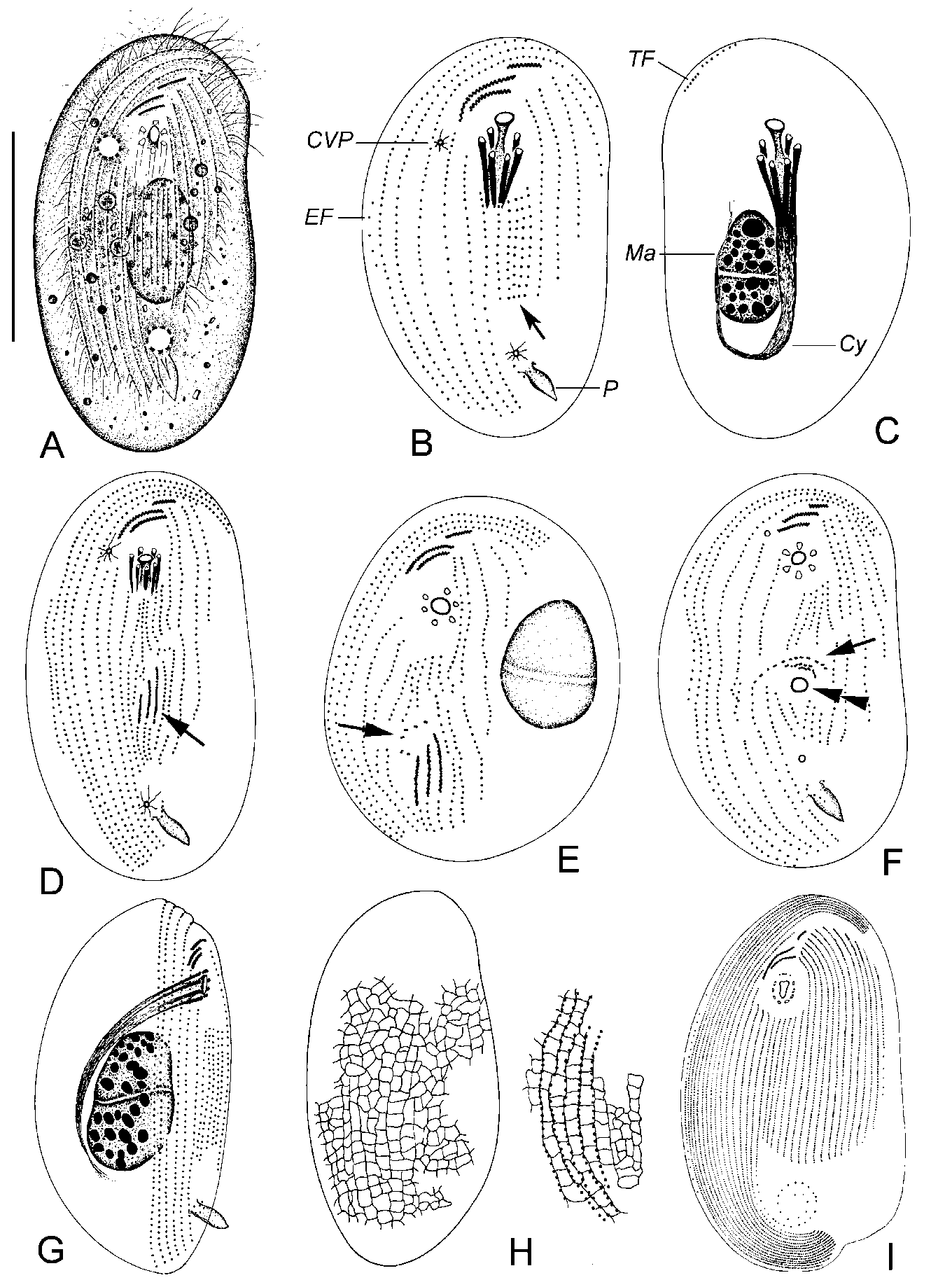
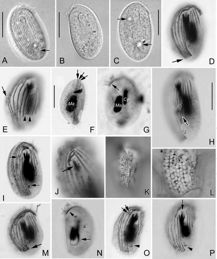
Brooklynella sinensis n. sp. ( Figs. 1 View FIGURE 1 A–H, 2A–P; Table 1)
Diagnosis: Marine freeliving Brooklynella size 40–50 × 20–30 m in vivo, body reniform to oval in outline; 15–17 ventral kineties, of which the rightmost three to four rows extend apically; five postoral kineties without cilia; about six nematodesmal rods; podite about 6 m long; two contractile vacuoles diagonally positioned.
Type location: Mesotrophic coastal water for scallopfarming near Qingdao (36 08’N; 120 43’E).
Type specimens: One holotype of protargol impregnated specimens is deposited in the Natural History Museum, UK (registration number 2005:10:21:1). Two paratype slides (HD2001080402, HD2001080403) are deposited in the Laboratory of Protozoology, Ocean University of China, China.
Etymology: Named after the country discovered.
Description: Size rather consistent, 40–50 × 20–30 m in vivo. Reniform to oval in shape outline ( Figs. 1 View FIGURE 1 A; 2A–C). Ventral surface flattened, dorsal side hunched, width to thickness ratio approximately 2:1. Cytostome rounded, about 5 m in diameter, positioned at anterior 1/4 of body length. Cyrtos composed of 4–7 (on average 6) toothed nematodesmal rods, which extend into endoplasm rightposteriorly. Endoplasm colorless, containing several small food granules (2–5 m in diameter) and refrangible crystals (1–2 m across). Macronucleus ovoid, located near body center. Cilia about 5 m long in vivo, cover ventral surface except for postoral and leftposterior regions ( Figs. 1 View FIGURE 1 A; 2A, C). Podite about 6 m long, subcaudally positioned. Two contractile vacuoles, each about 4 m in diameter, diagonally positioned: one right of cytostome; one meridian and anterior of the base of podite ( Figs. 1 View FIGURE 1 A; 2A, C); pulsing interval on average 2 mins. Movement moderately rapid, sliding on substrates, sometimes swimming with rotation.
Infraciliature as shown in Figs. 1 View FIGURE 1 B, C, G and Figs. 2 View FIGURE 2 D–G, I, J, M–P. A total of 15–17 somatic kineties on ventral side ( Table 1). Right kineties 6–7 rows, of which the rightmost 3–4 extend anteriorly and bend to left; 5 postoral kineties almost equal in length (about 1/3 of body length), terminate at posterior 1/3 of body length; 4–5 left rows progressively shortened from right to left, of which the innermost one posteriorly terminates at the same level as postoral rows. Terminal fragment anteriordorsally positioned, composed of about 10 basal bodies; equatorial fragment usually present, composed of about seven basal bodies. Podite located in body meridian, at level of posterior 1/5–1/4 of body length. Often about five kinetosomelike dots presented near the base of podite ( Figs. 1 View FIGURE 1 B; 2 O, P). Macronucleus juxtaposed heteromerous, size about 11 × 5 m after protargol impregnation. One to three micronuclei, positioned close to macronucleus.
Oral ciliature composed of one preoral and two circumoral kineties. Outer and inner circumoral kineties positioned posterior of preoral kinety, composed of about 10 and six dikinetids, respectively. Preoral kinety shorter than circumoral ones, comprises circa five dikinetids. Cyrtos tapering posteriorly, hookshaped in protargol impregnated specimens.
m.
Abbreviations: CV = coefficient of variation in %, Max = maximum, Mean = arithmetic mean, Min = minimum, n = number of individuals examined, SD = standard deviation.
Silverline system ladderlike, with many tiny argentophilic granules on silverlines ( Figs. 1 View FIGURE 1 H; 2K, L).
Morphogenesis: Three specimens in divisional stages have been observed. These showed that three of the five postoral kineties join to form the oral primordium ( Figs. 1 View FIGURE 1 D, E; 2H). The oral primordium turns horizontallyoriented thus forms the oral kineties in the proter ( Fig. 1 View FIGURE 1 F).
No known copyright restrictions apply. See Agosti, D., Egloff, W., 2009. Taxonomic information exchange and copyright: the Plazi approach. BMC Research Notes 2009, 2:53 for further explanation.