
Megabeleses tsurugiensis Togashi, 2008
|
publication ID |
https://doi.org/ 10.5281/zenodo.200135 |
|
DOI |
https://doi.org/10.5281/zenodo.6200890 |
|
persistent identifier |
https://treatment.plazi.org/id/03928F52-0C73-FFCA-FF56-5354FA8A8750 |
|
treatment provided by |
Plazi |
|
scientific name |
Megabeleses tsurugiensis Togashi, 2008 |
| status |
|
Megabeleses tsurugiensis Togashi, 2008
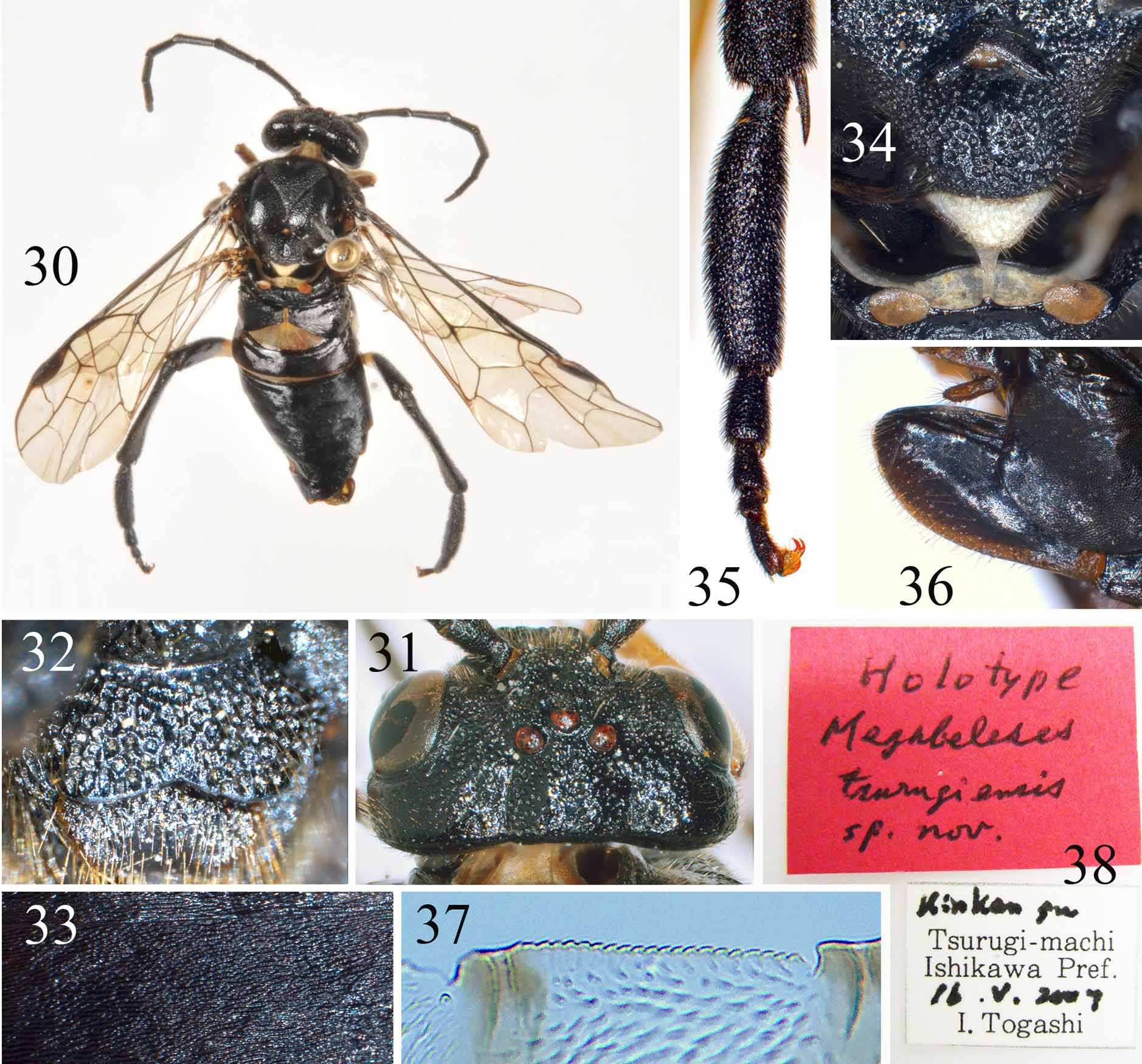
( Figs. 30–38 View FIGURES 30 – 38 )
Megabeleses tsurugiensis Togashi, 2008: 10 , Ƥ, type locality: Japan, Honshu, Ishikawa Prefecture, Hakusan-shi, Tsurugi-hizumecho, foot of Mt. Uwanai.
Description. Holotype: Ƥ.
Body length 13 mm ( Fig. 30 View FIGURES 30 – 38 ). Black, without any metallic tinge; fore tibia and tarsus largely, extreme base of middle tibia, middle tarsus largely pale brown; posttergite, a quadrate macula on outer side of hind coxa, hind trochanter partly, basal 3/7 of hind femur yellow white; palpi blackish brown. Wings subhyaline, faintly infuscate, stigma and veins black brown. Body hairs and setae on sheath silver brown.
Head coarsely punctured, bottoms of middle and lateral fovea feebly polished, temple and postocellar area somewhat sparsely punctured, interspaces somewhat shiny; lateral lobe of pronotum coarsely punctured; mesonotum including posttergite densely punctured, posterior 2/3 of prescutum, top of scutum, depressed part of mesoscutellum less densely punctured, interspaces shiny, parapsis microsculptured; elevated area of metanotum behind cenchri coarsely punctured, metascutellum densely microsculptured, without isolated punctures; lateral part of propleuron very sparsely and ventral part densely punctured; upper half of mesepisternum and dorsal margin of mesepimeron coarsely punctured, ventral half of mesepisternum sparsely punctured with distinct shiny interspaces, other part of mesepimeron microsculptured; ventral part of metepisternum very sparsely punctured, strongly shiny, upper margin of metepisternum and of metepimeron densely punctured, central area of metepimeron coriaceous; outer side of hind coxa densely punctured; posterior side of each coxa impunctate, strongly shiny. Abdominal tergites impunctate, distinctly microsculptured ( Fig. 33 View FIGURES 30 – 38 ); lateral sides of ovipositor sheath polished, shiny.
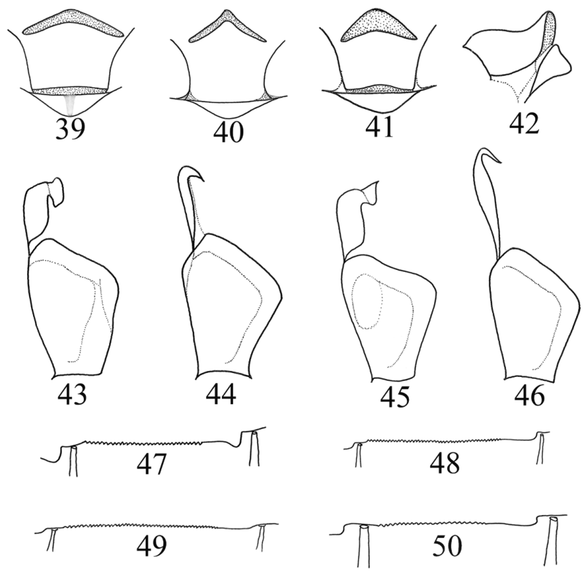
Anterior margin of clypeus shallowly and angularly incised for about 1/6 clypeus length ( Fig. 32 View FIGURES 30 – 38 ); malar space hardly shorter than radius of ocellus; closest distance between eyes 1.4× height of eye; distance between antennal sockets as long as distance between antennal socket and eye; middle fovea round, distinct, clearly deeper than frontal basin; frontal walls obtuse; circular furrow of anterior ocellus distinct, postocellar and interocellar furrows obscure; postocellar area distinctly elevated, 1.3× broader than long, middle furrow faint; lateral furrows broad and shallow, distinctly divergent backwards; lateral margins of head behind eyes in dorsal view about 0.6× length of eye, distinctly enlarged ( Fig. 31 View FIGURES 30 – 38 ). Antenna about as long as thorax and abdomen together, third antennomere 1.05× length of fourth antennomere. Mesoscutellum slightly broader than long, anterior corner roundly protruding, obtuse, anterior furrow of mesoscutellum triangularly bent, posterior margin without distinct carina ( Fig. 34 View FIGURES 30 – 38 ); posttergite without middle carina, apical margin round; distance between cenchri about 1.65× width of a cenchrus; dorsal lobe of metepimeron long and narrow (cf. Fig. 46 View FIGURES 39 – 50 ). Length of inner margin of 1st abdominal tergite about 1/3 length of widest lateral length. Apex of hind coxa reaching anterior margin of fourth sternite, hind tibia with a long and distinct longitudinal furrow on outer side, apex distinctly thickened; inner spur of hind tibia about 1/4 length of hind basitarsus; hind basitarsus 4× longer than broad, strongly thickened with constricted base, 1.3× as long as tarsomeres 2–5 together ( Fig. 35 View FIGURES 30 – 38 ), outer side with a shallow but distinct furrow. Vein cu-a close to but not interstitial to vein 1M, R+M distinct, 2r weakly bent at middle and meeting cell 2Rs at apical 3/7, 2m-cu not interstitial to 1rm; petiole of hind anal cell about half length of cu-a. Ovipositor sheath about 0.92× lengths of middle tibia, apical sheath 1.4× lengths of basal plate, apex narrowly rounded in lateral view ( Fig. 36 View FIGURES 30 – 38 ); apical sheath in dorsal view triangular, basal width 3.5× width of cercus; setae on lateral sides of apical sheath about as long as diameter of middle ocellus; cercus 3× as long as broad. Lancet with 24 flat serrulae, middle serrulae each with about 20 subbasal teeth ( Fig. 37 View FIGURES 30 – 38 ); medial breadth of membranous lobe of lance 0.65× medial width of lance.
Male: Unknown.
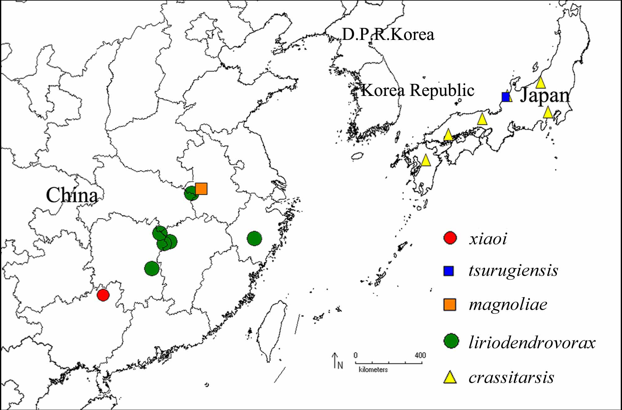
Distribution. Japan (Ishikawa) ( Fig. 51 View FIGURE 51 ).
Specimen examined. 1Ƥ, holotype, “Kin Ken gu, Tsurugi-machi, Ishikawa Pref., 16. V. 2007, I. Togashi leg.”; “ Holotype, Megabeleses tsurugiensis sp. nov. ” ( Fig. 38 View FIGURES 30 – 38 , NMNS, lance and lancet removed from specimen into a slide, no. NSMT I-Hym 62144).
Host plant. Unknown.
Remarks. See the above key for differences between M. tsurugiensis and other known species of the genus.
No known copyright restrictions apply. See Agosti, D., Egloff, W., 2009. Taxonomic information exchange and copyright: the Plazi approach. BMC Research Notes 2009, 2:53 for further explanation.
|
Kingdom |
|
|
Phylum |
|
|
Class |
|
|
Order |
|
|
Family |
|
|
Genus |
Megabeleses tsurugiensis Togashi, 2008
| Wei, Meicai 2010 |
Megabeleses tsurugiensis
| Togashi 2008: 10 |