
Penghou, Ruan, Yong-Ying, Konstantinov, Alexander S., Prathapan, K. D., Ge, Si-Qin & Yang, Xing-Ke, 2015
|
publication ID |
https://doi.org/ 10.11646/zootaxa.3973.2.5 |
|
publication LSID |
lsid:zoobank.org:pub:5851DACD-8AD3-44AB-B289-8F7368E90B8E |
|
DOI |
https://doi.org/10.5281/zenodo.6100195 |
|
persistent identifier |
https://treatment.plazi.org/id/0391554C-FF83-1124-FF13-FF1CFEF3898C |
|
treatment provided by |
Plazi |
|
scientific name |
Penghou |
| status |
gen. nov. |
Penghou new genus
( Figs 1–25 View FIGURES 1, 2 View FIGURES 3 – 6 View FIGURES 7 – 12 View FIGURES 13 – 16 View FIGURES 17 – 20 View FIGURES 21 – 25 )
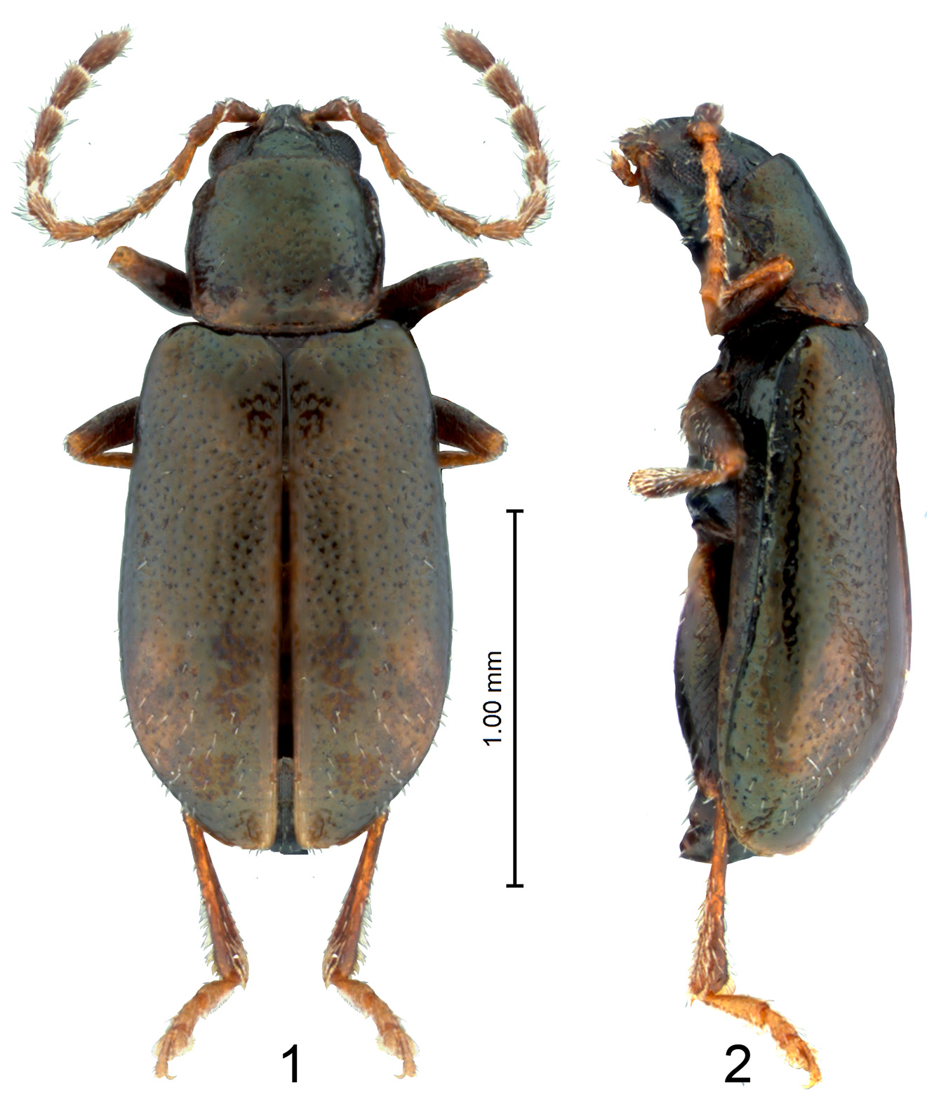
Body length: 1.90–2.20 mm; body width (widest point of elytra): 0.80–1.10 mm. Pronotum width to length: 1.08– 1.12. Width of elytra at base (in middle of humeral calli) to width of pronotum at base: 1.30–1.40.
Head, pronotum except base, ventral side of body, and femora dark brown to black with light bronzish tint. Elytral disc, basal part of pronotum, tibiae, tarsi, and antennae light brown. Elytral surface and frontal part of pronotum with sparse, erect hairs ( Figs 1, 2 View FIGURES 1, 2 ).
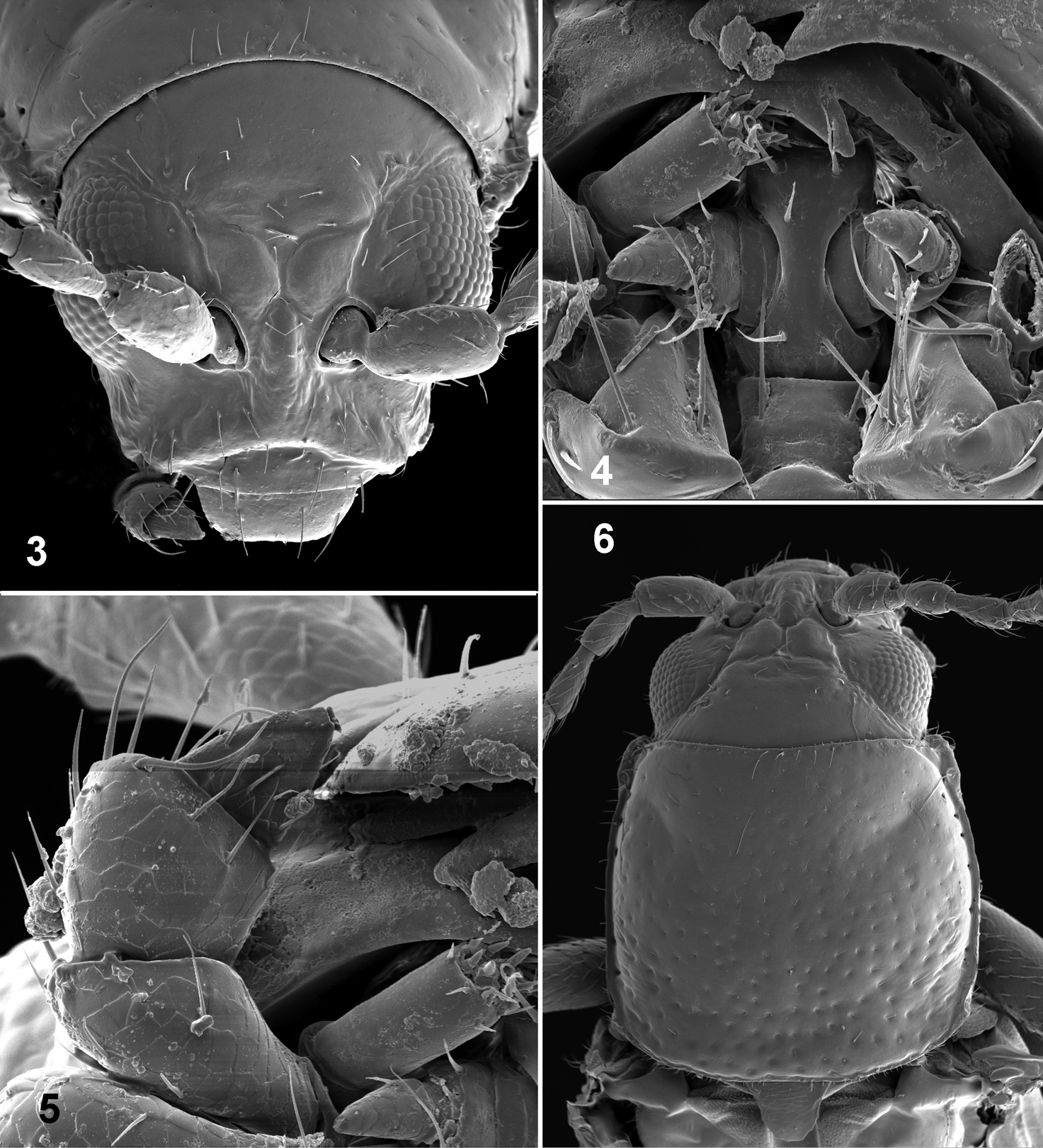
Head ( Figs 3, 6 View FIGURES 3 – 6 ) with midcranial and frontal sutures absent. Supraorbital sulcus shallow, sometimes consisting of few long and parallel wrinkles. Orbital sulcus and supracallinal sulci well developed. Supraantennal sulcus shallow, poorly developed. Supracallinal sulcus oblique and slightly convex. Midfrontal sulcus well developed, long; antennal calli completely separated. Suprafrontal and frontolateral sulci well developed. Antennal callus long, oblique, nearly trapezoidal, entering interantennal space. Surface of antennal callus above surface of vertex. Frontal ridge and vertex separated by antennal calli. Width of frontal ridge to width of antennal socket (counting surrounding ridges) 0.70–0.75. Frontal ridge in lateral view moderately convex. Area below antennal socket concave. Orbit normally wide, nearly as wide as transverse diameter of antennal socket. Distance between eyes above antennal sockets to transverse diameter of eye in frontal view 3.25–3.32. Sides of head below eyes converging ventrally. Labrum flat, trapezoid, with 2 pairs of setae; anterior margin slightly emarginate. Apical maxillary palpomere conical ( Fig. 5 View FIGURES 3 – 6 ). Preapical maxillary palpomere much wider than apical palpomere. Antennal sockets situated below middle of eye. Antenna filiform, with 11 antennomeres. Length of antenna over pronotum reaching middle of elytron.
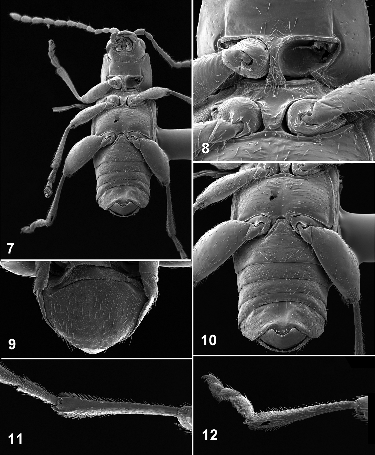
Pronotum ( Fig. 6 View FIGURES 3 – 6 ) about as long as wide, with sides only slightly convex. Pronotal surface near base slightly transversely impressed, impression without distinct borders. Pronotal surface near apex with two oblique, shallow impressions forming in middle an even shallower impression. Pronotal base straight. Lateral margin of pronotum slightly explanate, with short setae. Anterolateral callosity relatively long, nearly parallel to lateral margin. Posterolateral callosity short, not protruding beyond lateral margin. Pronotal punctures larger and denser at base than at apex. A few short setae situated on apical margin and on basal margin near corners. Procoxal cavities open ( Fig. 8 View FIGURES 7 – 12 ). Intercoxal prosternal process thin, extends beyond procoxae, lateral sides concave, posterior end slightly widened and straight.
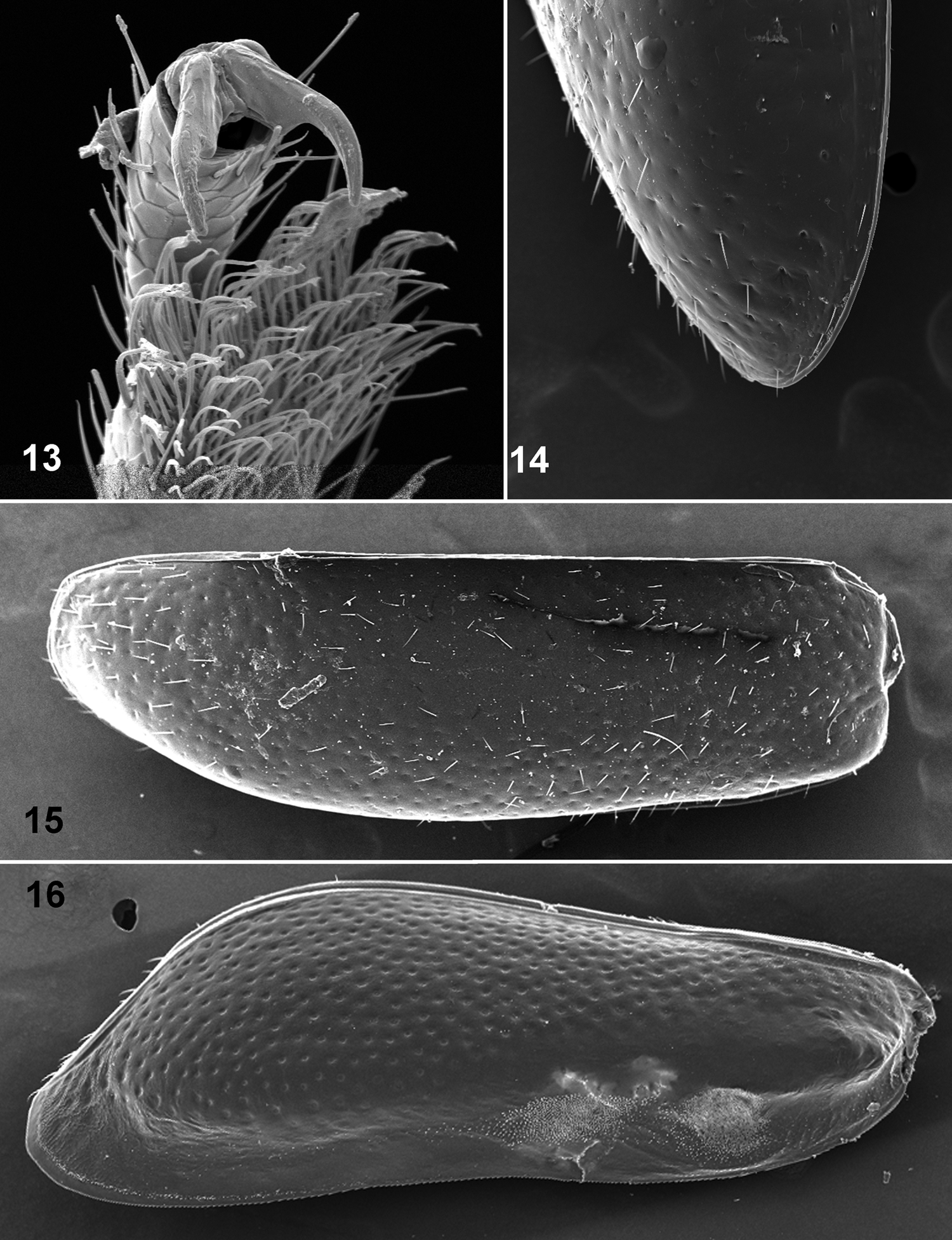
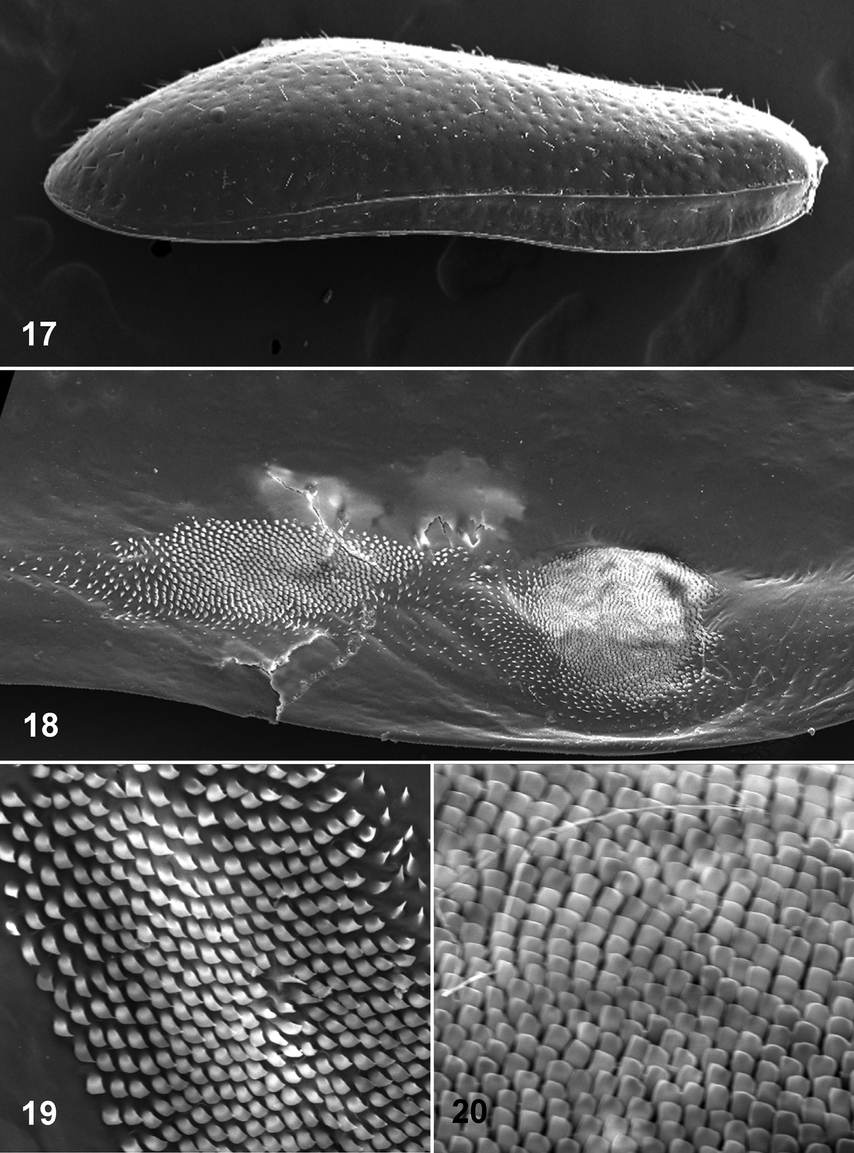
Scutellum present. Elytron ( Fig. 15 View FIGURES 13 – 16 ) with punctures confused, dorsal surface covered with sparse erected setae. Elytra at base wider than base of pronotum. Humeral calli well developed. Basal calli poorly developed with shallow impression posteriad. Epipleura ( Fig. 17 View FIGURES 17 – 20 ) oblique outwardly, gradually narrowing from base to apex, reaching end of side of elytron, but not apex. Width of epipleura in middle equal to width of metafemur at apex. Internal surface of elytron ( Figs 16 View FIGURES 13 – 16 , 18–20 View FIGURES 17 – 20 ) with 2 binding patches, spicules in middle of basal patch shovel shaped ( Fig. 20 View FIGURES 17 – 20 ), spicules on more distant patch ogival in shape, slightly bent anteriorly ( Fig 19 View FIGURES 17 – 20 ). Mesosternum ( Fig. 8 View FIGURES 7 – 12 ) without elevated projection in middle. Metasternum ( Fig. 10 View FIGURES 7 – 12 ) anteriorly without elevated projection in middle and not projecting forward hiding mesosternum. Metepisternum anteriorly wider than posteriorly.
Abdominal ventrites 1 and 2 not fused ( Fig. 10 View FIGURES 7 – 12 ). Abdominal ventrite 1 as long as ventrites 2, 3 and 4 together. Abdominal ventrite 5 longer than ventrites 4 and 3 together, evenly convex in female, in male obliquely cut on sides with lobe in middle. First abdominal ventrite beween coxae without longitudinal ridges, with apex narrowly truncate. Last visible tergite without longitudinal groove in middle.
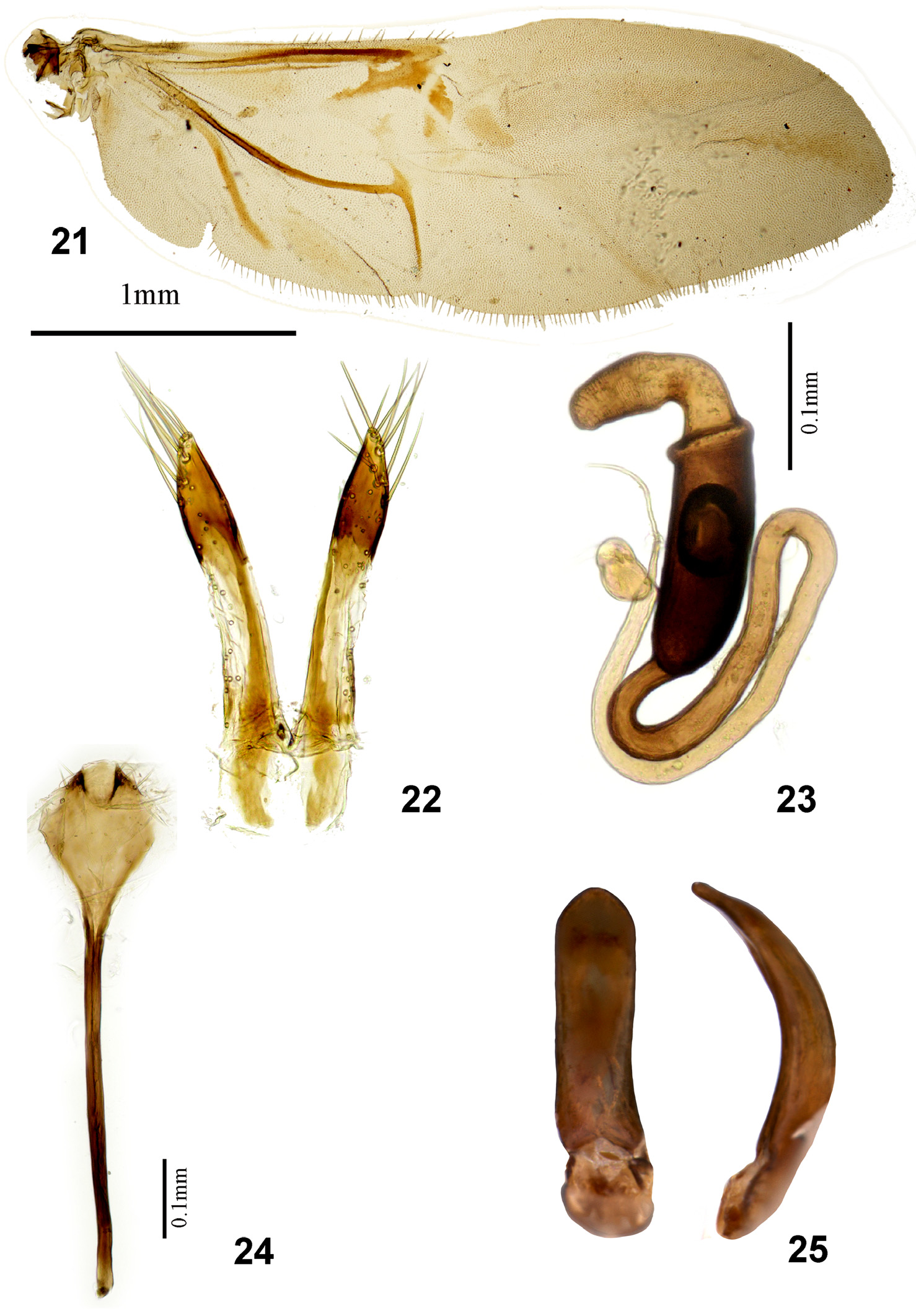
Protibial and mesotibial spurs absent. Metatibia straight ( Figs 7, 11, 12 View FIGURES 7 – 12 ). Metatibia in crossection around its middle more or less cylindrical. Middle part of metatibia dorsally convex. Bristles present on lateral and mesal sides of metatibiae. Metatarsomere 1 attached to apex of metatibia. Apical spur of metatibia simple, much shorter than maximum width of metatibial tip, narrow, ending in one tooth, situated medially. Tarsus with tarsomere 3 round, wider than long, with small incision in middle. First metatarsomere relatively long, but shorter than those in Longitarsus Latreille , convex dorsally, flat ventrally. Claws slightly appendiculate ( Fig. 13 View FIGURES 13 – 16 ). Wing with generally reduced veins. Radial vein slender, rp-mp2 short ( Fig. 21 View FIGURES 21 – 25 ).
Median lobe of male genitalia in cross section somewhat oval ( Fig. 25 View FIGURES 21 – 25 ).
Spermatheca with distinct border between receptacle and pump. Receptacle longer than wide, much wider and longer than pump, straight. Spermathecal duct curved, without coils ( Fig. 23 View FIGURES 21 – 25 ). Vaginal palpi not fused medially, with length many times greater than width ( Fig. 22 View FIGURES 21 – 25 ). Tignum with narrow base and dilated apex ( Fig. 24 View FIGURES 21 – 25 ).
Etymology. We named the genus after the Pénghoú () , a tree spirit from Chinese folklore. The name is masculine.
Type species. Penghou yulongshan Ruan, Konstantinov, Prathapan, Ge, Yang , new species.
Distribution. China (Yunnan province.).
Host plant. Unknown.
Remarks. Penghou is markedly different from all known genera of Chinese and broadly Oriental flea beetles. However, based on the general shape of the beetle body, setose elytra and general shape of the legs, Penghou is similar to Hespera and related genera ( Hesperomorpha Ogloblin and Taiwanohespera Kimoto ), Laotzeus Chen , Luperomorpha , Mandarella Duvivier , Omeiana Chen , and Stenoluperus Ogloblin. Penghou may be easily distinguished from the latter genera based on the following characters: smaller body size (maximum— 2.2 mm, while average body size of Hespera — Stenoluperus is 3.6 mm); pronotum clearly elongate, about as long as wide, pronotum in Hespera — Stenoluperus generally much wider than long; pronotum with weak antebasal transverse impression and two oblique impression near apex, pronotum in Hespera — Stenoluperus (except for Laotzeus ) lacks impressions, respectively; anterolateral callosity of pronotum elongate, barely extending beyond pronotal margin, the callosity is rounded, extending far beyond pronotal margin in Hespera — Stenoluperus (except some species of Luperomorpha ); supracallinal sulci oblique, they are generally perpendicular to mid of the head in Hespera — Stenoluperus .
No known copyright restrictions apply. See Agosti, D., Egloff, W., 2009. Taxonomic information exchange and copyright: the Plazi approach. BMC Research Notes 2009, 2:53 for further explanation.