
Hyalomma (Hyalommina) hussaini Sharif, 1928
|
publication ID |
https://doi.org/10.5281/zenodo.186557 |
|
DOI |
https://doi.org/10.5281/zenodo.6213755 |
|
persistent identifier |
https://treatment.plazi.org/id/039087FB-6074-FFBA-4BAC-A2153BF6FEF6 |
|
treatment provided by |
Plazi (2016-04-19 08:04:59, last updated 2024-11-24 23:00:28) |
|
scientific name |
Hyalomma (Hyalommina) hussaini Sharif, 1928 |
| status |
|
Hyalomma (Hyalommina) hussaini Sharif, 1928 View in CoL
( Figs. 7–12 View FIGURE 7 View FIGURE 8 View FIGURE 9 View FIGURE 10 View FIGURE 11 View FIGURE 12 )
Type specimens. Syntypes (sex not indicated; not quantified) ex cattle from Akola Town in the Central Provinces [now Maharashtra State, India]; deposited in the Indian Museum, reg. № 57/18 (Kolkata, India) (p. 317, Sharif 1928).
Material studied. A total of 31 males, 26 females, 7 nymphs and 32 larvae that originated from India, Myanmar and Pakistan were examined in the current study.
Synonym. Hyalomma hussaini forma typica Sharif, 1928
Description. Male ( Figs. 7 View FIGURE 7 , 8 View FIGURE 8 )
Conscutum ( Fig. 7 View FIGURE 7 ): length 2.21–2.93 (2.65 ± 0.17, n = 25), width 1.34–1.82 (1.65 ± 0.12, n = 25), ratio length:width 1.51–1.76 (1.61 ± 0.06, n = 25); dark red-brown; sparse large punctations mainly on anterior part of conscutum, lateral and caudal fields. Spiracular plate ( Fig. 8 View FIGURE 8 C): perforated portion of prolongation very broad.
Female ( Figs. 9 View FIGURE 9 , 10 View FIGURE 10 )
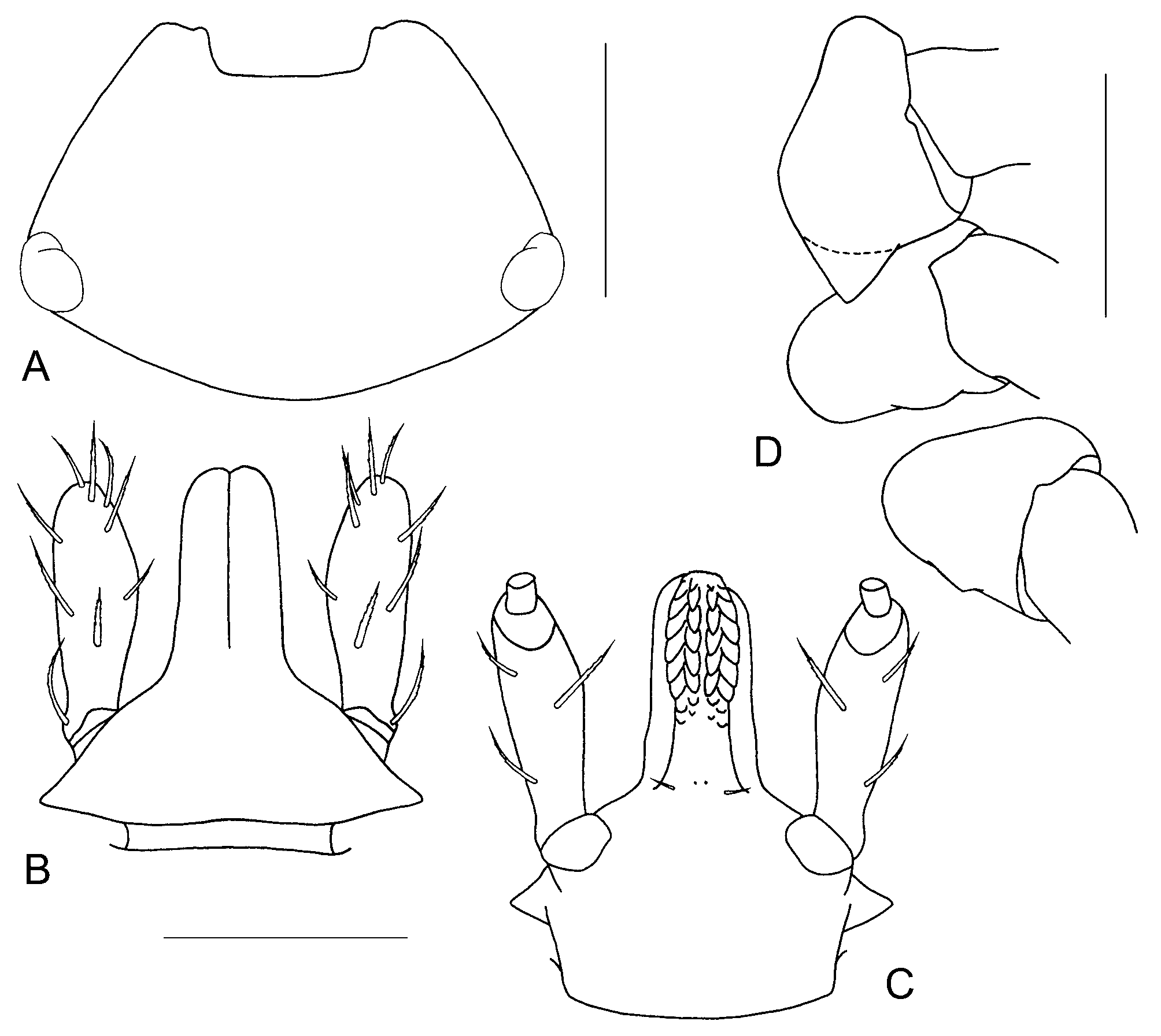
Scutum ( Fig. 9 View FIGURE 9 ): length 1.62–1.88 (1.75 ± 0.08, n = 19), width 1.52–1.85 (1.69 ± 0.09, n = 19), ratio length:width 1.00–1.06 (1.03 ± 0.02, n = 19); usually dark red-brown; large, deep punctations sparse, evenly distributed over scutum. Genital structures ( Fig. 10 View FIGURE 10 A): genital aperture very wide, arcuate (U-shaped); vestibular portion of vagina markedly bulging; preatrial fold of genital aperture bulging anteriorly and sloping sharply posteriorly ( Fig. 10 View FIGURE 10 B).
Nymph ( Fig. 11 View FIGURE 11 )
Scutum ( Fig. 11 View FIGURE 11 A): length 449–525 (487±27.51, n=6), width 453–533 (494±27.59, n=6), ratio length:width 0.88–1.06 (0.99±0.07, n=6), distance between posterior margin of eyes and posterior margin of scutum 151–175 (156±9.63, n=6), width:length of posterior portion of scutum 2.82–3.39 (3.16±0.22, n=6); slight posterolateral depressions on either side of scutal extremity.
Basis capituli ( Figs. 11 View FIGURE 11 D, E): length 312–376 (351±21.97, n=6); width 292–332 (313±16.33, n=6), ratio length:width 1.07–1.15 (1.12±0.03, n=6); ventrally lateral saliences with moderate spur directed posteriorly. Palpi (segment II) ( Figs. 11 View FIGURE 11 D, E): length 140–176 (158±11.48, n=6), width 45–48 (47±1.09, n=6), ratio length:width 3.04–3.83 (3.37±0.26, n=6). Hypostome ( Fig. 11 View FIGURE 11 E): length 144–178 (165±11.64, n=6), width 50–54 (52±1.26, n=6), ratio length:width 2.77–3.42 (3.18±0.23, n=6); rounded at apex; 7 or 8 large denticles in median file.
Coxae ( Fig. 11 View FIGURE 11 F): coxae II with large triangular spur.
Larva ( Fig. 12 View FIGURE 12 )
Scutum ( Fig. 12 View FIGURE 12 A): length 208–232 (223±5.69, n=32), width 300–344 (325±9.21, n=32), ratio length:width 0.66–0.72 (0.69±0.01, n=32), distance from posterior margin of eyes to posterior margin of scutum 48–58 (53±2.32, n=32), ratio width:length of posterior portion 5.57–6.92 (6.15±0.31, n=32). Portion of scutum posterior to eyes nearly equal to 1/4 of scutal length.
Basis capituli ( Figs. 12 View FIGURE 12 B, C): length 136–160 (147±5.60, n=32), width 150–164 (158±3.42, n=32), ratio length:width 0.89–1.00 (0.93±0.03, n=32). Palpi (segments II and III) ( Figs. 12 View FIGURE 12 B, C): length 104–114 (109±2.18, n=32), width 32–34 (33±0.83, n=32), ratio length:width 3.12–3.44 (3.26±0.08, n=32). Hypostome ( Fig. 12 View FIGURE 12 C): length 80–90 (86±2.33, n=32), width 22–24 (22±0.62, n=32), ratio length:width 3.58–4.00 (3.80±0.10, n=32); 6 large denticles in median file.
Genu I: length 114–128 (120±2.44, n=32), width 42–46 (44±1.40, n=7), ratio length:width 2.56–2.86 (2.69±0.10, n=7).
Hosts. The main hosts of the adults are large and medium-sized domestic and wild ungulates: cattle, buffaloes, camels, goats, sheep, pigs, horses and nilgai, B. tragocamelus . The adults have also been recorded from domestic dogs, a bear and humans (our data; Sharif 1928; Kaiser & Hoogstraal 1964; Miranpuri & Naithani 1978; Geevarghese & Dhanda 1987; Robbins et al. 2002).
The chief hosts of the immature stages are small mammals such as rodents and shrews. Larvae and nymphs have been recorded from: Asian house shrew, S. murinus , house rat, R. rattus, Indian bush rat, Golunda ellioti Gray, Indian desert jird, Meriones hurrianae Jordon and rock-loving mouse, M. saxicola ( Singh & Dhanda 1965; Wattal & Srivastva 1967; Geevarghese & Dhanda 1987).
Geographic distribution. Asia: India, Myanmar and Pakistan (our data; Sharif 1928; Kaiser & Hoogstraal 1964; Geevarghese & Dhanda 1987; Robbins et al. 2002).
Geevarghese, G. & Dhanda, V. (1987) The Indian Hyalomma ticks (Ixodoidea: Ixodidae). Indian Council of Agricultural Research, New Delhi, India, 119 pp.
Kaiser, M. N. and Hoogstraal, H. (1964) The Hyalomma ticks (Ixodoidea, Ixodidae) of Pakistan, India, and Ceylon, with keys to subgenera and species. Acarologia, 6, 257 - 286.
Miranpuri, G. S. & Naithani R. C. (1978) A check list of Indian ticks (Ixodoidea: Acarina). Indian Veterinary Research Institute, Izatnagar, India, 50 pp.
Robbins, R. G., Platt, S. G. & Keirans, J. E. (2002) First report of Hyalomma marginatum isaaci Sharif (Acari: Ixodida: Ixodidae) from the Union of Myanmar, with a concurrent collection of H. hussaini Sharif. Proceedings of the Entomological Society of Washington, 104, 1061 - 1063.
Sharif, M. (1928) A revision of the Indian Ixodidae with special reference to the collection in the Indian Museum. Records of the Indian Museum, 30, 217 - 344.
Singh, K. R. P. & Dhanda, V. (1965) Description and keys of immature stages of some species of Indian Hyalomma Koch, 1844 (Ixodoidea, Ixodidae). Acarologia, 7, 636 - 651.
Wattal, B. L. & Srivastva, S. P. (1967) Ectoparasite fauna of small mammals and domestic animals in the neighbourhood of Alwar city (Rajasthan). Bulletin of the Indian Society for Malaria and other Communicable Diseases, 4, 191 - 202.
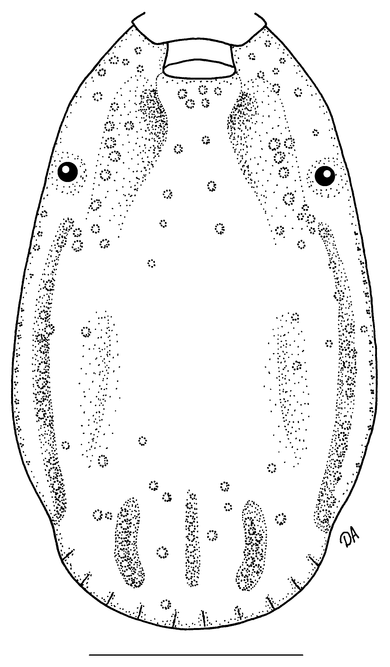
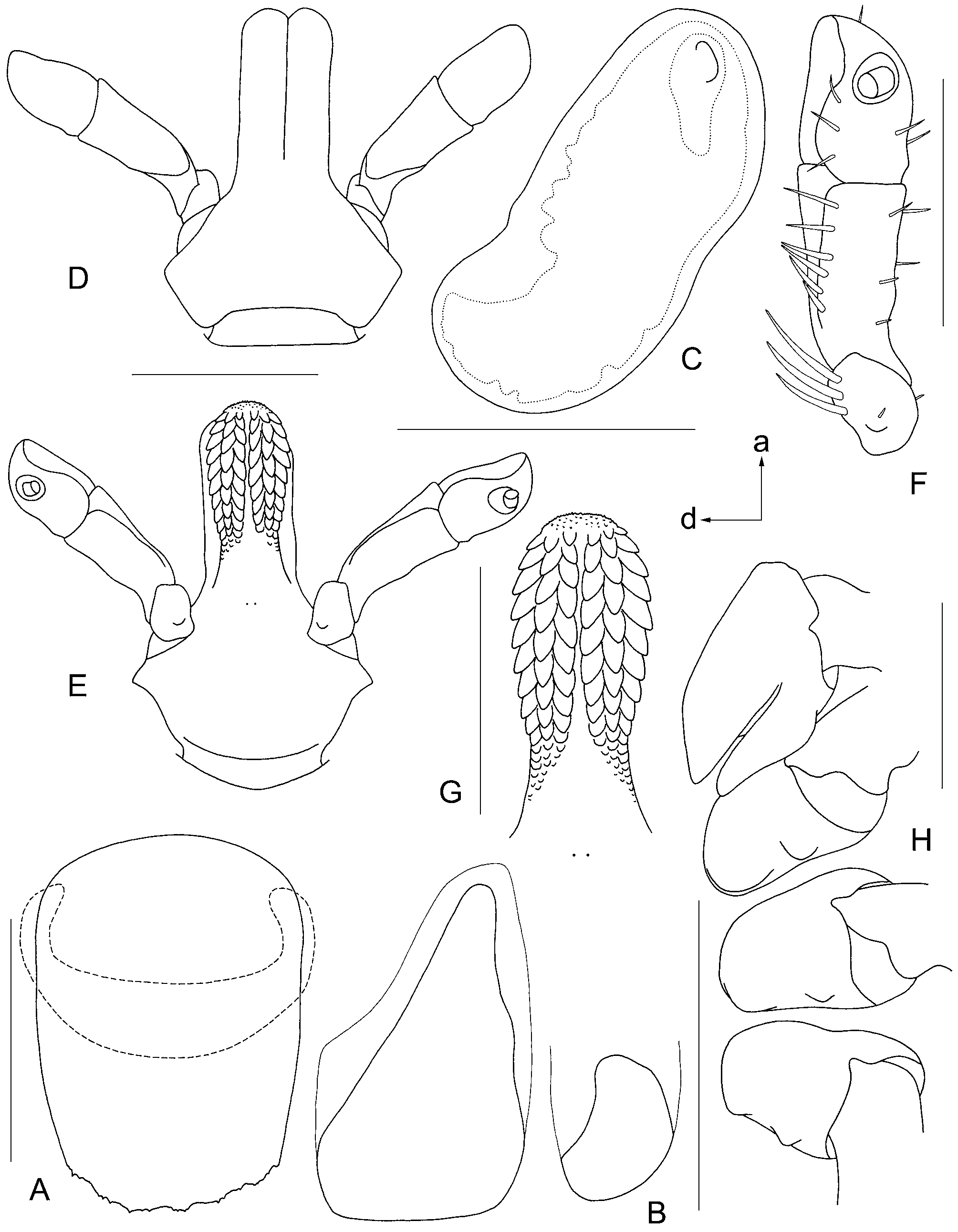
FIGURE 8. Hyalomma hussaini, male. A—genital structures; B—anal plates; C—spiracular plate (a—anterior, d—dorsal); D—gnathosoma dorsally; E—gnathosoma ventrally; F—palp ventrally; G—hypostome; H—coxae. Scale bars: A = 200 μm; B, D, E, H = 500 μm; C, F, G = 400 μm. All setae are omitted except drawing F where only setae of palpal segment IV are omitted.
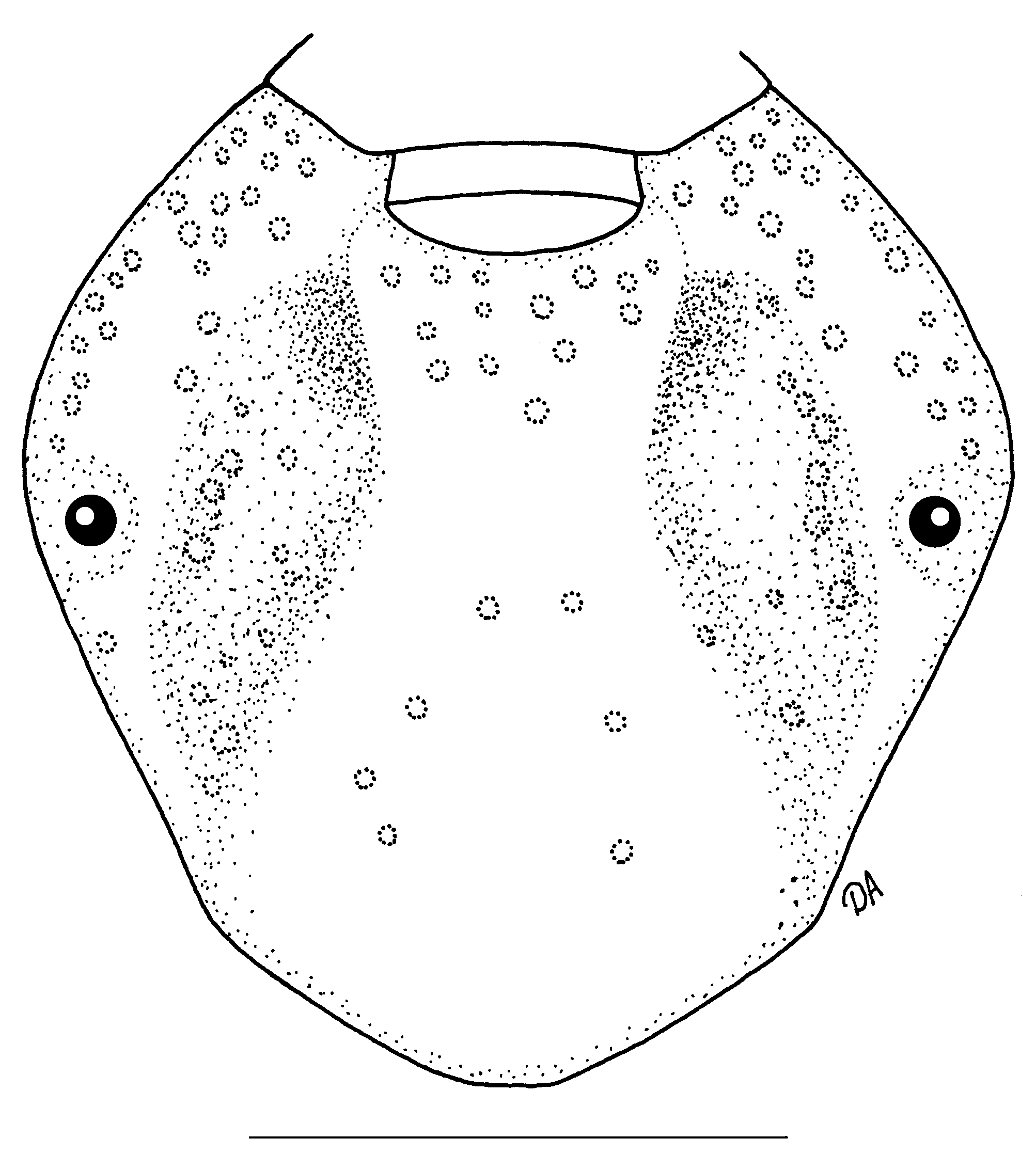
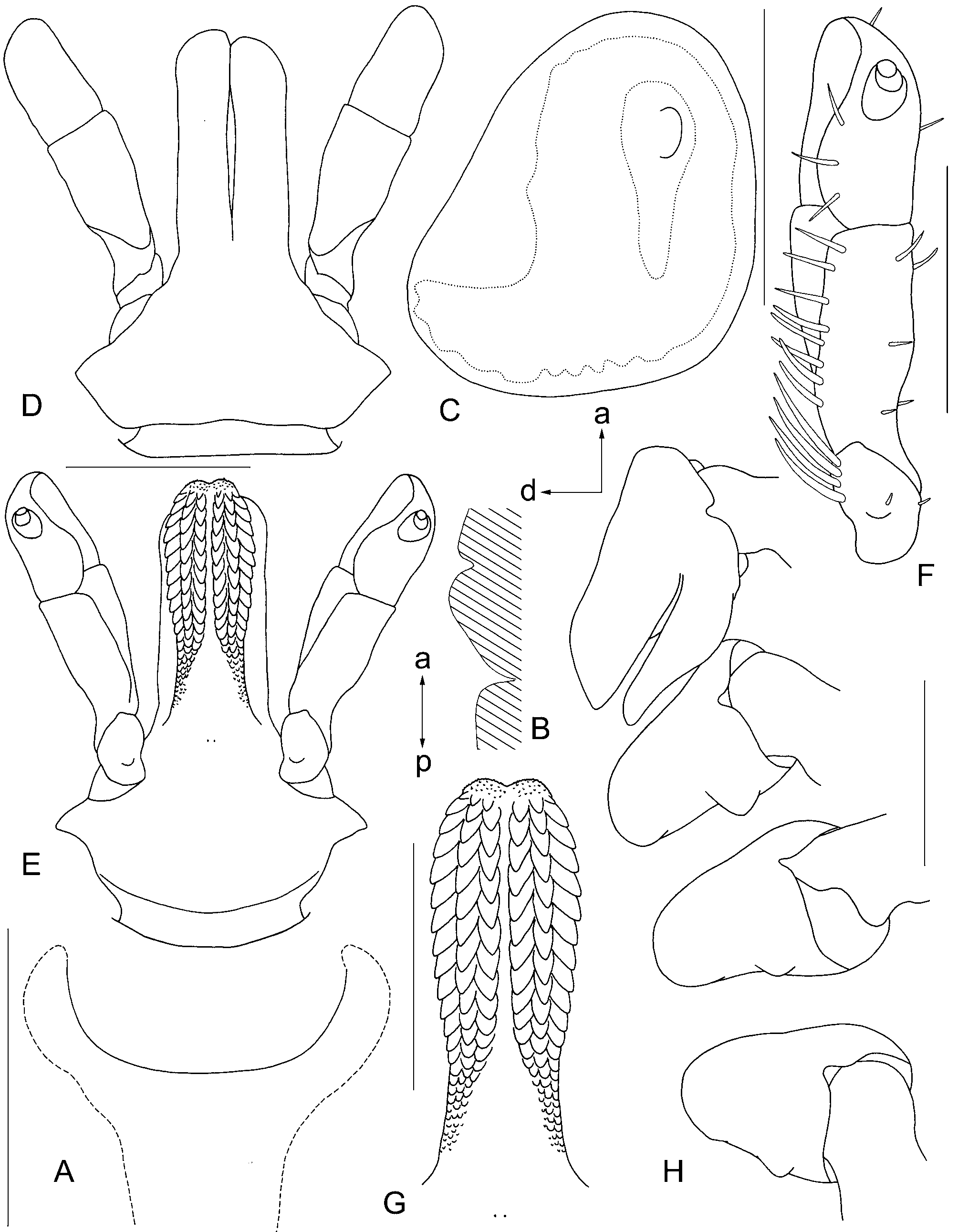
FIGURE 10. Hyalomma hussaini, female. A—genital structures; B—longitudinal cut through preatrial fold of genital aperture schematically (a—anterior, p—posterior); C—spiracular plate (a—anterior, d—dorsal); D—gnathosoma dorsally; E—gnathosoma ventrally; F—palp ventrally; G—hypostome; H—coxae. Scale bars: A = 200 μm; C, F, G = 400 μm; E, D, H = 500 μm. All setae are omitted except drawing F where only setae of palpal segment IV are omitted.
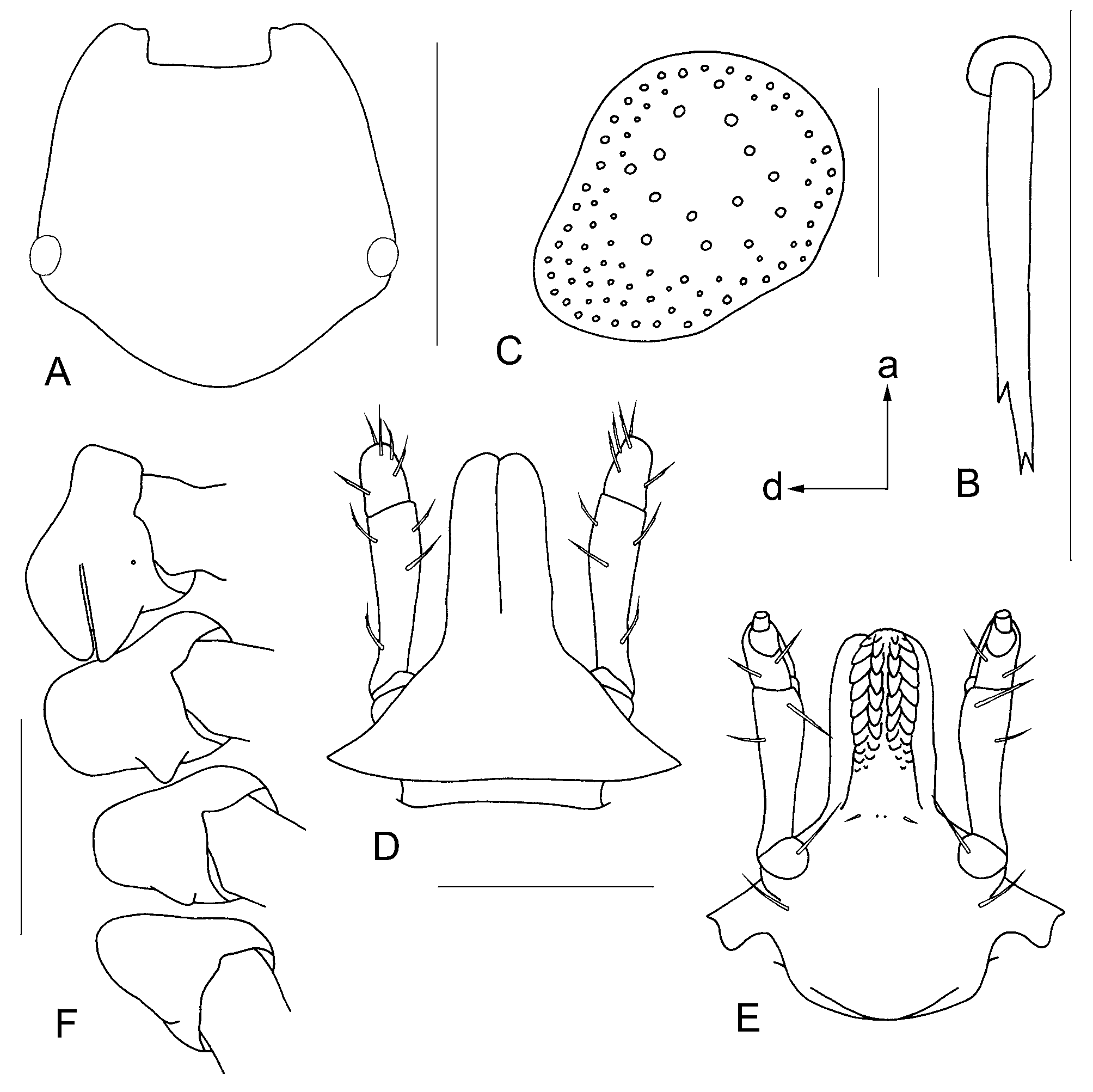
FIGURE 11. Hyalomma hussaini, nymph. A—scutum; B—seta of alloscutum; C—spiracular plate (a—anterior, d—dorsal); D—gnathosoma dorsally; E—gnathosoma ventrally; F—coxae. A = 400 μm; B, C = 50 μm; D, E, F = 200 μm. All setae are omitted except drawings D and E where only setae of palpal segment IV are omitted.
No known copyright restrictions apply. See Agosti, D., Egloff, W., 2009. Taxonomic information exchange and copyright: the Plazi approach. BMC Research Notes 2009, 2:53 for further explanation.
1 (by plazi, 2016-04-19 08:04:59)
2 (by ImsDioSync, 2016-11-30 05:02:05)
3 (by ImsDioSync, 2016-11-30 05:03:27)
4 (by ImsDioSync, 2017-06-13 17:55:45)
5 (by ExternalLinkService, 2019-09-26 12:27:25)
6 (by ExternalLinkService, 2022-01-30 17:39:29)
7 (by ExternalLinkService, 2022-02-22 00:22:31)
8 (by plazi, 2023-10-29 11:50:05)