
Scolytocis Blair, 1928
|
publication ID |
https://doi.org/10.11646/zootaxa.1832.1.1 |
|
publication LSID |
lsid:zoobank.org:pub:45A82AD6-F369-4A74-A280-048BAE6E2DA5 |
|
persistent identifier |
https://treatment.plazi.org/id/038E87B5-FFB8-CE7D-FF6A-4C58FE3CFACB |
|
treatment provided by |
Felipe (2021-07-23 11:52:45, last updated by Plazi 2023-11-03 12:38:44) |
|
scientific name |
Scolytocis Blair, 1928 |
| status |
|
Scolytocis Blair, 1928 ( Figs 1–4 View FIGURES 1–4 , 6 View FIGURES 5–8 , 10 View FIGURES 9–12 , 27–135 View FIGURES 27–29 View FIGURES 30–32 View FIGURES 33–35 View FIGURES 36–38 View FIGURES 39–41 View FIGURES 42–44 View FIGURES 45–47 View FIGURES 48–50 View FIGURES 51–53 View FIGURES 54–56 View FIGURES 57–59 View FIGURES 60–62 View FIGURES 63–65 View FIGURES 66–68 View FIGURES 69–71 View FIGURES 72–73 View FIGURES 74–76 View FIGURES 77–79 View FIGURE 80 View FIGURE 81 View FIGURES 82–84 View FIGURES 85–87 View FIGURES 88–90 View FIGURES 91–93 View FIGURES 94–95 View FIGURES 96–97 View FIGURES 98–100 View FIGURE 101 View FIGURES 102–104 View FIGURES 105–107 View FIGURES 108–110 View FIGURES 111–113 View FIGURES 114–116 View FIGURES 117–119 View FIGURES 120–122 View FIGURES 123–125 View FIGURES 126–127 View FIGURES 128–129 View FIGURES 130–133 View FIGURE 134 View FIGURE 135 )
Included species
Scolytocis bouchardi sp. nov.
Scolytocis cariborum sp. nov.
Scolytocis danielssoni sp. nov.
Scolytocis difficillimus sp. nov.
Scolytocis fritzplaumanni sp. nov.
Scolytocis furieriae sp. nov.
Scolytocis howdeni sp. nov.
Scolytocis indecisus sp. nov.
Scolytocis kiskeyensis sp. nov.
Scolytocis lawrencei sp. nov.
Scolytocis malayanus sp. nov.
Scolytocis novaezelandiae sp. nov.
Scolytocis panamensis sp. nov.
Scolytocis paschoali sp. nov.
Scolytocis philippinensis sp. nov.
Scolytocis samoensis Blair, 1928 (type species, by monotypy)
Scolytocis thayerae sp. nov.
Scolytocis werneri sp. nov.
Scolytocis zimmermani sp. nov.
Diagnosis
Scolytocis may be distinguished from the other Ciidae by the combination of the diagnostic characters of Xylographellini , mainly the compact antennal club in which each antennomere bears more than four sensillifers, and the longitudinally fissured prementum with labial palpi inserted at its middle. Scolytocis differs from the other Xylographellini genera by the combination of the following characters: (i) antennal funicle with four antennomeres; (ii) lateral pronotal margins smooth; (iii) prosternal process laminate; (iv) procoxae subconical. Among the Xylographellini , character i is restricted to Scolytocis . Character ii separates Scolytocis from Xylographella . Characters i, iii and iv separate Scolytocis from the other Xylographellini .
Redescription
Measurements in mm (n = 204; including all species, excepting Scol. samoensis Blair ): TL 1.00–2.05; PL 0.32–0.79; PW 0.42–0.89; EL 0.58–1.26; EW 0.42–0.95; GD 0.42–0.89. Body light brown to black, shiny to dull on dorsum, subglabrous, vestiture consisting of minute, inconspicuous fine setae, sometimes with moderately long setae on the ventral surface. Head strongly declined, barely visible from above. Frontoclypeal region simple in both genders. Labium ( Figs 3 View FIGURES 1–4 , 6 View FIGURES 5–8 ) with prementum elongate, subpentagonal, apex subacute, external surface bearing a longitudinal sulcus at midline, with labial palpi inserted at its middle and apical palpomere widely expanded. Antennae inserted in front of eye ( Fig. 1 View FIGURES 1–4 ); fossa deep; funicle always bearing four antennomeres; club compact (with three antennomeres; Figs 2 View FIGURES 1–4 , 63A–65B View FIGURES 63–65 , 94 View FIGURES 94–95 , 126–127 View FIGURES 126–127 ) or with the antennomeres completely fused to each (so that it appears to be one large antennomeres; 65C); apical antennomere of the club bearing at least five sensillifers ( Fig. 2 View FIGURES 1–4 , arrows) formed by a group of short and cylindrical sensilla. Pronotum with anterior edge simple in both sexes, broadly rounded, anterolateral angles obtuse, not produced; anterolateral margins straight or slightly arched inwards; posterolateral angles broadly rounded or angulate; lateral pronotal carinae smooth, not visible for their entire lengths from above; posterior margin sometimes bearing a rugose border along it. Scutellum conspicuous, triangular, glabrous, punctate or not; basal width more than 0.10X the EW. Elytra subquadrate to suboval; apex truncate or acute, convex or slightly concave, sometimes bearing small cuticular globules ( Figs 82–84 View FIGURES 82–84 , small arrows; each globule bearing a small fine seta); posterolateral corners bending dorsally, lateral margins usually not visible from above; suture margined or not, but never deflexed at apex; punctation single, seriate to confuse. Hindwings always fully developed, sometimes extremely membranous; radial cell present and conspicuous; apical area always longer than basal area, without pigmented patches or with one to two pigmented lines/patches (complete or incomplete; distinct or vague). Metaventrite suture (discrimen) absent, or inconspicuously short to long, usually restricted to the basal region ( Fig. 4 View FIGURES 1–4 ). Prosternum very short; concave, biconcave or triconcave; with or without longitudinal carina at midline; prosternal process laminate and slightly curved. All tibiae expanded to middle or to apex, outer margin bearing a row of several spines ( Figs 66–68 View FIGURES 66–68 , 95 View FIGURES 94–95 , 128–129 View FIGURES 128–129 ). Outer face of protibiae setose along the inner margin; inner face of meso and metatibiae with a setose region near the inner margin, restricted to apical third or extending to base. Femorae subglabrous on internal and external surfaces. Each tarsus formed by four tarsomeres. Male without abdominal setose patch or with a very small, inconspicuous setose patch in the midline of the first abdominal ventrite, barely visible even in high magnifications (around 100X) or in slide preparations. Male genitalia. Ninth segment Y-shaped ( Figs 69 View FIGURES 69–71 , 73 View FIGURES 72–73 , 76 View FIGURES 74–76 , 78–79 View FIGURES 77–79 , 96–99 View FIGURES 96–97 View FIGURES 98–100 , 130–132 View FIGURES 130–133 ). Tegmen and median lobe elongate, cylindrical (69–75, 77–79, 96–100, 130–133). Tegmen with sclerotized apex and membranous basal portion; median lobe as long as tegmen or longer, membranous to barely sclerotized. Female terminalia. ( Fig. 10 View FIGURES 9–12 ) Eighth sternite broadly rounded or slightly arched inwards at apex. Spiculum ventrale slightly longer to twice as long as ovipositor. Gonostyli of ovipositor always absent.
Distribution
Scolytocis is found on all the regions where the subtribe Xylographellina occurs, except on Japan. However, it is most diversified in the neotropics, with 12 known species (all these species being described here).
Comments
Six morphological species groups are recognizable, and are arbitrarily named here as follows: (i) the bouchardi group, comprising the species with triconcave prosternum and larger hindwings (in comparison to the species of the lawrencei group), and including Scol. bouchardi sp. nov. and Scol. difficillimus sp. nov.; (ii) the lawrencei group, comprising the small species with tumid prosternum and small hindwings, and including Scol. cariborum sp. nov., Scol. kiskeyensis sp. nov., Scol. lawrencei sp. nov., Scol. malayanus sp. nov. and Scol. samoensis Blair ; (iii) the danielssoni group, comprising the large Central-American species with a conspicuous rugose border along the posterior pronotal margin and a biconcave prosternum bearing a narrow longitudinal carina at midline, including Scol. danielssoni sp. nov., Scol. howdeni sp. nov., Scol. indecisus sp. nov. and Scol. panamensis sp. nov.; (iv) fritzplaumanni group, comprising the species with smooth posterior pronotal border and a biconcave prosternum, and including the Brazilian Scol. fritzplaumanni sp. nov., Scol. furieriae sp. nov., and Scol. paschoali sp. nov.; (v) the werneri group, comprising the species with a concave prosternum and a rugose border along the posterior pronotal margin, including Scol. novaezelandiae sp. nov. and Scol. werneri sp. nov.; (vi) the zimmermani group, comprising the species with each metatibia expanded to the middle, with its outer margin broadly rounded and bearing several spines very close to each other at the apical half, and including Scol. philippinensis sp. nov., Scol. thayerae sp. nov. Scol. zimmermani sp. nov. The species-groups are just taxonomic tools, and they cannot be considered a priori to be monophyletic taxa ( Lopes-Andrade et al. 2003a).
Blair, K. G. (1928) Heteromera, Bostrychoidea, Malacodermata and Buprestidae. In: Insects of Samoa and Other Samoan Terrestrial Arthropoda, Part 45. Coleoptera. Fasc. 2. British Museum (Natural History), London, 67 - 109.
Lopes-Andrade, C., Gumier-Costa, F. & Zacaro, A. A. (2003 a) Cis leoi, a new species of Ciidae (Coleoptera: Tenebrionoidea) from the Neotropical Region. Zootaxa, 161, 1 - 7.
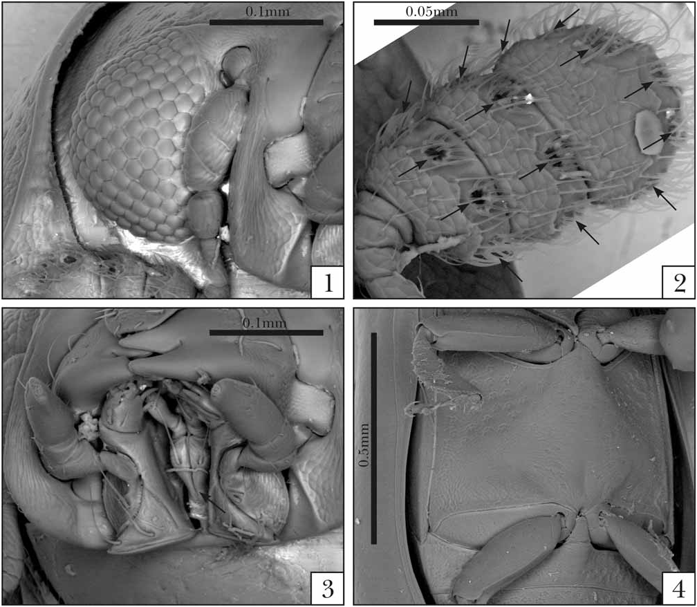
FIGURES 1–4. Scolytocis paschoali sp. nov. 1. Part of the head, frontal view showing the eye. 2. Antennal club, showing the sensillifers (arrows). Note that the club is compact, with antennomeres not freely articulated. 3. Head, ventral view showing the mouthparts. Note the longitudinal sulcus of prementum (arrow). 4. Ventral view showing the metaventrite.
FIGURES 5–8. Prementum (pm) with labial palpi (l.p). Note that the apical labial palpomeres are enlarged (small arrows), and that the apex of prementum is acute in Xylographella and Scolytocis, and rounded in Syncosmetus and Tropicis (see big arrows). 5. Xyl. punctata Miyatake, lateral view. 6. Scol. paschoali sp. nov. 7. Sync. reticulatus Miyatake. 8. Trop. sexcarinatus (Waterhouse).

FIGURES 9–12. Female terminalia, showing the gonostyli (gs), gonocoxites (gc), baculi of basal gonocoxites (small arrows), paraprocts (pp), baculi of paraprocts (b.pp), proctiger (pt), and spiculum ventrale (sv). The baculi of proctiger are barely sclerotized, so they are not indicated. 9. Xylographella punctata Miyatake. 10. Scolytocis furieriae sp. nov. 11. Syncosmetus japonicus Sharp. 12. Tropicis sexcarinatus (Waterhouse).


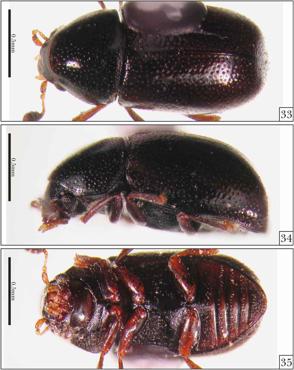

FIGURES 36–38. Scolytocis difficillimus sp. nov, holotype. 36. Dorsal view. 37. Lateral view. 38. Ventral view.


FIGURES 45–47. Scolytocis kiskeyensis sp. nov, holotype. 45. Dorsal view. 46. Lateral view. 47. Ventral view.

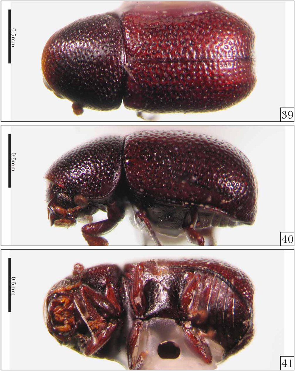
FIGURES 48–50. Scolytocis lawrencei sp. nov, holotype. 48. Dorsal view. 49. Lateral view. 50. Ventral view.


FIGURES 54–56. Hindwings of Northern Neotropical Scolytocis Blair. 54. Scol. bouchardi sp. nov. 55. Scol. cariborum sp. nov. 56. Scol. danielssoni sp. nov.

FIGURES 57–59. Hindwings of Northern Neotropical Scolytocis Blair. 57. Scol. difficillimus sp. nov. 58. Scol. howdeni sp. nov. 59. Scol. indecisus sp. nov.

FIGURES 60–62. Hindwings of Northern Neotropical Scolytocis Blair. 60. Scol. kiskeyensis sp. nov. 61. Scol. lawrencei sp. nov. 62. Scol. panamensis sp. nov.
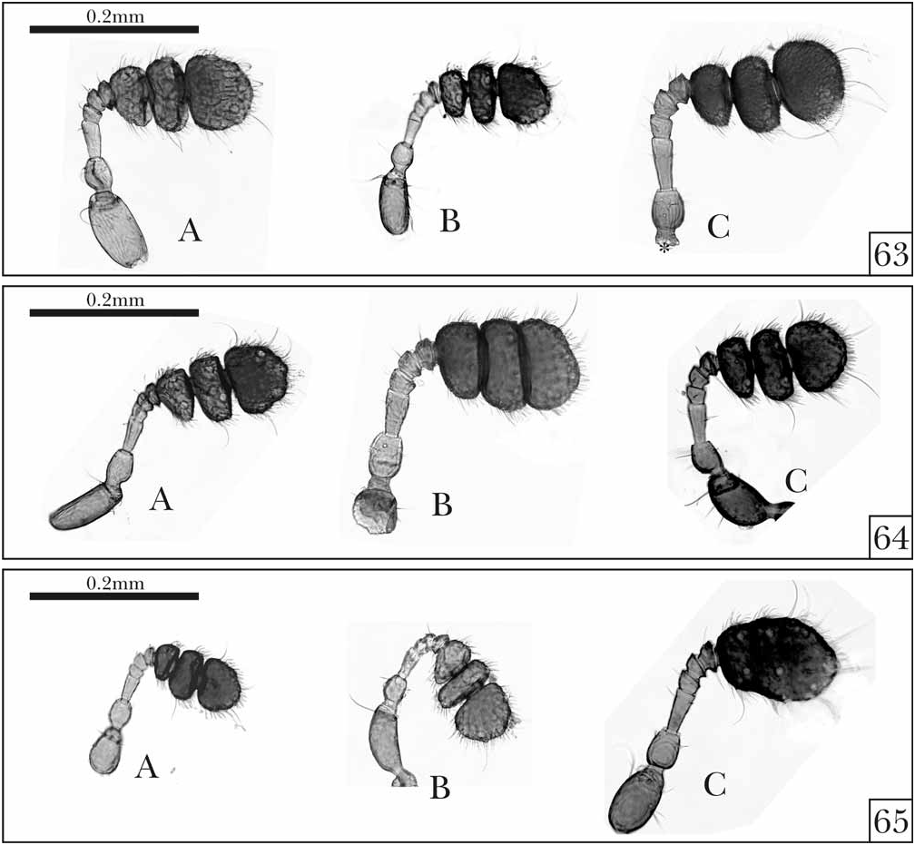
FIGURES 63–65. Antennae of the Northern Neotropical Scolytocis Blair. 63. Scol. bouchardi sp. nov. (A), Scol. cariborum sp. nov. (B), Scol. danielssoni sp. nov. (C). 64. Scol. difficillimus sp. nov. (A), Scol. howdeni sp. nov. (B), Scol. indecisus sp. nov. (C). 65. Scol. kiskeyensis sp. nov. (A), Scol. lawrencei sp. nov. (B), Scol. panamensis sp. nov. (C). First antennomere not shown in Scol. danielssoni sp. nov. (63C, asterisk).

FIGURES 66–68. Metatibiae of the Northern Neotropical Scolytocis Blair. 66. Scol. bouchardi sp. nov. (A), Scol. cariborum sp. nov. (B), Scol. danielssoni sp. nov. (C). 67. Scol. difficillimus sp. nov. (A), Scol. howdeni sp. nov. (B), Scol. indecisus sp. nov. (C). 68. Scol. kiskeyensis sp. nov. (A), Scol. lawrencei sp. nov. (B), Scol. panamensis sp. nov. (C).

FIGURES 69–71. Male genitalia of Scolytocis Blair showing the Y-shaped ninth segment (ix), basal piece (b.p), tegmen (teg) and penis (pen). 69. Scol. bouchardi sp. nov. 70. Scol. cariborum sp. nov. (ninth segment broken; see asterisks). 71. Scol. difficillimus sp. nov.
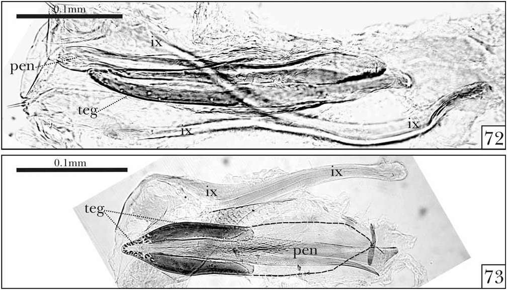
FIGURES 72–73. Male genitalia of Scolytocis Blair showing the Y-shaped ninth segment (ix), tegmen (teg) and penis (pen). 72. Scol. howdeni sp. nov. 73. Scol. indecisus sp. nov. The dashed line indicates the membranous basal portion of tegmen. Note that the apical portion of tegmen is extremely sclerotized.
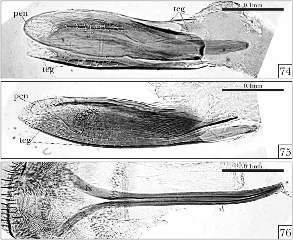
FIGURES 74–76. Male genitalia of Scolytocis kiskeyensis sp. nov. showing the Y-shaped ninth segment (ix), tegmen (teg) and penis (pen). 74. Tegmen and penis of specimen from the Dominican Republic (type locality). 75. Tegmen and penis of specimen from Puerto Rico. 76. Ninth segment (pregenital segment) of specimen from Puerto Rico.
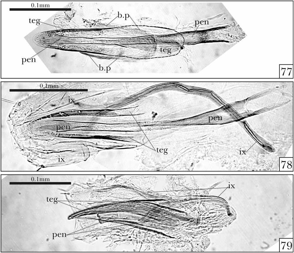
FIGURES 77–79. Male genitalia of Scolytocis Blair showing the Y-shaped ninth segment (ix), basal piece (b.p), tegmen (teg) and penis (pen). 77. Scol. lawrencei sp. nov., specimen from Costa Rica. The dashed line indicates the membranous basal portion of tegmen. 78. Scol. lawrencei sp. nov., specimen from Panama. 79. Scol. panamensis sp. nov.
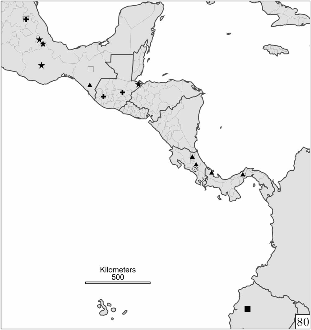
FIGURE 80. Distribution map of the continental Northern Neotropical species of Scolytocis Blair, showing the known distributions of Scol. bouchardi sp. nov. (stars), Scol. danielssoni sp. nov. (double circles), Scol. difficillimus sp. nov. (fulfilled square), Scol. howdeni sp. nov. (open squares), Scol. indecisus sp. nov. (plus symbols), Scol. lawrencei sp. nov. (fulfilled triangles), Scol. panamensis sp. nov. (open circle).
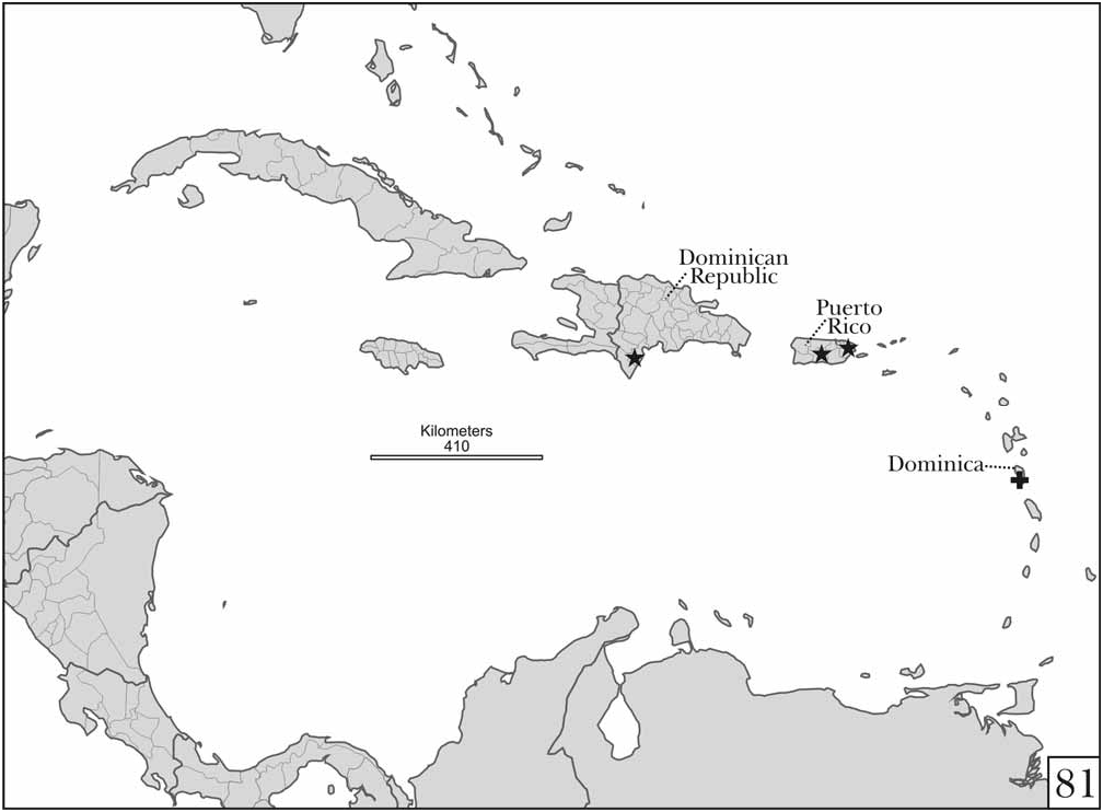
FIGURE 81. Distribution map of the insular Northern Neotropical species of Scolytocis Blair, showing the known distributions of Scol. cariborum sp. nov. (plus symbol) and Scolytocis kiskeyensis sp. nov. (stars).
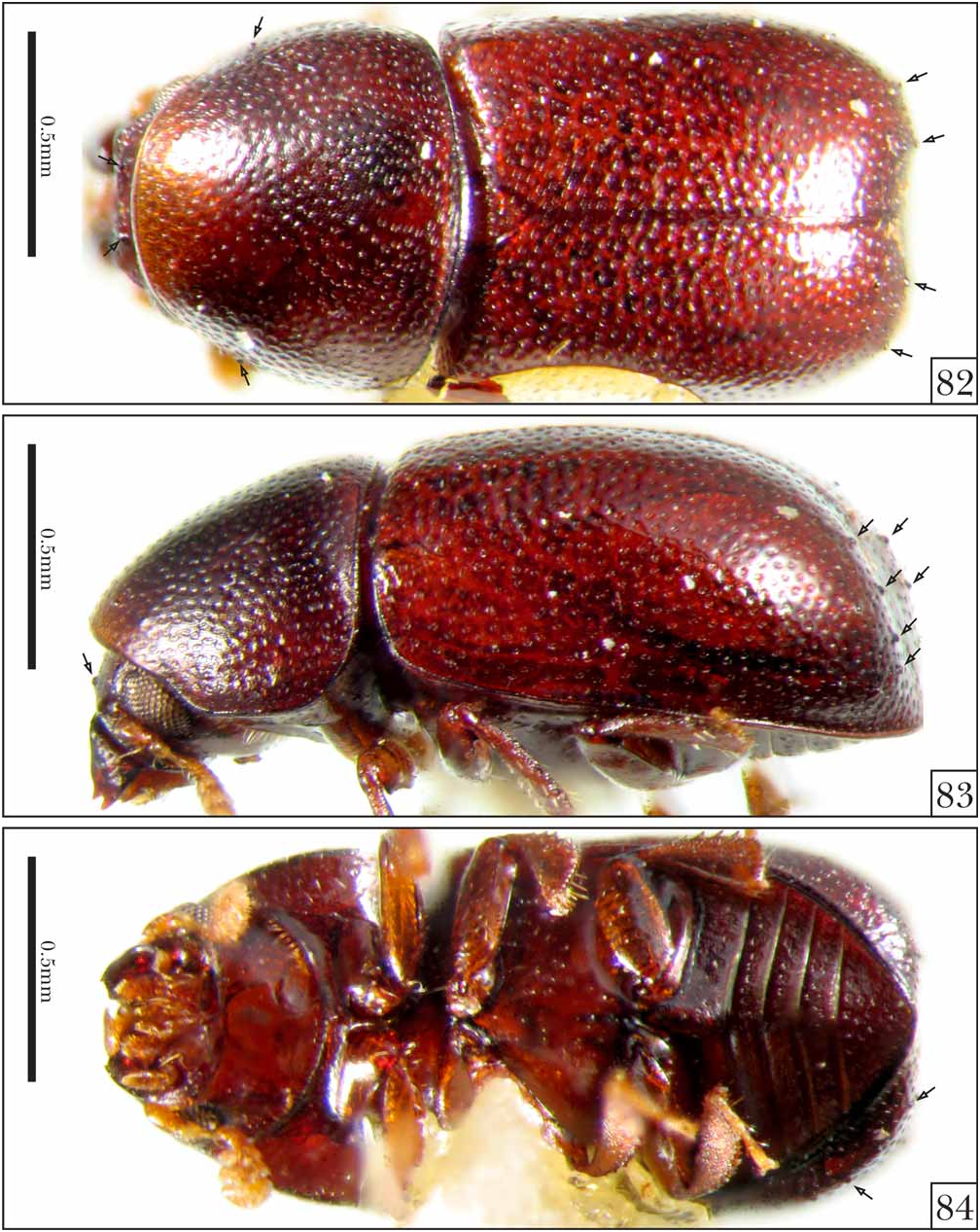
FIGURES 82–84. Scolytocis fritzplaumanni sp. nov, holotype. Note the presence of small cuticular globules (protuberances) on the head, pronotum and elytra (small arrows). 82. Dorsal view. 83. Lateral view. 84. Ventral view.

FIGURES 85–87. Scolytocis furieriae sp. nov, holotype. 85. Dorsal view. 86. Lateral view. 87. Ventral view.

FIGURES 88–90. Scolytocis paschoali sp. nov, holotype. 88. Dorsal view. 89. Lateral view. 90. Ventralview.

FIGURES 91–93. Hindwings of the Southern Neotropical Scolytocis Blair. 91. Scol. fritzplaumanni sp. nov. 92. Scol. furieriae sp. nov. 93. Scol. paschoali sp. nov.

FIGURES 94–95. Southern Neotropical Scolytocis Blair. 94. Antennae of Scol. fritzplaumanni sp. nov. (A), Scol. furieriae sp. nov. (B) and Scol. paschoali sp. nov. (C). 95. Metatibiae of Scol. fritzplaumanni sp. nov. (A), Scol. furieriae sp. nov. (B) and Scol. paschoali sp. nov. (C). First antennomere not shown in Scol. furieriae sp. nov. (94B, asterisk).
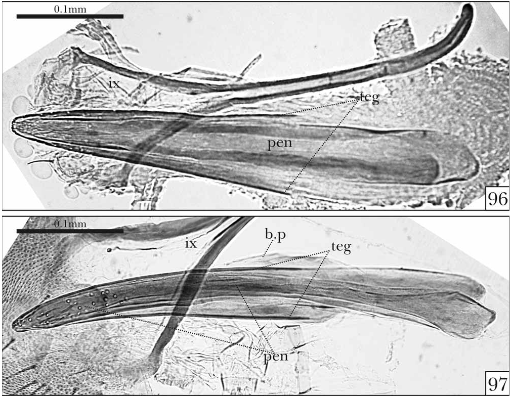
FIGURES 96–97. Male genitalia of Scolytocis fritzplaumanni sp. nov. showing the Y-shaped ninth segment (ix), basal piece (b.p), tegmen (teg) and penis (pen). 96. Specimen from Nova Teutônia, Santa Catarina, Brazil (type locality). 97. Specimen from Guaratuba, Rio Grande do Sul, Brazil.

FIGURES 98–100. Male genitalia of Scolytocis Blair showing the Y-shaped ninth segment (ix), basal piece (b.p), tegmen (teg) and penis (pen). 98. Scol. furieriae sp. nov. from Jussari, Bahia, Brazil (type locality). 99–100. Scol. paschoali sp. nov., specimens from Macaé de Cima, Rio de Janeiro, Brazil (type locality). 99. Dorsal-ventral view. 100. Lateral view.

FIGURE 101. Map showing the distribution of the Southern Neotropical species of Scolytocis. Scol. fritzplaumanni sp. nov. (circles), Scol. furieriae sp. nov. (triangles), Scol. paschoali sp. nov. (squares).

FIGURES 102–104. Scolytocis malayanus sp. nov., holotype. 102. Dorsal view. 103. Lateral view. 104. Ventral view.

FIGURES 105–107. Scolytocis novaezelandiae sp. nov., holotype. 105. Dorsal view. 106. Lateral view. 107. Ventral view.
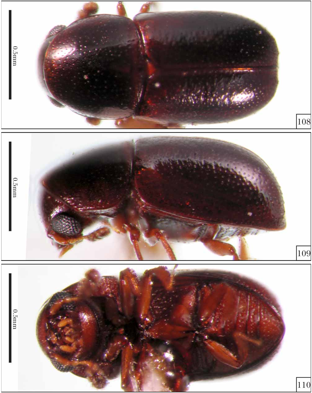
FIGURES 108–110. Scolytocis philippinensis sp. nov., holotype. 108. Dorsal view. 109. Lateral view. 110. Ventral view.

FIGURES 111–113. Scolytocis thayerae sp. nov., holotype. 111. Dorsal view. 112. Lateral view. 113. Ventralview.

FIGURES 114–116. Scolytocis werneri sp. nov., holotype. 114. Dorsal view. 115. Lateral view. 116. Ventral view.

FIGURES 117–119. Scolytocis zimmermani sp. nov., holotype. 117. Dorsal view. 118. Lateral view. 119. Ventral view.
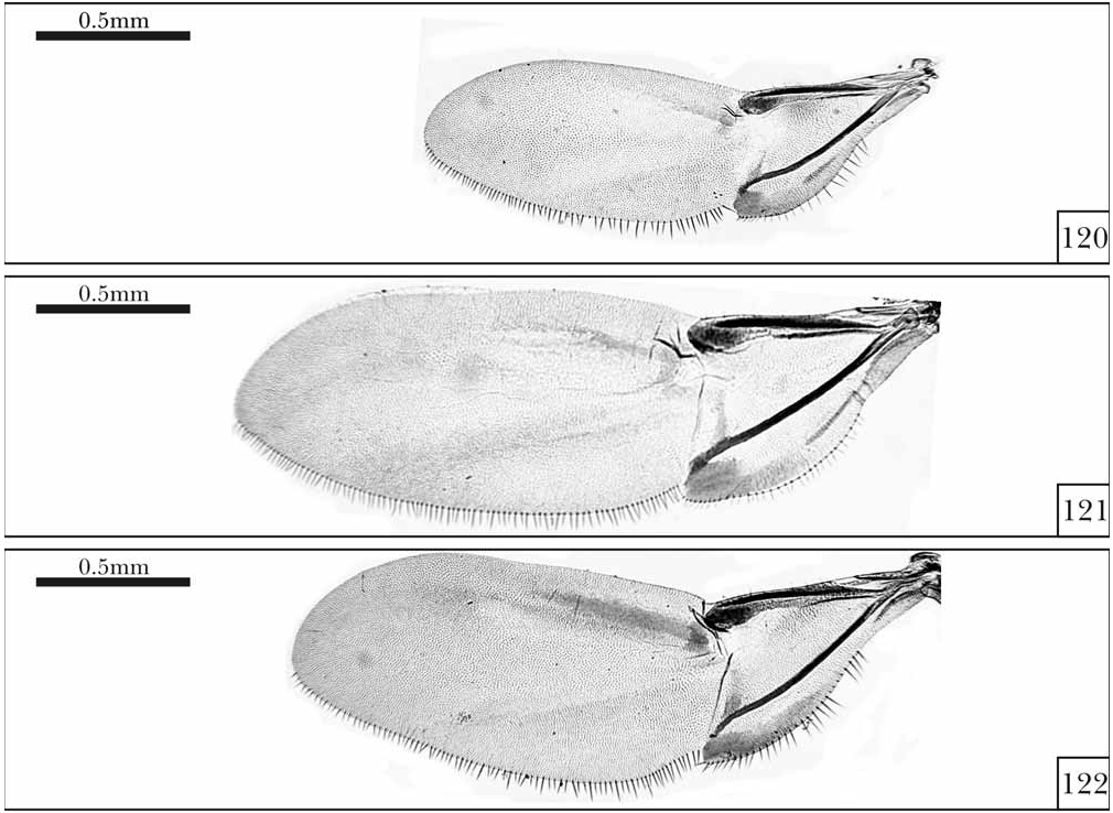
FIGURES 120–122. Hindwings of the new Indo-Malayan (in part) and Australasian Scolytocis Blair. 120. Scol. malayanus sp. nov. 121. Scol. novaezelandiae sp. nov. 122. Scol. philippinensis sp. nov.

FIGURES 123–125. Hindwings of the new Indo-Malayan (in part) and Oceanic Scolytocis Blair. 123. Scol. thayerae sp. nov. 124. Scol. werneri sp. nov. 125. Scol. zimmermani sp. nov.

FIGURES 126–127. Antennae of the Indo-Malayan, Australasian and Oceanic species of Scolytocis Blair. 126. Scol. malayanus sp. nov. (A), Scol. novaezelandiae sp. nov. (B) and Scol. philippinensis sp. nov. (C). 127. Scol. thayerae sp. nov. (A), Scol. werneri sp. nov. (B) and Scol. zimmermani sp. nov. (C).

FIGURES 128–129. Metatibiae of the Indo-Malayan, Australasian and Oceanic species of Scolytocis Blair. 128. Scol. malayanus sp. nov. (A), Scol. novaezelandiae sp. nov. (B) and Scol. philippinensis sp. nov. (C). 129. Scol. thayerae sp. nov. (A), Scol. werneri sp. nov. (B) and Scol. zimmermani sp. nov. (C).

FIGURES 130–133. Male genitalia of the Indo-Malayan species of Scolytocis Blair showing the Y-shaped ninth segment (ix), tegmen (teg) and penis (pen). 130. Scol. malayanus sp. nov. 131. Scol. philippinensis sp. nov. 132. Scol. thayerae sp. nov. 133. Scol. werneri sp. nov.
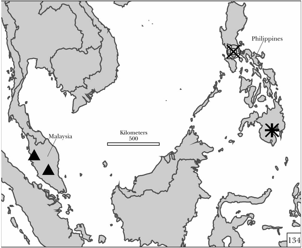
FIGURE 134. Distributional map of the Indo-Malayan species of Scolytocis Blair, showing the distribution of Scol. malayanus sp. nov. (triangles), Scol. philippinensis sp. nov. (double circle), Scol. thayerae sp. nov. (letters “X”) and Scol. werneri sp. nov. (plus symbol).

No known copyright restrictions apply. See Agosti, D., Egloff, W., 2009. Taxonomic information exchange and copyright: the Plazi approach. BMC Research Notes 2009, 2:53 for further explanation.
|
Kingdom |
|
|
Phylum |
|
|
Class |
|
|
Order |
|
|
Family |
Scolytocis Blair, 1928
| Lopes-Andrade, Cristiano 2008 |
Scolytocis thayerae
| Lopes-Andrade 2008 |
Scolytocis samoensis
| Blair 1928 |