
Asteroporpa (Astrohelix) paucidens ( Mortensen, 1933 ) Mortensen, 1933
|
publication ID |
https://doi.org/ 10.11646/zootaxa.4227.4.4 |
|
publication LSID |
lsid:zoobank.org:pub:D71F8124-BD24-4FDC-A659-9ECC496150BD |
|
DOI |
https://doi.org/10.5281/zenodo.5688480 |
|
persistent identifier |
https://treatment.plazi.org/id/038E87A0-4E7F-D362-FF7F-FD7AE566FCF3 |
|
treatment provided by |
Plazi |
|
scientific name |
Asteroporpa (Astrohelix) paucidens ( Mortensen, 1933 ) |
| status |
comb. nov. |
Asteroporpa (Astrohelix) paucidens ( Mortensen, 1933) View in CoL comb. nov.
( Figs 3–4 View FIGURE 3 View FIGURE 4 )
Astrohelix bellator var. paucidens Mortensen, 1933 View in CoL , 26–27, pl. 5, figs 18–19.
Type material. ZMUC OPH- 225 , holotype of Astrohelix bellator var. paucidens , north off Goto Islands , western Japan, 32˚10’N, 128˚20’E, 190 m deep, 23 May 1898.
Diagnosis. Epidermal ossicles are plate-shaped and cone-shaped on radial shields; plate-shaped on oral plate; cone-shaped with one or two relatively short terminal projections, on the lateral interradial disc; hooklet-bearing plates on the basal portion of arms are separated.
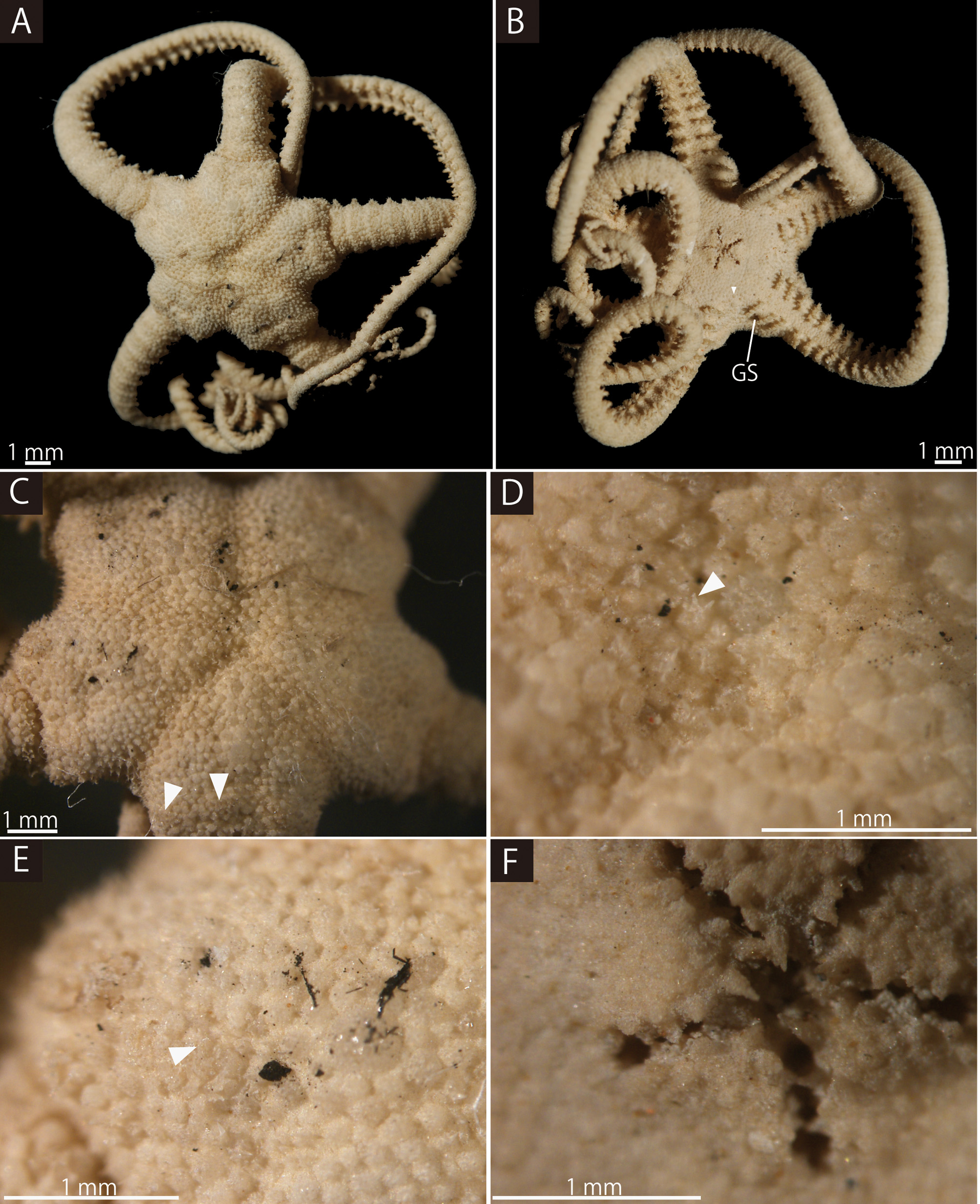
Redescription of holotype. Size is 6.8 mm in disc diameter, at least 40 mm in arm length ( Fig. 3 View FIGURE 3 A–B).
Disc. Disc is five-lobed in shape with notched interradial edges ( Fig. 3 View FIGURE 3 A–B). On the aboral side, radial shields and their surrounds are slightly tumid ( Fig. 3 View FIGURE 3 A, C). There are no conspicuous raised rows of hooklet-bearing plates and two hooklet-bearing plates are scattered on periphery of each radial shield ( Fig. 2 View FIGURE 2 C). Each hooklet has two inner teeth. Except for the hooklet-bearing plates, the aboral periphery of the disc is covered both by flat, round plate-shaped epidermal ossicles and by cone-shaped epidermal ossicles ( Fig. 3 View FIGURE 3 D–E). The flat and round plateshaped epidermal ossicles are approximately 150–240 µm in length at center of the disc and 110–190 µm in length at the periphery of the disc ( Fig. 3 View FIGURE 3 D–E). The cone-shaped epidermal ossicles have typically two, rarely three terminal projections, which are approximately half the height of the cone-shaped epidermal ossicles. The coneshaped epidermal ossicles are approximately 160–190 µm in length at center of the disc ( Fig. 3 View FIGURE 3 D), and approximately 180 µm in length at periphery of the disc ( Fig. 3 View FIGURE 3 E). Radial shields are completely concealed by oval epidermal ossicles, approximately 3 mm in length and 1.5 mm in width, that do not reach the disc center ( Fig. 3 View FIGURE 3 C).
The oral surface of the disc is covered both by flat, polygonal plate-shaped epidermal ossicles and by coneshaped epidermal ossicles with one to three terminal projections, similar to those on the aboral periphery of the disc ( Fig. 3 View FIGURE 3 F). The cone-shaped epidermal ossicles are scattered on the periphery of the disc ( Fig. 3 View FIGURE 3 B). The plate-shaped epidermal ossicles are approximately 95–130 µm ( Fig. 3 View FIGURE 3 F). The cone-shaped epidermal ossicles are approximately 110 µm in length ( Fig. 3 View FIGURE 3 B). Oral shields, adoral shields, oral plates and ventral arm plates are concealed by epidermal ossicles ( Fig. 3 View FIGURE 3 B). Uniformly acute and spiniform teeth are situated on the jaws. The length of the teeth vary depending on their location on the jaw. On the top of the jaws, they are approximately 170 µm in length, and gradually decrease in length toward the basal part of the jaws ( Fig. 3 View FIGURE 3 F).
The lateral interradial surface of the disc is slightly inclined toward the oral side and covered both by coneshaped epidermal ossicles with one or two terminal projections, similar to those on oral surface of the disc ( Fig. 4 View FIGURE 4 A). The cone-shaped epidermal ossicles are approximately 110–190 µm in length. The terminal projections of the cone-shaped epidermal ossicles are approximately the same length as the height of the ossicles ( Fig. 4 View FIGURE 4 A). Two pore like genital slits (0.3 mm long and 0.13 mm wide) are present in each interradius ( Fig. 3 View FIGURE 3 B). One hemispherical madreporite is situated on an oral interradius.
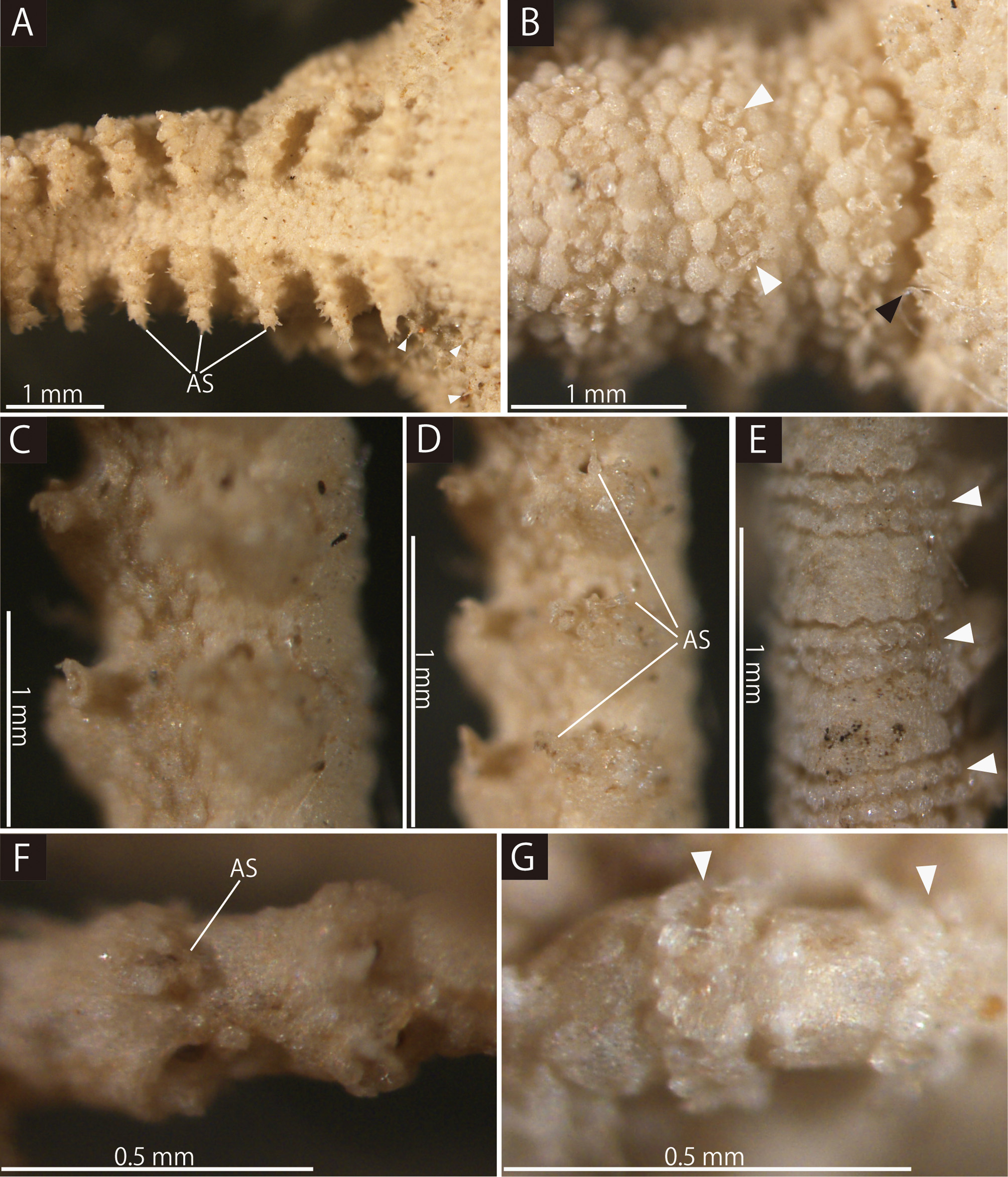
Arms. Arms are simple, five in number, and have no abrupt change in thickness ( Fig. 3 View FIGURE 3 A–B). The basal portion of the arm is 1.6 mm wide and almost the same height as the width, with an arched aboral surface and flattened oral surface. Arms taper gradually toward the arm tip ( Fig. 4 View FIGURE 4 ).
On the aboral and lateral surface, each arm segment is ringed by single transverse rows of hooklet-bearing plates ( Fig. 4 View FIGURE 4 B, E, G). On the basal three or four arm segments, each hooklet-bearing plate is separated by domed and polygonal plate-shaped epidermal ossicles ( Fig. 4 View FIGURE 4 B). On the fifth and subsequent distal segments, those plates are fully in contact with each other ( Fig. 4 View FIGURE 4 E, G). Each hooklet on the basal one fourth of the arm bears two inner teeth. Toward the distal portion of the arm, the number of the inner teeth decreases to one. The aboral and lateral surface of the basal arm, except for the hooklet-bearing plates, is completely covered by polygonal plate-shaped epidermal ossicles of approximately 110–200 µm in length ( Fig. 4 View FIGURE 4 B). The oral surface is covered by plate-shaped epidermal ossicles similar to those on the oral surface of the disc of approximately 90–190 µm in length ( Fig. 4 View FIGURE 4 A). In the middle portion of the arm, the aboral and lateral surface is covered by slightly domed and round plate-shaped epidermal ossicles, approximately 70–90 µm in length ( Fig. 4 View FIGURE 4 E). The oral surface is covered by flat and polygonal plate-shaped epidermal ossicles, approximately 110–140 µm in length ( Fig. 4 View FIGURE 4 C). In the distal portion of the arm, aboral and lateral surface is covered by flat and round plate-shaped epidermal ossicles, approximately 70 µm in length ( Fig. 4 View FIGURE 4 G). The oral surface is covered by flat and slightly round plate-shaped epidermal ossicles approximately 60 µm in length ( Fig. 4 View FIGURE 4 F). Along the entire arm, lateral arm plates and ventral arm plates are completely concealed by epidermal ossicles ( Fig. 4 View FIGURE 4 ).
First tentacle pores have no arm spine; from the second pore to the middle portion of the arm there are five arm spines. The number of arm spines decrease gradually to a single spine toward the arm tip. Throughout the arm, all the arm spines are approximately half the length of the corresponding arm segment, and are covered by a thin integument ( Fig. 4 View FIGURE 4 A, D, F). Arm spines on the basal one-fourth of the arm are ovoid, carrying three terminal projections. In the middle to distal portion, the arm spines change to being hook-shaped and the inner most spines possess one inner tooth ( Fig. 4 View FIGURE 4 D, F).
Color. In ethanol specimen, body color is uniform creamy white. No living color information for original description ( Mortensen, 1933).
Distribution. Japan: north of Goto Island, 190 m ( Mortensen, 1933), western Japan.

| ZMUC |
Zoological Museum, University of Copenhagen |
No known copyright restrictions apply. See Agosti, D., Egloff, W., 2009. Taxonomic information exchange and copyright: the Plazi approach. BMC Research Notes 2009, 2:53 for further explanation.
|
Kingdom |
|
|
Phylum |
|
|
Class |
|
|
Order |
|
|
Family |
|
|
Genus |
|
|
SubGenus |
Asteroporpa |
Asteroporpa (Astrohelix) paucidens ( Mortensen, 1933 )
| Okanishi, Masanori 2017 |
Astrohelix bellator var. paucidens
| Mortensen 1933 |