
Haematotropis callyi, De & Bueno-Villegas & Rafael, 2021
|
publication ID |
https://doi.org/10.11646/zootaxa.5064.1.1 |
|
publication LSID |
lsid:zoobank.org:pub:65B0A21A-8B8D-4B55-B6F0-8BE60EB8D3BC |
|
DOI |
https://doi.org/10.5281/zenodo.5653738 |
|
persistent identifier |
https://treatment.plazi.org/id/03876671-FFEE-151D-13C5-C9CDFDD6A2E3 |
|
treatment provided by |
Plazi (2021-11-08 12:14:55, last updated 2024-11-29 12:38:10) |
|
scientific name |
Haematotropis callyi |
| status |
sp. nov. |
Haematotropis callyi sp. nov.
Figs 22 View FIGURE 22 , 23 View FIGURE 23 , 48B View FIGURE 48 , 52 View FIGURE 52
Diagnosis. Adult males of H. callyi sp. nov. differ from other Haematotropis species based on the following combination of characters: gonopod with LP1 long and wide, projected towards the lateral region of the gonopod ( Fig. 23A–D View FIGURE 23 ), aculeate margins; solenomere tapered and hooked, projected towards the acropodital apical region ( Fig. 23A, B View FIGURE 23 ).
Material examined. Holotype ♂, French Guiana, Camopi , Monte Itoupé (3.016730, −53.108130), 446 m a.s.l, 1.VII.2016, 95 % EtOH, EDB 4 R1 , collected in rotting wood, S. Cally leg. ( INPA). GoogleMaps
Paratypes: 1 ♂, Brasil, Amapá, Serra do Navio , 1/3, 27. VI .1994, nº de campo: 34, Diversitas Neotropical ( MZUSP 970 View Materials ) ; 1 ♂, French Guiana, Awala-Yalimapo, Simili (5.74439, −53.93197), collected under dead wood on the ground, 95% EtOH, EDB 4 R1 GoogleMaps , 01.VII.2016, S. Cally leg. ( MNRJ) ; 1 ♂, French Guiana, Camopi, Monte Itoupé (3.016690, −53.108090), collected under dead wood on the ground, 442 m a.s.l, 95% EtOH, EDB 4 R1 GoogleMaps , 07.I.2016, S. Cally leg. ( MNRJ) ; 1 ♂, French Guiana, Camopi, Monte Itoupé (3.016640, −53.108040), collected under dead wood on the ground, 437 m a.s.l, 95% EtOH, EDB 4 R1 GoogleMaps , 07.I.2016, S. Cally leg. ( INPA) ; 1 ♂, French Guiana, Camopi, Monte Itoupé (3.016640, −53.108070), collected under dead wood on the ground, 440 m a.s.l, 95% EtOH, EDB 4 R1 GoogleMaps , 07.I.2016, S. Cally leg. ( MNRJ) ; 1 ♂, French Guiana, Camopi, Monte Itoupé (3.016650, −53.108050), collected under dead wood on the ground, 438 m a.s.l, 95% EtOH, EDB 4 R1 GoogleMaps , 07.I.2016, S. Cally leg. ( INPA) .
Description.
Size and form (holotype ♂). Body length = 43 and wide = 5 mm. TL/GW = 8.6.
Coloration (long preserved in 70% ethanol). Head, prozonite, metazonite and telson dark brown, antennomeres, sides of metazonite, paranota, posterior region of epiproct and legs orange ( Fig. 22A–D View FIGURE 22 ).
Head. Smooth and shiny, without microgranulations.
Trunk. Collum 2.1 mm long, 4.9 mm wide; dorsal surface of all rings smooth and shiny, without microgranulation; posterolateral margins slightly concave ( Fig. 22A, B View FIGURE 22 ). Rings 2–19: prozonite and metazonite smooth and shiny, without microgranulation in ventral region of all rings. Anterolateral teeth on paranota of rings 2–4 ( Fig. 2G View FIGURE 2 ). Lateral margins of ring 2 not projected posteriad ( Fig. 22A, B View FIGURE 22 ). Gonopodal aperture elliptical, approximately 1.7 mm wide and 0.9 mm long at midpoints. Telson ( Fig. 22D View FIGURE 22 ) smooth and shiny, without microgranulations.
Legs. Smooth and shiny, without microgranulations.
Gonopods. Right gonopod structure as follows: coxa with lateral swelling at midlength absent ( Fig. 23A, C View FIGURE 23 ); acropodite elongated, about four times as long as prefemur; median region expanded, cup-shaped in ventral view, with a concavity and cavity ( Fig. 23D View FIGURE 23 ); distal region of acropodite strongly sinuous, bifurcate, curved ventrally at a 75° angle ( Fig. 23A–D View FIGURE 23 ); VP1 emarginated on posterior region, evident, exceeding the width of ventral region of acropodite, in lateral view ( Fig. 23A, C View FIGURE 23 ); VP2 absent; LP1 long and wide, projected towards the lateral region of gonopod, aculeate margins; LP2, LP3 and DP absent ( Fig. 23A–D View FIGURE 23 ). Opening of solenomere located at distal end of acropodite, at bifurcation. Solenomere tapering, hooked, projected towards the acropodital apical region ( Fig. 23A, B View FIGURE 23 ).
Remarks. Similar in colour to some specimens such as H. disjuncta ( Fig. 9A–E View FIGURE 9 ) and H. octocentra ( Fig. 15I, J, O, P View FIGURE 15 ). Tegument dark brown with appendages and paranota orange. Acropodital distal region bifurcate, with the solenomere originating at the beginning of the bifurcation ( Fig. 23A–D View FIGURE 23 ), thus differing from the non-bifurcate condition in H. disjuncta and H. octocentra . DP absent, unlike in H. disjuncta ( Fig. 10A–C View FIGURE 10 ). Gonopods similar to those of H. callipa ( Fig. 4A–E View FIGURE 4 ) and H. mosaica sp. nov. ( Fig. 37A–D View FIGURE 37 ). These species have the acropodite distal region bifurcate, but are differentiated mainly by the solenomere, which is narrow and curved in H. callyi sp. nov. ( Fig. 23A, B View FIGURE 23 ), narrow and projected towards the ventral region of the body in H. mosaica sp. nov. ( Fig. 37A, C View FIGURE 37 ), and wide in H. callipa ( Fig. 4A, B View FIGURE 4 ). Gonopod conformation similar to H. mosaica sp. nov. due to the sinuosity of the distal region of the acropodite. Distinguished by LP1 being curved ( Fig. 23A–D View FIGURE 23 ), versus not curved in H. mosaica sp. nov. ( Fig. 37A–D View FIGURE 37 ).
Distribution. Brazil: Amapá: Serra do Navio; French Guiana: Camopi ( Fig. 52 View FIGURE 52 ).
Etymology. The specific epithet is dedicated to the collector, Sébastien Cally, from the French National Centre for Scientific Research (CNRS), Paris.

FIGURE 2. General somatic features of Haematotropis Jeekel, 2000. A. H. jurutiensis sp. nov., non-type ♂ (MPEG), body ring 7, ventral view. B. H. jurutiensis sp. nov., non-type ♂ (MPEG), body ring 9 (highlighting the microgranulations), ventral view. C. H. jurutiensis sp. nov., non-type ♂ (MPEG), body ring 7, red arrow indicates the wrinkles close to paranota, ventral view. D. H. jurutiensis sp. nov., non-type ♂ (MPEG), body ring 8, red arrow indicates rounded projections on ventral surfaces, ventral view. E. H. jurutiensis sp. nov., non-type ♂ (MPEG), metazonite of body ring 8, red arrow indicates the microgranulations on anterior and posterior margins, dorsal view. F. H. megalcensis sp. nov. non-type ♂ (IBSP), body ring 10, red arrow indicates the microgranulations on anterior and posterior margins, ventral view. G. H. callipa, holotype ♂ (MfN), head and body rings 1–4, red arrow indicates a depression on collum or anterolateral teeth, dorsalateral view. Scale bars: 300 μm (A, E), 200 μm (C, D, F), 100 μm (B), 1 mm (G).
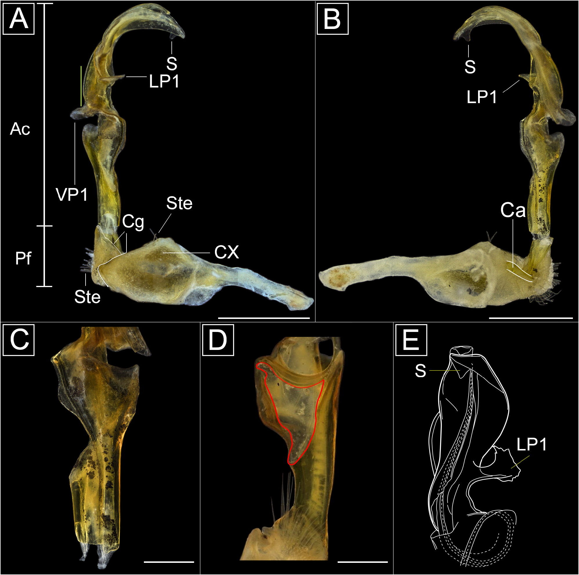
FIGURE 4. Left gonopods of Haematotropis callipa (Peters, 1864). A. Holotype ♂ (MfN), lateral view. B, C. Holotype ♂ (MfN), mesal views. D. Holotype ♂ (MfN), ventral view. E. ♂ from Kabelstation, Suriname, dorsal view, modified from Jeekel (2000), without scale. Yellow scale indicates when VP1 exceeds the width of ventral region of acropodite. Red line shows a concavity and cavity in ventral region of the acropodite. Abbreviations: Ac = acropodite; Ca = cannula; Cg = cingulum; Cx = coxa; LP1 = Lateral Process 1; Pf = Prefemur; S = Solenomere; Ste = setae; VP1 = Ventral Process 1. Scale bars: 1.0 mm.

FIGURE 9. Somatic features of Haematotropis disjuncta Golovatch, Hoffman & Spelda, 2004 (IEPA). A. Head and body rings 1–5, dorsal view, holotype ♂ (IEPA). B. Head and body rings 1–5, lateral view, holotype ♂ (IEPA). C. Body rings 9–11, dorsal view, holotype ♂ (IEPA). D. Body rings 18–20, dorsal view, holotype ♂ (IEPA). E. Habitus, lateral view, paratype ♀ (IEPA). Scale bars: 1.0 mm.

FIGURE 10. Right gonopod of Haematotropis disjuncta Golovatch, Hoffman & Spelda, 2004, holotype ♂ (IEPA). A. Mesal view. B. Dorsal view. C. Lateral view. D. Ventral view. Yellow scale indicates when VP1 exceeds the width of ventral region of acropodite. Abbreviations: Ac = acropodite; Ca = cannula; Cx = coxa; DP = Distal Process 1; LP1 = Lateral Process 1; Pf = Prefemur; S = Solenomere; Sg = Spermatic groove; Ste = setae; VP1 = Ventral Process 1. Scale bars: 1.0 mm.
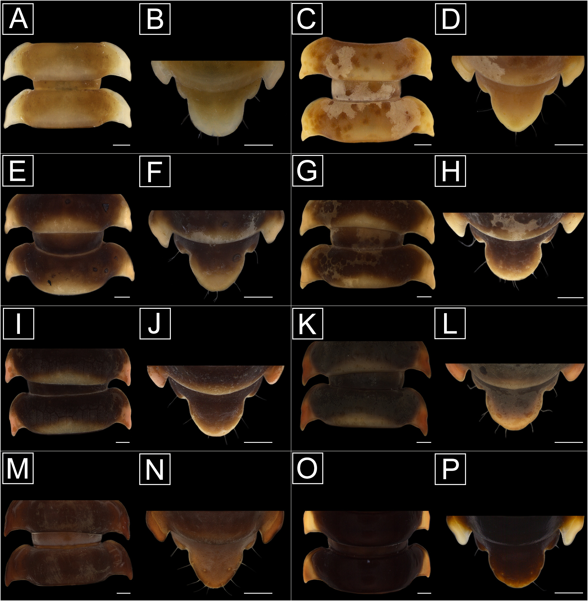
FIGURE 15. Somatic features of Haematotropis octocentra (Brölemann, 1905) (color and shape variation), non-types ♂ (INPA, MPEG, MZUSP, UFAM), dorsal views. Scale bars: 1.0 mm.
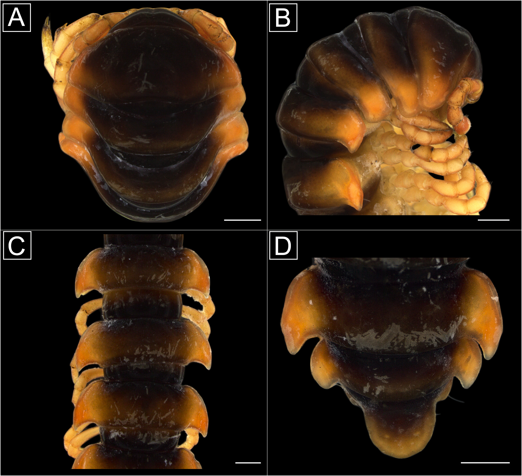
FIGURE 22. Somatic features of Haematotropis callyi sp. nov., holotype ♂ (INPA). A. Head and body rings 1–4, dorsal view. B. Head and body rings 1–5, dorsolateral view. C. Body rings 8–11, dorsal view. D. Body rings 18–20, dorsal view. Scale bars: 1.0 mm.

FIGURE 23. Right gonopod of Haematotropis callyi sp. nov., holotype ♂ (INPA). A. Mesal view. B. Dorsal view. C. Lateral view. D. Ventral view. Yellow scale indicates when VP1 exceeds the width of ventral region of acropodite. Abbreviations: Ac = acropodite; Ca = cannula; Cx = coxa; LP1 = Lateral Process 1; Pf = Prefemur; S = Solenomere; Sg = spermatic groove; VP1 = Ventral Process 1. Scale bars: 1.0 mm.
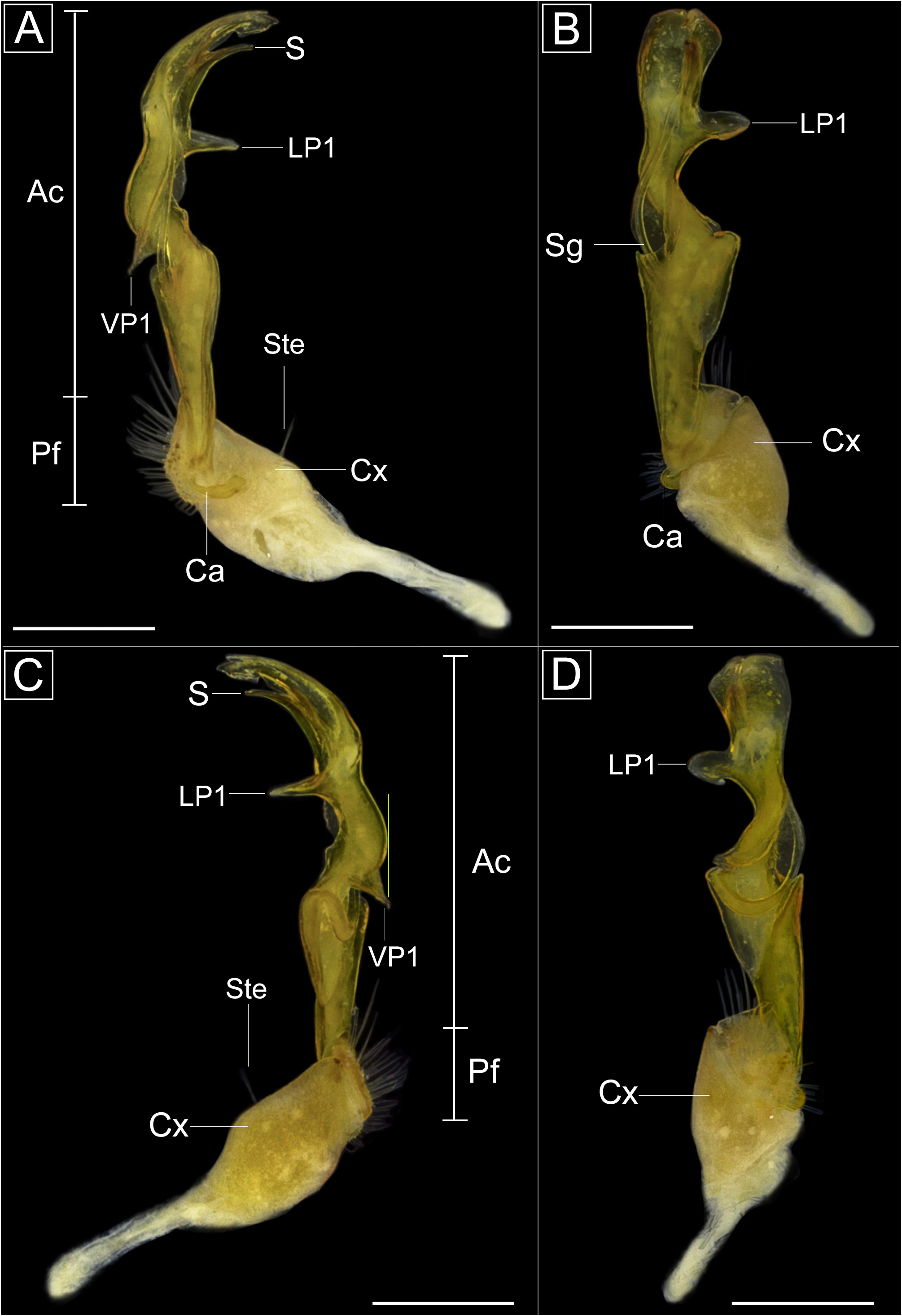
FIGURE 37. Right gonopod of Haematotropis mosaica sp. nov., holotype ♂ (MPEG). A. Mesal view. B. Dorsal view. C. Lateral view. D. Ventral view. Yellow scale indicates when VP1 exceeds the width of ventral region of acropodite. Abbreviations: Ac = acropodite; Ca = cannula; Cx = coxa; LP1 = Lateral Process 1; Pf = Prefemur; S = Solenomere; Sg = spermatic groove; Ste = setae; VP1 = Ventral Process 1. Scale bars: 1.0 mm.
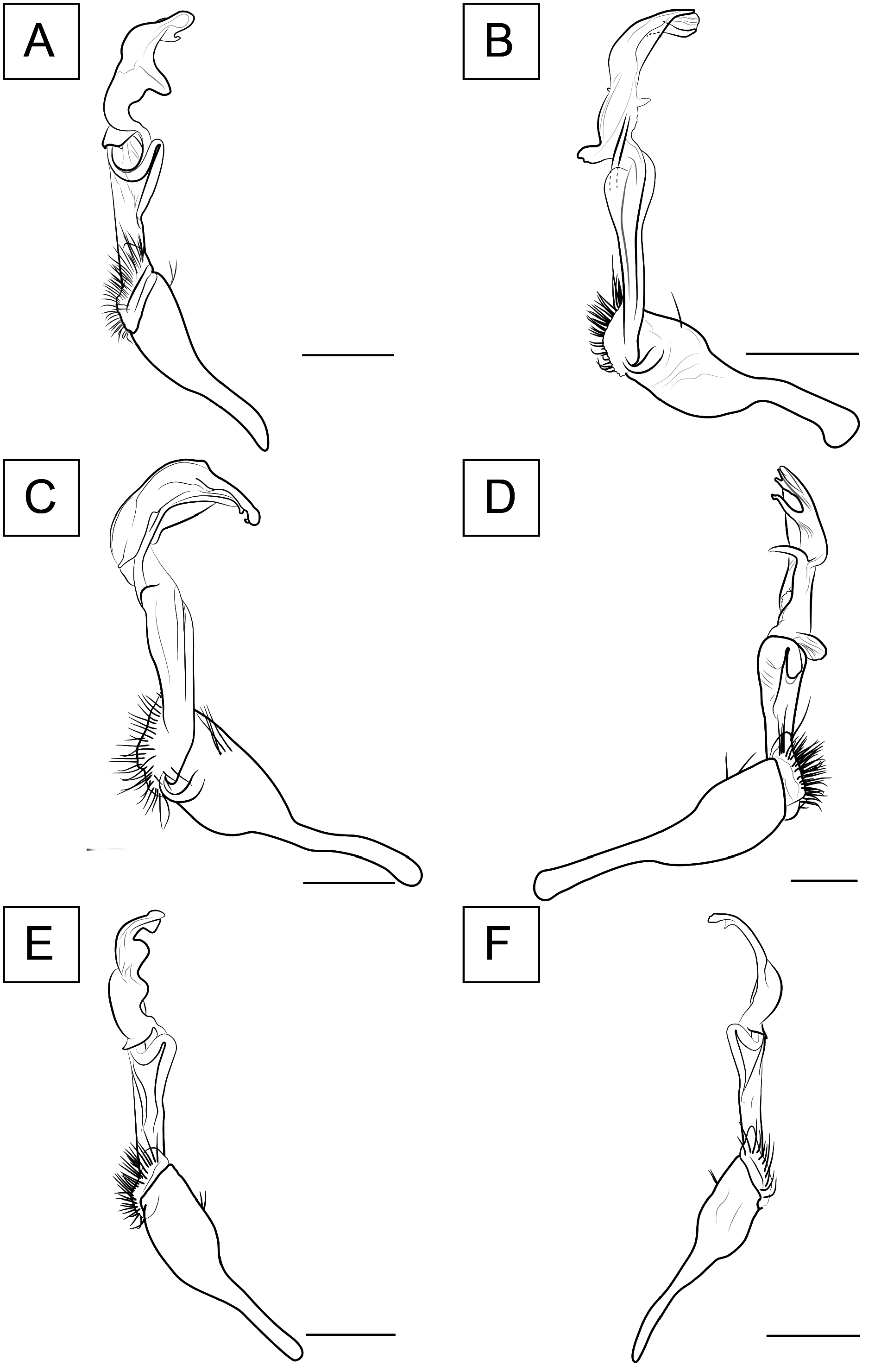
FIGURE 48. Gonopodal features of Haematotropis Jeekel, 2000 species. A. H. aripuanensis sp. nov., holotype (MZUSP), left gonopod, lateral view. B. H. callyi sp. nov., holotype (INPA), right gonopod, mesal view. C. H. dentata sp. nov., holotype (IBSP), right gonopod, mesal view. D. H. disjunctoides sp. nov., holotype (MPEG), right gonopod, lateral view. E. H. goeldii sp. nov., holotype (MPEG), left gonopod, lateral view. F. H. jurutiensis sp. nov., holotype (MPEG), right gonopod, lateral view. Scale bars: 1.0 mm.

FIGURE 52. Distribution of Haematotropis Jeekel, 2000. Black symbol = H. jurutiensis sp. nov., green symbol = H. dentata sp. nov., orange symbol = H. goeldii sp. nov., pink symbol = H. aripuanensis sp. nov., red symbol = H. amazonica sp. nov., white symbol = H. disjunctoides sp. nov., yellow symbol = H. callyi sp. nov.
No known copyright restrictions apply. See Agosti, D., Egloff, W., 2009. Taxonomic information exchange and copyright: the Plazi approach. BMC Research Notes 2009, 2:53 for further explanation.
|
Kingdom |
|
|
Phylum |
|
|
Class |
|
|
Order |
|
|
SubOrder |
Leptodesmidea |
|
SuperFamily |
Platyrhacoidea |
|
Family |
|
|
Genus |