
Styphrus corpulentus Motschulsky, 1845
|
publication ID |
https://doi.org/10.5281/zenodo.4272127 |
|
DOI |
https://doi.org/10.5281/zenodo.4342075 |
|
persistent identifier |
https://treatment.plazi.org/id/0385915E-FF4D-09F6-60CB-FB1CCD34FE2D |
|
treatment provided by |
Felipe (2020-11-13 20:09:42, last updated 2024-11-28 19:22:11) |
|
scientific name |
Styphrus corpulentus Motschulsky, 1845 |
| status |
|
Styphrus corpulentus Motschulsky, 1845 View in CoL
( Figs. 12 View Figs , 71 View Figs , 105 View Figs , 136 View Figs , 677–693 View Figs View Figs )
Styphrus corpulentus Motschulsky, 1845: 54 View in CoL .
Styphrus corpulentus: JAKOBSON (1911) View in CoL : 651; REICHARDT (1941): 174, Figs. 83, 84 View Figs ; KRYZHANOVSKIJ & REICHARDT (1976): 187, Figs. 367–372 View Figs View Figs ; MAZUR (1984): 78; MAZUR (1997): 245; MAZUR (2004): 101.
Saprinus akinini Schmidt, 1890: 19 View in CoL . Synonymized by JAKOBSON (1910): 264.
Saprinus mimulus Reitter, 1904: 29 View in CoL . Synonymized by JAKOBSON (1910): 263.
Xenonychus akinini: LEWIS (1905) : 78.
Pachylopus akinini: REITTER (1906) : 268.
Xenonychus laevidorsis Reitter, 1910: 13 View in CoL . Synonymized by BICKHARDT (1916): 103.
Styphrus laevidorsis: BICKHARDT (1910) : 107.
Saprinus rugosipennis Dahlgren, 1971: 50 View in CoL . Synonymized by Kryzhanovskij in KRYZHANOVSKIJ & REICHARDT (1976): 187.
Type locality. Uzbekistan, Buchara.
Type material examined. Styphrus corpulentus . NEOTYPE (here designated):♁, ‘ Buchara , Kyzyl Kum / Uzbekistan 28.–30.IV. / A. Olexa 1975 [printed] // D07-026 [pink label, written] // NEOTYPUS / Styphrus / corpulentus / Motschulsky 1845 / Des. T. Lackner 2009 [red label, written]’ ( ZIN).
Saprinus akinini . Par at ype: 1 spec., ‘ Saprinus / akinini / Schmidt / typ [written] // Paratypus [red label, written]’ ( ZIN).
Comment. Type specimen(s) of this species were not found in Motschulsky’s collection,housed at ZMUM.According to N. Nikitsky (ZMUM), a part of the type specimens described by Motschulsky in 1845 has been destroyed and lost. Perhaps the type specimen(s) of Styphrus corpulentus were among those. The Motschulsky’s collection is in a very poor state. Therefore, it is necessary to designate a neotype for this species to fix its identity as type species of Styphrus . The neotype will be housed at ZIN.
Additional material examined. UZBEKISTAN: Kyzylkum , Buchara, 28.–30.iv.1975, 1 ♁, A. Olexa lgt .; Kyzyk-Kum, Chiva [= Khiva], 25.iv.1972, 1 ♁ 1 ♀, A. Olexa lgt.; ditto, but 3.v.1978, A. Olexa lgt., 3 ♀♀ ; ditto, but 23.iv.1972, A. Olexa lgt. ( TLAN) .
Redescription. Body length: PEL: 2.50–3.45 mm; APW: 0.85–1.20 mm; PPW: 1.975–2.80 mm; EL: 1.55–2.25 mm; EW: 2.20–3.10 mm.
Body ( Figs. 677–678 View Figs ) roundly oval, ventrally strongly convex, moderately convex dorsally; cuticle dark brown with slight metallic tinge, elytral apex rufous; legs, mouthparts and antennae rufopiceous.
Antennal scape ( Fig. 681 View Figs ) substrigulate, with numerous well sclerotized long setae; club ( Figs. 679–680 View Figs ) large, flattened dorso-ventrally, round, without visible articulation, dorsal surface with large apical sensory area with dense short sensilla, intermingled with longer, much sparser erect sensilla; surface of club otherwise imbricate, with sparse long erect sensilla (except for basal fourth); sensory structures of antennal club ( Fig. 12 View Figs ) in form of one large apical sensory area and four rather large ventral sensory areas and one vesicle on internal distal margin of ventral side.
Mouthparts. Mandibles ( Fig. 104 View Figs ) with rounded outer margin, mandibular apex acute; subapical tooth of left mandible moderately large; labrum ( Fig. 70 View Figs ) with scattered microscopic punctation, convex, with a slight median depression; labral pits with two weakly sclerotized setae; terminal labial palpomere thickened, its width about half its length; mentum ( Fig. 683 View Figs ) square-shaped, imbricate, anterior margin medially with conspicuous notch ( Fig. 136 View Figs ), fringed with numerous long setae, lateral margins as well as disc of mentum with numerous much shorter setae; cardo of maxilla on lateral margin with several short setae; stipes triangular, with three much longer setae; terminal maxillary palpomere thickened, its width about half its length, approximately twice as long as penultimate.
Clypeus ( Fig. 681 View Figs ) rectangular, basally constricted, depressed laterally, with shallow irregular punctures; frontal stria well impressed, usually interrupted medially, shortly prolonged onto clypeus; supraorbital stria well impressed, carinate; frontal disc ( Fig. 681 View Figs ) smooth, only with scattered microscopic punctation; eyes convex, conspicuous from above.
Pronotal sides ( Fig. 677 View Figs ) moderately narrowed anteriorly, apical angles obtuse; pronotal foveae vaguely impressed, often absent; marginal pronotal stria complete, slightly weakened behind head; pronotal disc (except for inconspicuous scattered punctation along lateral margins and a double row of small ellipsoid punctures along pronotal base) entirely smooth; pronotal hypomeron setose; scutellum small, visible.
Elytral epipleura shiny, almost smooth, with scattered microscopic punctures; marginal epipleural stria weakly impressed, complete; marginal elytral stria well impressed, continued as weakly impressed complete apical elytral stria; inner subhumeral stria well impressed, rather long, apically reaching as far as four-fifths of elytral length, basally connected with weakly impressed humeral elytral stria; elytra with four rather thin dorsal elytral striae 1–4, apically reaching about elytral half, occasionally second dorsal elytral stria slightly longer; fourth dorsal elytral stria basally connected with complete sutural elytral stria; sutural elytral stria well impressed, complete, apically connected with apical elytral stria. Elytral disc on apical half with dense, shallow, often confluent punctures, basal half with much finer and sparser punctation; elytral flanks and elytral humeri smooth.
Propygidium almost completely exposed; punctation of propygidium and pygidium shallow but dense, punctures confluent, becoming more granulose apically.
Anterior margin of median portion of prosternum ( Fig. 685 View Figs ) almost straight, pre-apical foveae absent; marginal prosternal stria present laterally and as thin apical fragment; prosternal process flattened; surface between carinal prosternal striae smooth; laterally substrigulate, with sparse punctures fringed with setae; carinal prosternal striae on prosternal apophysis slightly divergent, approximate, sub-parallel, slightly divergent apically, not united; lateral prosternal striae well impressed, shortened, slightly carinate, anteriorly ‘open’.
Anterior margin of mesoventrite straight; discal marginal mesoventral stria well impressed, slightly carinate; disc of mesoventrite flat, with shallow scattered punctation, few punctures with microscopic setae; meso-metaventral sutural stria well impressed, undulate; intercoxal disc of metaventrite convex, almost smooth, along posterior margin with irregular sparse fine punctures. Lateral metaventral stria well impressed, slightly carinate, almost straight, shortened apically; lateral disc of metaventrite concave, with scattered shallow punctures fringed with long setae; metepisternum on basal three-fourths with similar punctation and setae, on apical fourth + fused metepimeron punctation and setae sparser and finer.
Intercoxal disc of the first abdominal sternite completely striate laterally; surface of disc with sparse scattered punctation, interspaces imbricate, apically punctation sparser and finer; lateral sides of all visible abdominal sternites setose.
Protibia ( Fig. 686 View Figs ) dilated, outer margin with three low teeth topped with rather long acute denticle, second and third teeth larger than first, followed by six short denticles diminishing in size in proximal direction; setae of outer row regular, rather sparse; protarsal groove deep; anterior protibial stria absent; setae of median row much shorter and sparser than those of outer row, present only on basal half; single tiny tarsal denticle present near tarsal insertion; protibial spur well developed, bent, growing out from apical margin of protibia; apical margin of protibia posteriorly with three short denticles; outer part of posterior surface of protibia ( Fig. 686 View Figs ) broadly strigate, interspaces smooth, separated from glabrous median part of posterior surface by clear-cut boundary; posterior protibial stria complete, apically terminating in two tiny inner ventral denticles; inner row of setae lamelliform, strongly sclerotized, long.
Mesotibia slender, outer margin with single row of short thin denticles, only slightly growing in size in proximal direction; setae of outer row situated approximately in middle of posterior mesotibial surface, setae regular, dense, about as long as denticles themselves; setae of median row absent; posterior mesotibial stria shortened apically; anterior surface of mesotibia microscopically punctate; anterior mesotibial stria almost complete; inner anterior denticles absent; mesotibial spur rather long and stout; apical margin of mesotibia anteriorly with two tiny denticles; inner margin of mesotibia with long dense well sclerotized setae; claws of apical tarsomere almost straight, thin, almost as long as apical-most tarsomere itself; each mesotarsomere anteriorly with single long well sclerotized seta; metatibia ( Fig. 684 View Figs ) similar to mesotibia, but slightly more thickened and another row of four sparse longer denticles appears on apical half of outer margin and posterior metatibial stria even shorter than posterior mesotibial stria.
Male genitalia. Eighth sternite ( Fig. 687–688 View Figs ) widely separated along their entire length, apically with large inflatable membrane (velum) mesally and laterally with several rows of long close-set setae creating a conspicuous apical brush; eighth tergite and eighth sternite fused laterally ( Fig. 689 View Figs ). Ninth tergite ( Figs. 690–691 View Figs ) typical for the subfamily; spiculum gastrale ( Fig. 690 View Figs ) expanded on both ends. Aedeagus ( Figs. 692–693 View Figs ) slender; basal piece of aedeagus rather short, ratio of its length: length of parameres 1: 3.50; parameres fused almost along their apical length, on apical tenth with opening for the median lobe (best seen from lateral view); aedeagus strongly curved ventrad ( Fig. 693 View Figs ).
BICKHARDT H. 1910: Histeridae. In: JUNK W. & SCHENKLING S. (eds.): Coleopterorum Catalogus, volume 24. W. Junk, Berlin, 137 pp.
BICKHARDT H. 1916 - 1917: Histeridae. In: WYTSMAN P. (ed.): Genera Insectorum, Fasc. 166 a, b. La Haye, 302 pp.
JAKOBSON G. 1910: Note synonymique sur quelques Coleopteres palearctiques [Histeridae et Malacodermata]. Bulletin de la Societe Entomologique de France 1910: 263 - 264.
JAKOBSON G. G. 1911: Zhuki Rosii i zapadnoy Evropy. Rukovodstvo k opredeleniyu zhukov. 9. [Beetles of Russia and western Europe. Manual for the beetles' determination. 9]. A. F. Devrien, St. Petersburg, pp. 1 + 641 - 720 (in Russian).
KRYZHANOVSKIJ O. L. & REICHARDT A. N. 1976: Zhuki Nadsemeystva Histeroidea (semeystva Sphaeritidae, Histeridae, Synteliidae). [Beetles of the superfamily Histeroidea (families Sphaeritidae, Histeridae, Syntelidae)]. Fauna SSSR, Zhestokrylye, Vyp. 4. Nauka, Leningrad, 434 pp (in Russian).
LEWIS G. 1905: A systematic catalogue of Histeridae. Kessinger, London, vi + 81 pp.
MAZUR S. 1984: A world catalogue of Histeridae. Polskie Pismo Entomologiczne 54 (3 - 4): 1 - 376.
MAZUR S. 1997: A world catalogue of the Histeridae (Coleoptera: Histeridae). Genus, Supplement, pp. 1 - 373.
MAZUR S. 2004: Family Histeridae. Pp. 68 - 102. In: LOBL I. & SMETANA A. (eds.): Catalogue of Palaearctic Coleoptera, Vol. 2, Hydrophiloidea-Histeroidea-Staphylinidea. Apollo Books, Stenstrup, 942 pp.
MOTSCHULSKY V. 1845: Remarques sur la collection de Coleopteres Russes. 1 er Article. Bulletin de la Societe Imperiale des Naturalistes de Moscou 18 (I): 3 - 127.
OLEXA A. 1975: Sur la variabilite de Saprinus perrisi Marseul, 1871 (Col. Histeridae). Bulletin de la Societe Entomologique de Mulhouse 31: 21.
REICHARDT A. 1941: Semeystva Sphaeritidae i Histeridae (Vol. 1). [Families Sphaeritidae and Histeridae]. Fauna SSSR, Nasekomye Zhestokrylye, V, 3. Nauka, Moskva-Leningrad, xiii + 419 pp.
REITTER E. 1904: Ueber neue und wenig bekannte Histeriden (Coleoptera). Wiener Entomologische Zeitung 23: 29 - 36.
REITTER E. 1906: Histeridae. In: HEYDEN L. V., REITTER E. & WEISE J. (eds.): Catalogus Coleopterorum Europae, Caucasi et Armeniae Rossicae. Editio secunda. Berlin, Paskau, Caen, vi + 775 pp.
REITTER E. 1910: Uebersicht der Arten der Coleopterengattung Xenonychus Woll. Entomologische Blatter 6: 13.
SCHMIDT J. 1890: Neun neue Saprinus aus der Gruppe des metallescens und rufipes. Deutsche Entomologische Zeitschrift 1890: 81 - 87.

Figs. 9–28. Saprininae, sensory structures of the antennal club (vesicles marked with grey; sensory areas shaded): 9 – Saprinus (Saprinus) semistriatus (Scriba, 1790); 10 – Saprinus (Hemisaprinus) subvirescens (Ménétries, 1832); 11 – Myrmetes paykulli Kanaar, 1979; 12 – Styphrus corpulentus Motschulsky, 1845; 13 – Saprinus (Phaonius) pharao (Marseul, 1855); 14 – Ammostyphrus cerberus Reichardt, 1924; 15 – Chalcionellus amoenus (Erichson, 1834); 16 – Chivaenius kryzhanovskii Olexa,1980; 17 – Ctenophilothis chobauti (Théry,1900); 18 – Eopachylopus ripae (Lewis, 1885); 19 – Exaesiopus grossipes grossipes (Marseul, 1855); 20 – Hypocaccus (Hypocaccus) rugiceps (Duftschmid, 1805); 21 – Hypocaccus (Baeckmanniolus) dimidiatus dimidiatus (Illiger, 1807); 22 – Hypocacculus (Hypocacculus) metallescens (Erichson, 1834); 23 – Hypocacculus (Colpellus) praecox (Erichson, 1834); 24 – Hypocacculus (Nessus) rubripes (Erichson, 1834); 25 – Paravolvulus lateristrius (Solskij, 1876); 26 – Pholioxenus phoenix (Reichardt, 1930); 27 – Reichardtiolus duriculus (Reitter, 1904); 28 – Xenonychus tridens (Jacquelin-Duval, 1852).

Figs. 47–73. Saprininae, labral structures, dorsal view (left half showing external and right half showing internal structures): 47 – Alienocacculus neftensis (Olexa, 1984); 48 – Ammostyphrus cerberus Reichardt, 1924; 49 – Chalcionellus amoenus (Erichson, 1834); 50 – Chivaenius kryzhanovskii Olexa, 1980; 51 – Eopachylopus ripae (Lewis, 1885); 52 – Eremosaprinus vlasovi (Reichardt, 1941); 53 – Euspilotus (Neosaprinus) perrisi (Marseul, 1872); 54 – Exaesiopus grossipes grossipes (Marseul, 55); 55 – Gnathoncus rotundatus (Kugelann, 1792); 56 – Hypocaccus (Hypocaccus) rugiceps (Duftschmid, 1805); 57 – Hypocacculus (Hypocacculus) metallescens (Erichson, 1834); 58 – Hypocacculus (Colpellus) praecox (Erichson, 1834); 59 – Hypocacculus (Nessus) rubripes (Erichson, 1834); 60 – Microsaprinus therondianus (Dahlgren, 1973); 61 – Myrmetes paykulli Kanaar, 1979; 62 – Paravolvulus lateristriatus (Solskij, 1876); 63 – Philothis (Philothis) arcanus Reichardt, 1930; 64 – Philothis (Atavinus) atavus (Reichardt, 1931); 65 – Philothis (Farabius) hexeris Reichardt, 1930; 66 – Pholioxenus phoenix (Reichardt, 1930); 67 – Reichardtiolus duriculus (Reitter, 1904); 68 – Saprinus (Saprinus) semistriatus (Scriba, 1790); 69 – Saprinus (Hemisaprinus) subvirescens (Ménétries, 1832); 70 – Saprinus (Phaonius) pharao (Marseul, 1855); 71 – Styphrus corpulentus Motschulsky, 1845; 72 – Xenonychus tridens (Jacquelin-Duval, 1852); 73 – Xenophilothis choumovitchi (Thérond, 1965).

Figs.99–107.Saprininae, mandibles,ventral view:99 – Philothis (Farabius) hexeris Reichardt, 1930; 100 – Pholioxenus phoenix (Reichardt,1930); 101 – Reichardtiolus duriculus (Reitter, 1904); 102 – Saprinus (Saprinus) semistriatus (Scriba, 1790); 103 – Saprinus (Hemisaprinus) subvirescens (Ménétries, 1832); 104 – Saprinus (Phaonius) pharao (Marseul, 1855); 105 – Styphrus corpulentus Motschulsky, 1845; 106 – Xenonychus tridens (Jacquelin-Duval, 1852); 107 – Xenophilothis choumovitchi (Thérond, 1965).
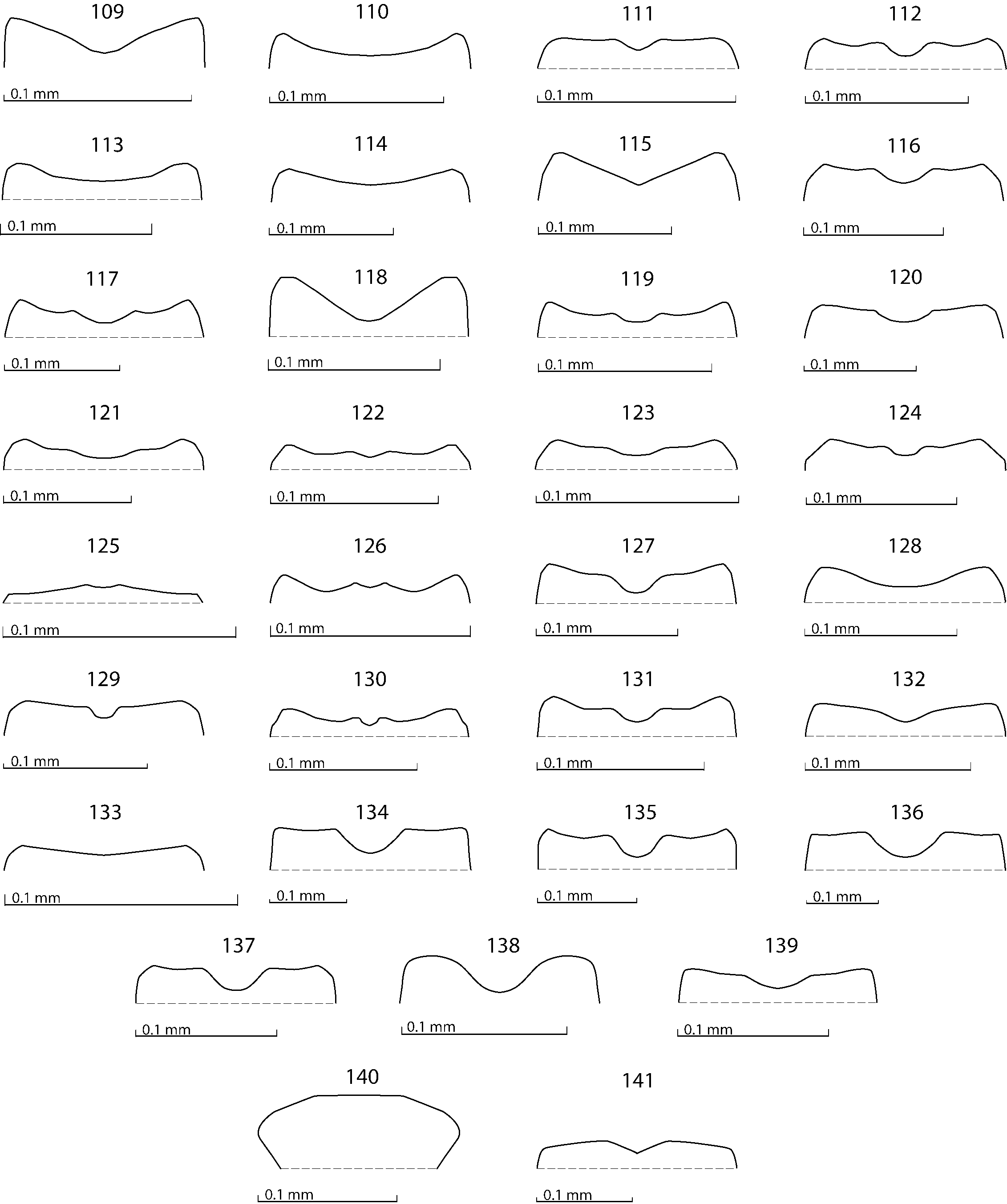
Figs. 109–141. Saprininae, anterior margin of mentum, ventral view: 109 – Alienocacculus neftensis (Olexa, 1984); 110 – Ammostyphrus cerberus Reichardt, 1924; 111 – Axelinus ghilarovi Kryzhanovskij, 1976; 112 – Chalcionellus amoenus (Erichson, 1834); 113 – Chivaenius kryzhanovskii Olexa, 1980; 114 – Ctenophilotis chobauti (Théry, 1900); 115 – Eopachylopus ripae (Lewis, 1885); 116 – Eremosaprinus vlasovi (Reichardt, 1941); 117 – Euspilotus (Neosaprinus) perrisi (Marseul, 1872); 118 – Exaesiopus grossipes grossipes (Marseul, 1855); 119 – Gnathoncus rotundatus (Kugelann, 1792); 120 – Hypocaccus (Hypocaccus) rugiceps (Duftschmid, 1805); 121 – Hypocaccus (Baeckmanniolus) dimidiatus dimidiatus (Illiger, 1807); 122 – Hypocacculus (Hypocacculus) metallescens (Erichson, 1834); 123 – Hypocacculus (Colpellus) praecox (Erichson, 1834); 124 – Hypocacculus (Nessus) rubripes (Erichson, 1834); 125 – Microsaprinus therondianus (Dahlgren, 1973); 126 – Myrmetes paykulli Kanaar, 1979; 127 – Paravolvulus ovillum (Solskij, 1876); 128 – Philothis (Philothis) arcanus Reichardt, 1930; 129 – Philothis (Atavinus) atavus (Reichardt, 1931); 130 – Philothis (Farabius) hexeris Reichardt, 1930; 131 – Pholioxenus phoenix (Reichardt, 1930); 132 – Reichardtiolus duriculus (Reitter, 1904); 133 – Saprinillus paromaloides Kryzhanovskij, 1974; 134 – Saprinus (Saprinus) semistriatus (Scriba, 1790); 135 – Saprinus (Hemisaprinus) subvirescens (Ménétries, 1832); 136 – Saprinus (Phaonius) pharao (Marseul, 1855); 137 – Styphrus corpulentus Motschulsky, 1845; 138 – Turanostyphrus ignoratus Tishechkin, 2005; 139 – Xenonychus tridens (Jacquelin-Duval, 1852); 140 – Xenophilotis choumovitchi (Thérond, 1965); 141 – Zorius funereus (Schmidt, 1890).
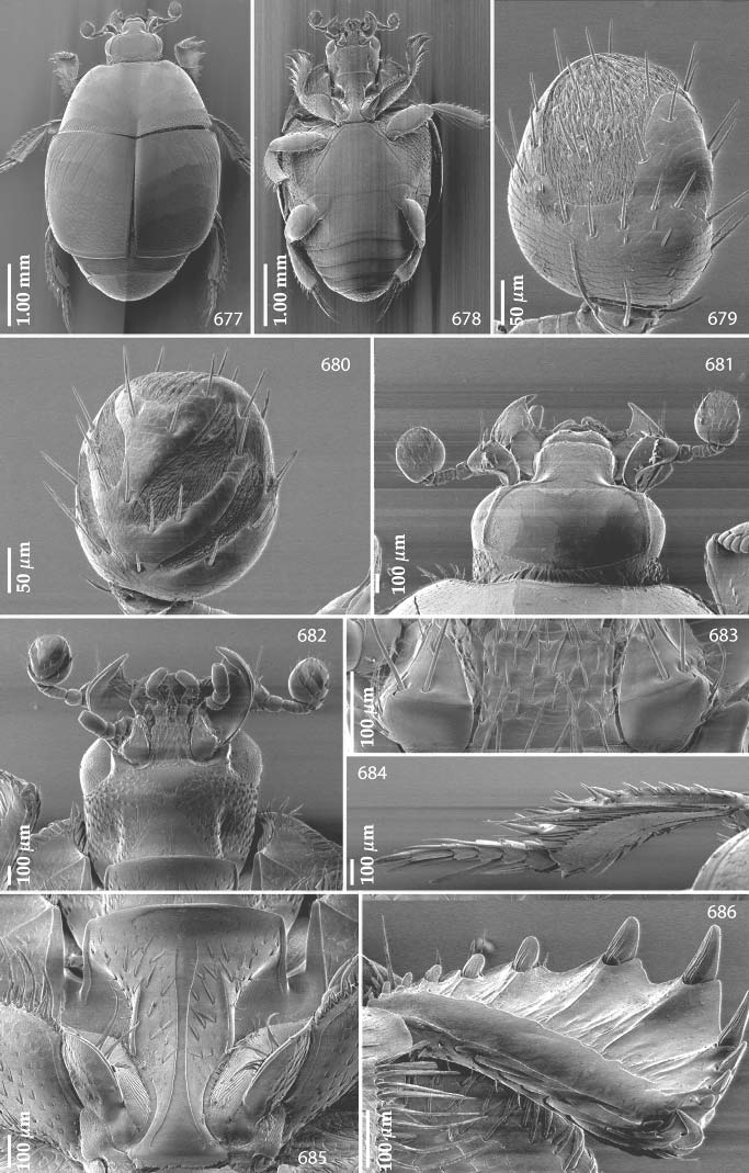
Figs.677–686. Styphrus corpulentus Motschulsky, 1845, SEM micrographs:677 – habitus, dorsal view; 678 – ditto, ventral view; 679 – antennal club, dorsal view; 680 – ditto, ventral view; 681 – head, dorsal view; 682 – ditto, ventral view; 683 – mentum, cardines and stipites of maxilla, ventral view; 684 – metatibia, dorsal view; 685 – prosternum; 686 – protibia, ventral view.
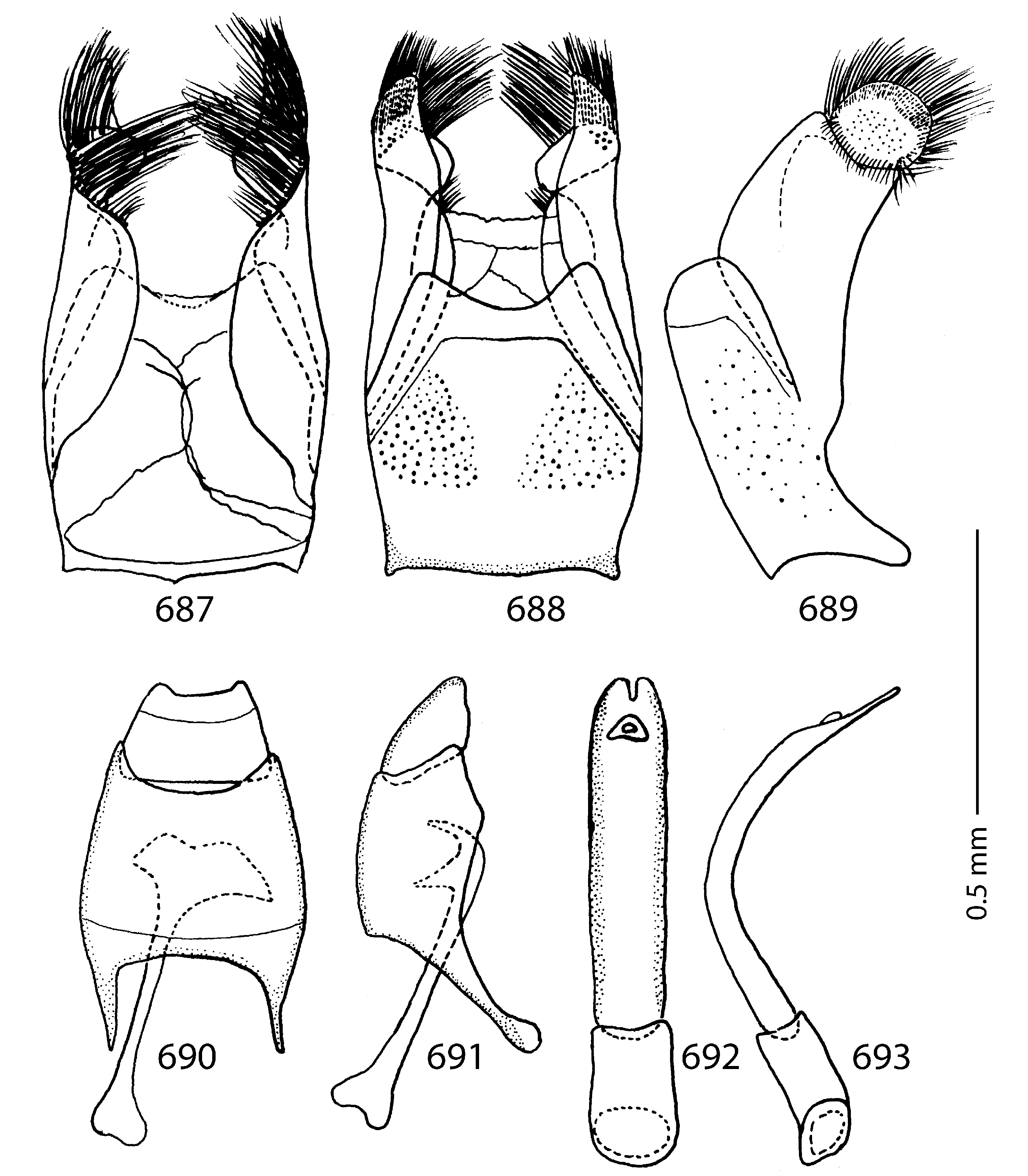
Figs.687–693.Styphrus corpulentus Motschulsky, 1845, male terminalia: 687 – 8th sternite and tergite, ventral view; 688 – ditto, dorsal view; 689 – ditto, lateral view; 690 – 9th tergite, 10th tergite (dorsal view) and spiculum gastrale (ventral view); 691 – 9th tergite, 10th tergite and spiculum gastrale, lateral view; 692 – aedeagus, dorsal view; 693 – ditto, lateral view.
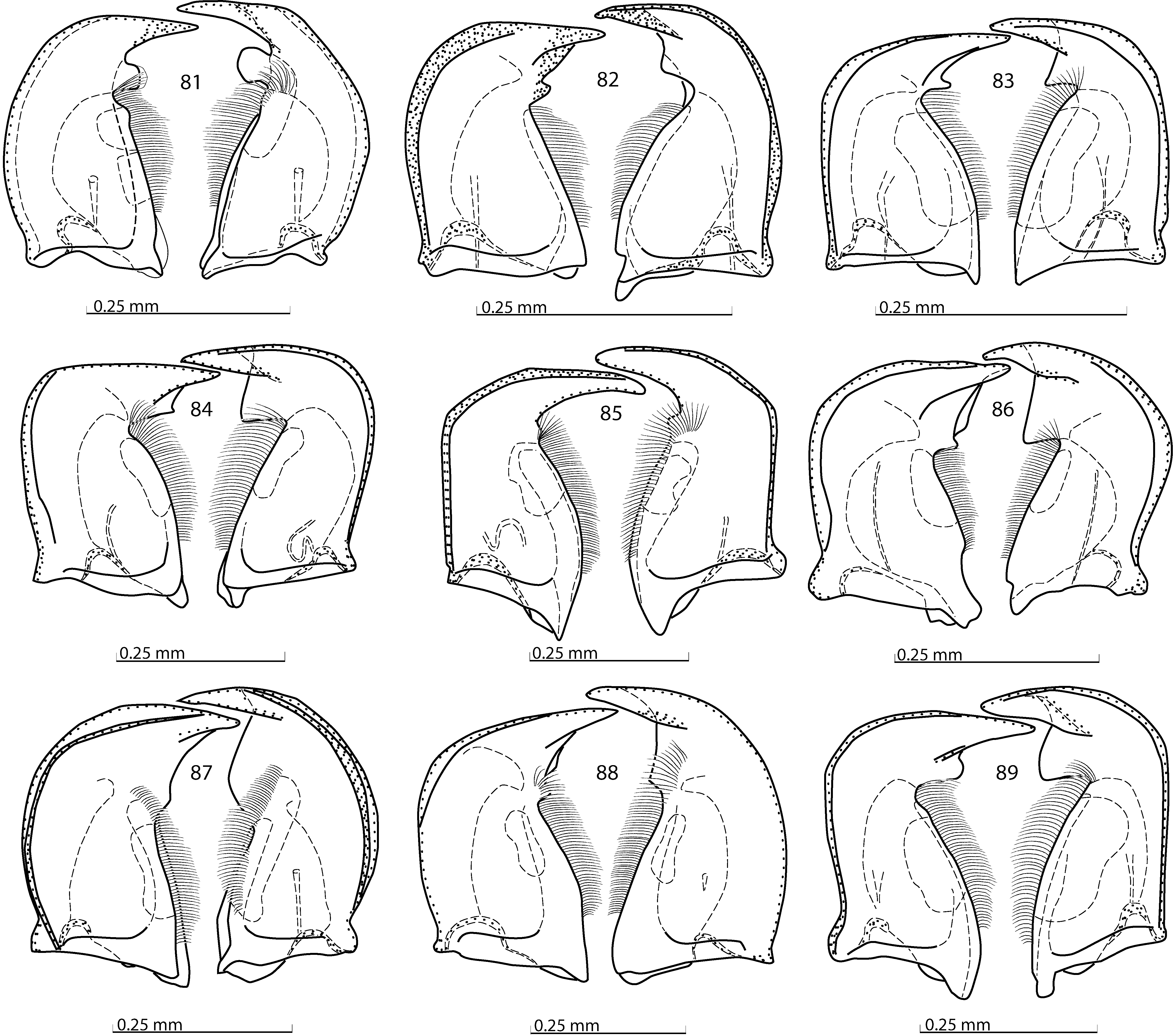
Figs. 81–89. Saprininae, mandibles, ventral view: 81 –Alienocacculus neftensis (Olexa, 1984); 82 – Ammostyphrus cerberus Reichardt, 1924; 83 – Chalcionellus amoenus (Erichson, 1834); 84 – Chivaenius kryzhanovskii Olexa, 1980; 85 – Ctenophilotis chobauti (Théry, 1900); 86 – Eopachylopus ripae (Lewis, 1885); 87 – Eremosaprinus vlasovi (Reichardt, 1941); 88 – Euspilotus (Neosaprinus) perrisi (Marseul, 1872); 89 – Exaesiopus grossipes grossipes (Marseul, 1855).

Figs. 360–370.Gnathoncus rotundatus (Kugelann, 1792), SEM micrographs:360 – habitus, dorsal view; 361 – ditto, ventral view; 362 – antennal club, ventral view; 363 – head, dorsal view; 364 – ditto, ventral view; 365 – mentum, ventral view; 366 – prosternum; 367 – mesoventrite and metaventrite; 368 – lateral disc of metaventrite, metepisternum and fused metepimeron; 369 – protibia, ventral view; 370 – mesotibia, ventral view.
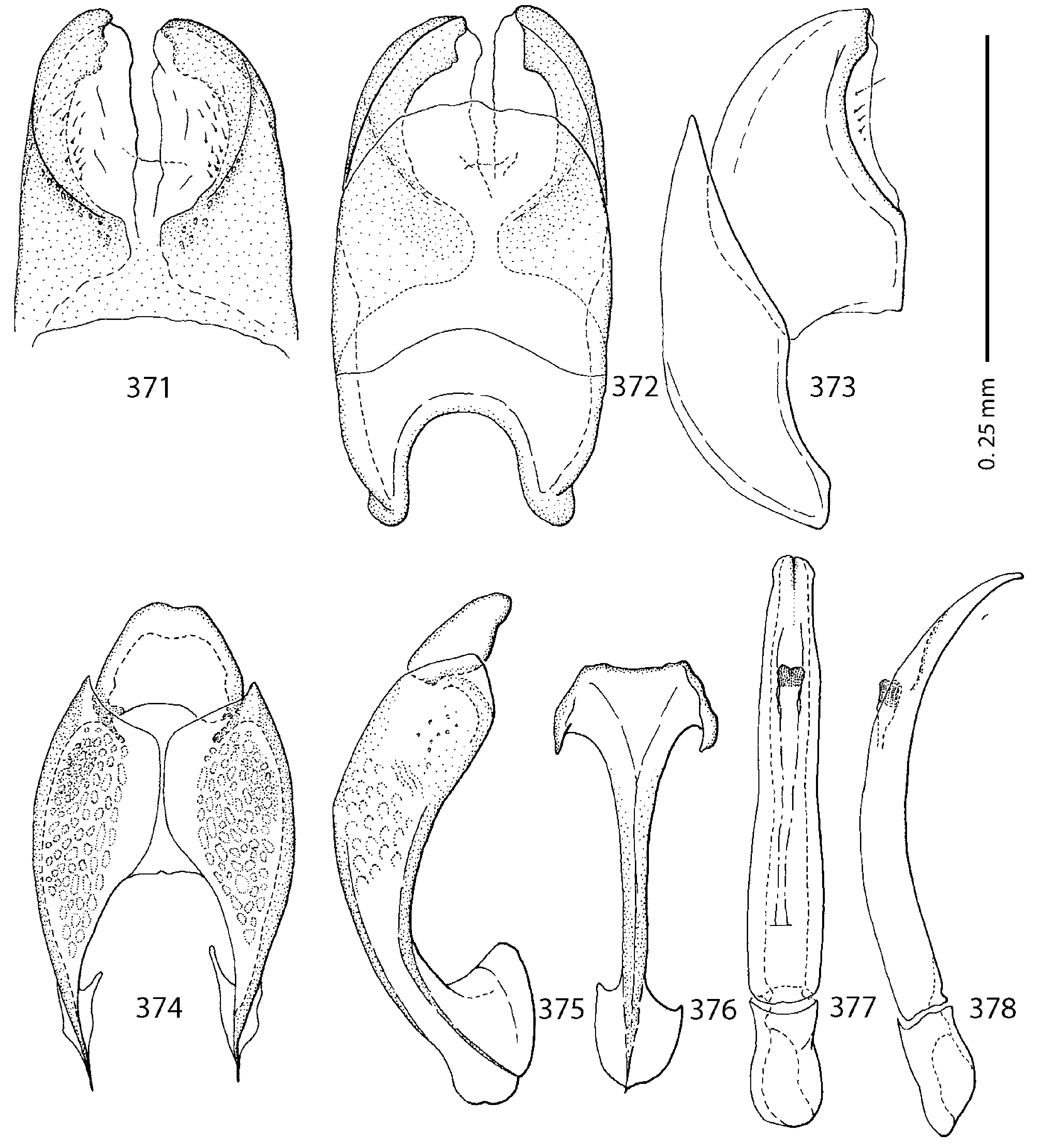
Figs. 371–378. Gnathoncus rotundatus (Kugelann, 1792), male terminalia (after ÔHARA 1994): 371 – 8th sternite and tergite, ventral view; 372 – ditto, dorsal view; 373 – ditto, lateral view; 374 – 9th tergite, 10th tergite, dorsal view; 375 – ditto, lateral view; 376 – spiculum gastrale, ventral view; 377 – aedeagus, dorsal view; 378 – ditto, lateral view.
| ZIN |
Russian Academy of Sciences, Zoological Institute, Zoological Museum |
No known copyright restrictions apply. See Agosti, D., Egloff, W., 2009. Taxonomic information exchange and copyright: the Plazi approach. BMC Research Notes 2009, 2:53 for further explanation.
|
Kingdom |
|
|
Phylum |
|
|
Class |
|
|
Order |
|
|
Family |
|
|
SubFamily |
Saprininae |
|
Genus |
Styphrus corpulentus Motschulsky, 1845
| Lackner, Tomáš 2010 |
Saprinus rugosipennis
| KRYZHANOVSKIJ O. L. & REICHARDT A. N. 1976: 187 |
Styphrus corpulentus: JAKOBSON (1911)
| MAZUR S. 2004: 101 |
| MAZUR S. 1997: 245 |
| MAZUR S. 1984: 78 |
| KRYZHANOVSKIJ O. L. & REICHARDT A. N. 1976: 187 |
| REICHARDT A. 1941: 174 |
| JAKOBSON G. G. 1911: 651 |
Xenonychus laevidorsis
| BICKHARDT H. 1916: 103 |
| REITTER E. 1910: 13 |
Styphrus laevidorsis:
| BICKHARDT H. 1910: 107 |
Pachylopus akinini:
| REITTER E. 1906: 268 |
Xenonychus akinini:
| LEWIS G. 1905: 78 |
Saprinus mimulus
| JAKOBSON G. 1910: 263 |
| REITTER E. 1904: 29 |
Saprinus akinini
| JAKOBSON G. 1910: 264 |
| SCHMIDT J. 1890: 19 |
Styphrus corpulentus
| MOTSCHULSKY V. 1845: 54 |
1 (by felipe, 2020-11-13 20:09:42)
2 (by ExternalLinkService, 2020-11-13 20:24:22)
3 (by valdenar, 2020-11-16 20:03:45)
4 (by valdenar, 2020-11-17 12:55:40)
5 (by ExternalLinkService, 2020-11-17 13:08:02)
6 (by valdenar, 2020-11-20 19:18:44)
7 (by valdenar, 2020-11-21 14:06:37)
8 (by valdenar, 2020-11-23 14:09:53)
9 (by valdenar, 2020-11-23 15:50:41)
10 (by valdenar, 2020-11-23 18:50:04)
11 (by valdenar, 2020-11-23 23:06:41)
12 (by valdenar, 2020-11-23 23:15:12)
13 (by felipe, 2020-11-24 13:08:56)
14 (by ExternalLinkService, 2020-11-24 13:14:15)
15 (by ExternalLinkService, 2020-12-17 20:42:14)
16 (by ExternalLinkService, 2021-09-20 00:47:40)
17 (by plazi, 2023-10-31 23:05:15)
18 (by ExternalLinkService, 2023-11-01 13:43:37)