
Zatypota percontatoria (Müller), Muller
|
publication ID |
https://doi.org/ 10.5281/zenodo.196324 |
|
DOI |
https://doi.org/10.5281/zenodo.5688933 |
|
persistent identifier |
https://treatment.plazi.org/id/03850372-CA0E-3F0C-32B5-FC954076B36C |
|
treatment provided by |
Plazi |
|
scientific name |
Zatypota percontatoria (Müller) |
| status |
|
Zatypota percontatoria (Müller) View in CoL
Ichneumon percontatorius Müller, 1776: 154 . Type destroyed, Neotype Ψ (Copenhagen), Horstmann (2000). Pimpla percontatoria phoenicea Haliday, 1839: 116 . Yu & Horstmann (1997). Polysphincta gracilis Holmgren, 1860: 32 . Townes & Townes (1960), Horstmann (2000). Polysphincta scutellaris Holmgren, 1860: 33 . Townes & Townes (1960), Horstmann (2000). Polysphincta pulchrator Thomson, 1877: 757 . Horstmann (2000).
Polysphincta theridii Howard, 1892: 292 . Townes & Townes (1951).
Polysphincta granulosa Davis, 1898: 369 . Townes & Townes (1951).
Polysphincta pulchratrix Schulz, 1906 . Yu & Horstmann (1997).
Lycorinopsis decorata Haupt, 1954: 112 . Oehlke (1966).
Lycorinopsis rhombifer Haupt, 1954: 112 . Oehlke (1966).
This species is new to Japan. The following description is based on Japanese specimens.
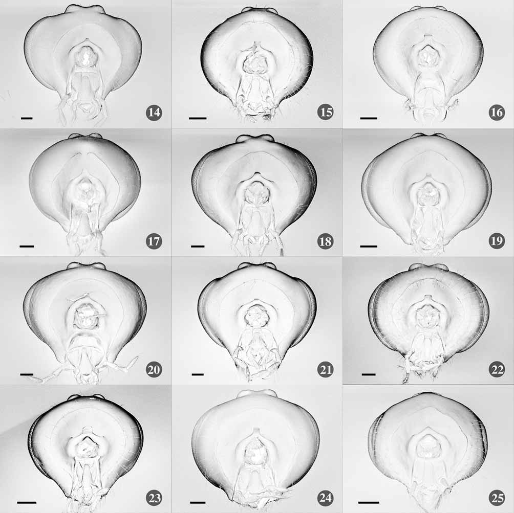
Female. Head ( Figs 9 View FIGURES 1 – 13 , 22 View FIGURES 14 – 25 ). Flagellum with 18–20 segments; frons impunctate and polished; face and clypeus almost impunctate, polished covered with pubescence, the former weakly separated from the latter by a very shallow supraclypeal suture, slightly convex in lateral view; face between antennal socket and supraclypeal suture 1.4 x its minimum width between eyes, with a pair of shallow vertical furrows arising from supraclypeal suture; clypeus 1.6 x its length, with apical margin rounded and weakly truncate medially; eye bare, its inner margin almost straight to slightly convergent downward; ocelli of moderate size, eye remote from lateral ocellus by slightly more than the maximum diameter of the latter; mandible with upper tooth longer and wider than the lower, moderately tapered, about 0.6 x as wide as the basal width at the middle, outer face of mandible covered with some rather long pubescence; palpi formula 4: 3; malar space about 0.5 x basal width of mandible, granulate between lower end of eye and mandible base; hypostomal carina narrowly lamellate between the lower articulation of mandible and junction of occipital and hypostomal carinae; vertex with inter-ocellar area weakly raised; outline of gena weakly rounded in dorsal view; occipital carina complete throughout, in dorsal view weakly bowed forward.
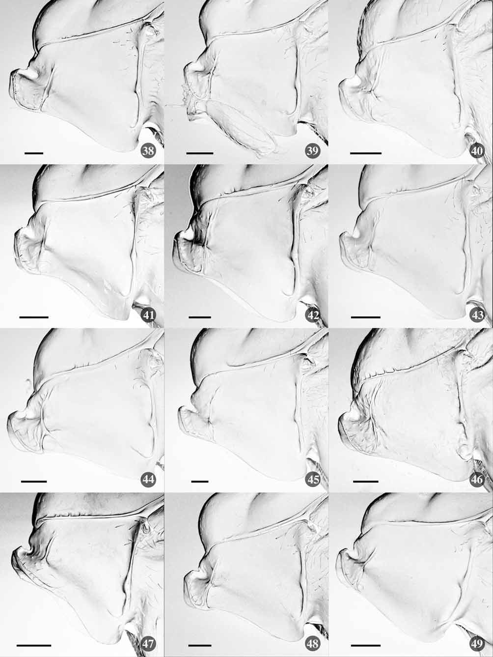
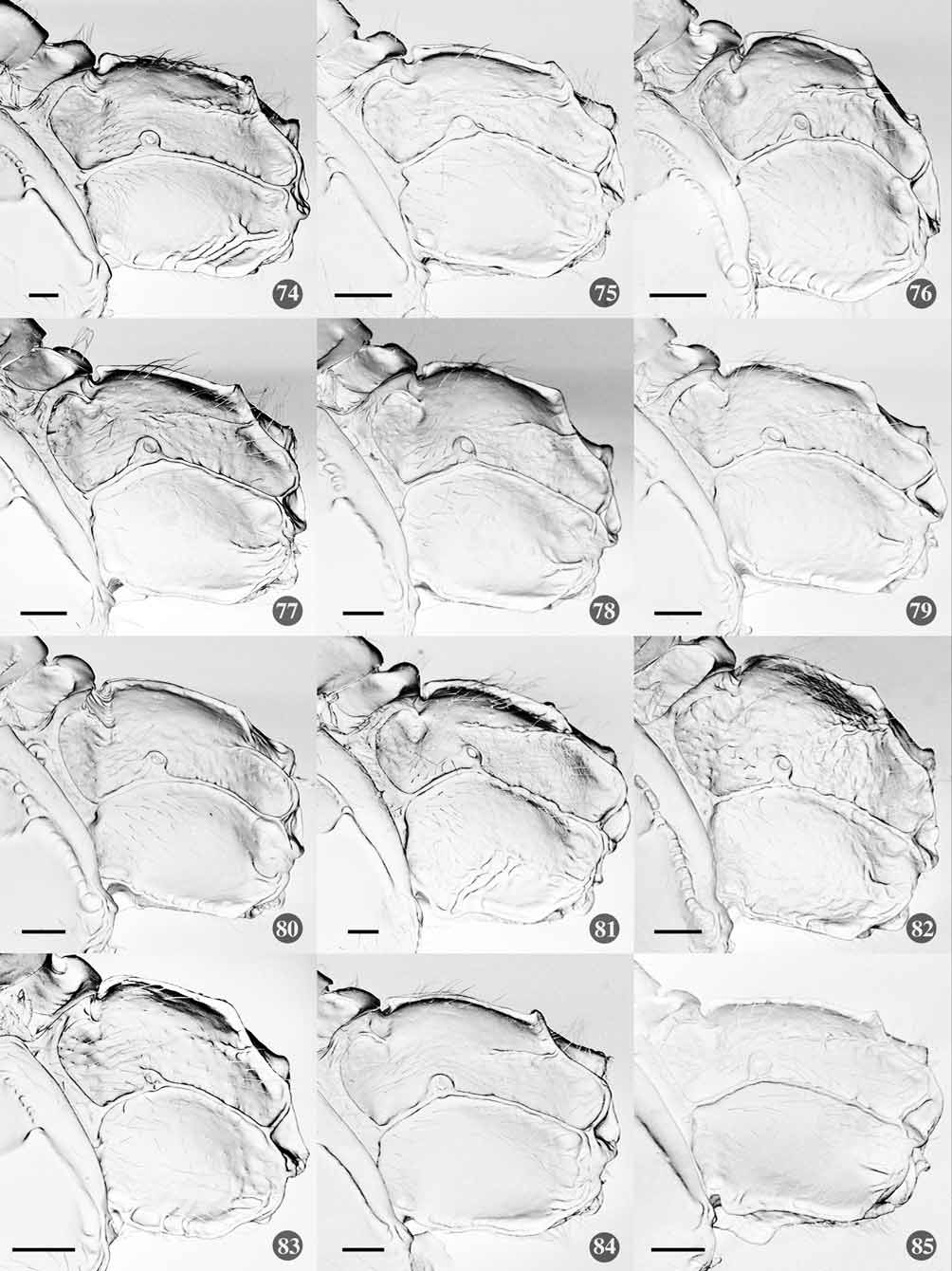
Mesosoma . Pronotum ( Figs 34 View FIGURES 26 – 37 , 46 View FIGURES 38 – 49 ) with anterior margin weakly reflexed, with a median low bridge between anterior and posterior margin, polished, with rather distinct epomia accompanied with some irregular wrinkles near upper and lower ends; mesoscutum in front of scuto-scutellar groove 1.2 x as long as wide in dorsal view, almost bare and polished all over except some short pubescence on median lobe of mesoscutum, with some very short vertical carinae arising from antero-lateral margin above epomia; notauli shallow, almost straight and convergent to a shallow hollow at the middle of mesoscutum; slope of mesoscutum in lateral view rather gradual; mesopleuron ( Fig. 58 View FIGURES 50 – 61 ) with a distinct sinuate epicnemial carina, its upper end surpassing the level of lower corner of pronotum and rather far from posterior margin of pronotum, often slightly coriaceous with dense pubescence ventrally, with a short groove below speculum, mesopleural suture foveolate; scutellum convex; propodeum ( Figs 70 View FIGURES 62 – 73 , 82 View FIGURES 74 – 85 ) with rather dense pubescence laterally, coriaceous all over; areas basalis and superomedia, and areas posterialis and postero-externa fused respectively, carinae delimiting these combined areas distinct; pleural carina complete; propodeal spiracle touching or almost touching pleural carina; metapleuron ( Fig. 82 View FIGURES 74 – 85 ) coriaceous, weakly rugose ventrally and posteriorly; submetapleural carina complete, strong but with no projection anteriorly.
Wings. Fore wing ( Fig. 94 View FIGURES 86 – 97 ) with vein Rs+M basad of cu-a by 2– 3 x the width of the vein, 2rs-m about 0.1– 0.2 x the length of M between 2rs-m and 2m-cu; vein cu-a moderately inclined; vein Cu1a separated from 1mcu by the length of Cu1b or slightly more. Hind wing ( Fig. 106 View FIGURES 98 – 109 ) with vein M+ Cu moderately bowed; distal abscissa of Cu absent; vein cu-a slightly inclivous to vein 1A.
Legs. Legs relatively slender; fore femur 3.9–4.8 x as long as maximum width; hind femur 4.6–4.8 x as long as maximum width: hind tibia 9.5–10.0 x as long as apical width; first tarsal segment of hind leg as long as second and third segments combined; fifth segment as long as the third.
Metasoma ( Fig. 118 View FIGURES 110 – 121 ). T1 distinctively coriaceous, with pair of dostero-lateral oblique impressions; with dorsomedian longitudinal carinae extending to 0.6 of its length; carinae convergent posteriorly in dorsal view but rather separated at posterior end; dorsolateral longitudinal carina usually distinct, reaching to the posterior margin, or sometimes indistinct posteriorly, with spiracle located on it; T2 distinctively coriaceous except smooth posterior margin, with very sharply impressed anterolateral and posterolateral oblique grooves, delimiting a central evenly convex rhombic area; T3–5 similar to T2, with anterior oblique groove more transverse, making the central areas triangular; posterolateral oblique groove of T5 weak; T1 1.1–1.2 x as long as apical width; T2 0.9–1.0 x as long as T1 and 0.7–0.8 x as long as apical width. Ovipositor straight, rather short projecting beyond apex of metasoma by 0.3 x length of hind tibia; upper valve thickened medially, rather strongly tapered to a very sharp point; lower valve without distinct basal swelling ventrally, weakly thinned around basal 0.4.
Coloration. Head mostly black with following parts yellowish white: pedicel and scape below, lower half of clypeus, mandible except tip, palpi and lower margin of gena; flagellum light brown. Mesosoma reddish brown; propleuron and pronotum anteriorly dark brown; latero-dorsal corner of pronotum, tegula and subalar prominence yellowish white; mesopleuron darkened ventrally; mesepimeron, axilla, postscutellum and propodeum dark brown; metapleuron darkened along submetapleural carina. Legs pale brown; coxa, trochanter and trochantellus of fore and mid leg paler; hind leg with trochanter, trochantellus, tip of femur and basal 0.6–0.7 of tibia paler, apical 0.3 of hind tibia dark brown. Wings hyaline, pterostigma brown. Metasomal tergites dark brown; T1–5 reddish brown behind posterior transverse groove; extreme margin of T2–4 narrowly blackish; Ovipositor brown, sheath black.
Length. Fore wing 3.0– 3.8 mm.
Male. Similar to female but slightly smaller; flagellum with 18 segments, lacks short vertical carinae arising from antero-lateral margin of mesoscutum; legs stouter, hind femur 4.2–4.5 x as long as maximum width.
Length. Fore wing 2.9 mm.
Cocoon ( Fig. 137 View FIGURES 122 – 141 ). Diaphanous, white, sub-cylindrical, with a springy and very open construction of sparse loose whorls, attached to the substrate at the capital wider end. The cocoon of this species and Z. brachycera is very similar to those of Hemerobiidae (Neuroptera) . Some of other European species, namely Z. discolor , Z. bohemani , have also similar cocoon and this resemblance accounts for the several incorrect host records in which the wasp of the Polysphincta -group were reared from hemerobiids ( Fitton et al. 1988).
Variation. Body color of one female from Hokkaido darker. The reddish brown part of other specimens dark brown. Some European specimens also dark in colour in both sexes. One Japanese male specimen reddish almost all over. T2 slightly more transverse in Japanese specimens than European ones, the ratio of length to posterior width centered around 0.8 in the former and around 0.9 in the latter. But the ranges of ratio of each population partly overlaps.
Host. Theridion pinastri (L. Koch) * ( Theridiidae ) in Japan. Theridion varians Hahn , T. simile C. L. Koch and T. tinctum (Walckenaer) are recorded as a host in Europe ( Fitton et al. 1988).
Biological notes. The host spider bearing larvae of Z. percontatoria appears in May. The larva ( Fig. 136 View FIGURES 122 – 141 ) is located on dorso-lateral to lateral surface near the base of host abdomen. The host spider builds an irregular spatial three-dimensional web in the concavity of tree trunk, for example those of Celtis sinensis var. japonica , or on the underside of balustrades in a park. The web of host spider containing parasitoid cocoon was seemingly unmodified.
Comments. This species has Holarctic distribution and is recorded from Japan for the first time. From other Japanese species it can be distinguished by distinctively coriaceous T1–2.
Distribution records. Japan: Hokkaido*, Honshu*; Russian Far East, Europe, Nearctic.
Specimens examined. Note that all Japanese reared specimens were from T. pinastri . [Hokkaido] 1Ψ, 29.IX–3.X.1993, Shimo-go-ku, Furano, (K.Suzuki et al.) (NIAES). [Honshu] 1Ψ, 29.VI.2009 (cocoon, emer. 2.VII.), Heijo Palace site, Nara, Nara Pref., (R.M.); 1ɗ, 4.VI.2006 (larva on host, emer. 15.VI.), Osaka-jo Park, Osaka, Osaka Pref., (Y.Shimizu); 2ΨΨ ( Fig. 122 View FIGURES 122 – 141 ), 20.V.2007 (larva on host, emer. 26–30.V.), same locality (R.M.); 2ΨΨ, 20.V.2007 (larva on host, emer. 26–30.V.), same locality, (R.M.); 2ΨΨ, 23.V.2007 (larva on host, cocooned 26.V. emer. 3.VI.), same locality (R.M.); 1Ψ, 23.V.2007 (cocoon, emer. 29.V.), same locality and date (R.M.); 1ɗ2ΨΨ, 7.V.2008 (cocoon, emer. 8–15.V.), same locality and date (R.M.); 5ɗɗ1Ψ, 13.V.2008 (cocoon, emer. 13–20.V.), same locality and date (R.M.); 2ɗɗ, 9.VI.2008 (cocoon, emer. VI.), same locality and date (R.M.); 3ɗɗ, 26.IV.2009 (cocoon, emer. 1– 2.V.), same locality and date (R.M.); 1ɗ2ΨΨ, 16.VIII.2009 (cocoon, emer. 21.VIII.), same locality and date (R.M.).


No known copyright restrictions apply. See Agosti, D., Egloff, W., 2009. Taxonomic information exchange and copyright: the Plazi approach. BMC Research Notes 2009, 2:53 for further explanation.
|
Kingdom |
|
|
Phylum |
|
|
Class |
|
|
Order |
|
|
Family |
|
|
Genus |
Zatypota percontatoria (Müller)
| Matsumoto, Rikio & Takasuka, Keizo 2010 |
Lycorinopsis decorata
| Haupt 1954: 112 |
Lycorinopsis rhombifer
| Haupt 1954: 112 |
Polysphincta granulosa
| Davis 1898: 369 |
Polysphincta theridii
| Howard 1892: 292 |
Ichneumon percontatorius Müller, 1776 : 154
| Thomson 1877: 757 |
| Holmgren 1860: 32 |
| Holmgren 1860: 33 |
| Haliday 1839: 116 |
| Muller 1776: 154 |