
Ischnopelta marginella Rosso & Campos
|
publication ID |
https://doi.org/10.11646/megataxa.6.2.3 |
|
publication LSID |
lsid:zoobank.org:pub:0A8F7E38-8D11-44EE-8840-F8DE241306D0 |
|
DOI |
https://doi.org/10.5281/zenodo.5753409 |
|
persistent identifier |
https://treatment.plazi.org/id/03828787-2C07-FFA5-FF77-FA36FD9C061E |
|
treatment provided by |
Plazi (2021-12-03 08:25:52, last updated 2024-11-24 23:35:14) |
|
scientific name |
Ischnopelta marginella Rosso & Campos |
| status |
sp. nov. |
Ischnopelta marginella Rosso & Campos , sp. n. ( Figs. 5T View FIGURE 5 , 34–35 View FIGURE 34 View FIGURE 35 )
Etymology. Epithet proposed by Dr. H. Ruckes as registered in the manuscripts found with the specimens used in the study. He considered this species having both the head and scutellum broadly rounded. In this species the apex of the head, and in some specimens also the apex of the scutellum, is emarginated, from which the epithet is inferred. Latin: margino = margin, emarginated + ellus = diminutive.
Type locality. ARGENTINA, Salta, Urundel [- 23.5562, -64.3978] GoogleMaps .
Holotype. Male. ARGENTINA, Salta, Urundel, 2.XII.1934, Birabén Coll. Deposited at Museo de La Plata, Universidade Nacional de La Plata ( MLPA), La Plata, Argentina.
Paratypes. 9 males and 18 females. BOLÍVIA, La Paz, Nor Yungas , 2 males and 1 female, 4. V.1931, Dernier Coll., [-16.182222, -55.383333], ( MLPA) GoogleMaps ; Santa Cruz, Colorado River ( Amboró National Park ), 1 female, VII.1921 – III.1922, Mulford Bio. Expl., [-17.675, - 63.875], ( USNM) GoogleMaps ; ARGENTINA, Salta, Orán, 2 females, 22. V.1947, Birabén Coll., [-23.1340, -64.3246], ( MLPA) GoogleMaps ; 1 female, [-23.1340, -64.3246], ( MACN) GoogleMaps ; Urundel , 4 females, 2.XII.1934, Birabén Coll., [-23.5562, -64.3978], ( MLPA) GoogleMaps ; Jujuy , La Mendieta, 1 females, 27.XII.1939, Dernier Coll., [-26.8241, -65.2226], ( MLPA) GoogleMaps ; Tucumán , 1 female, 1956, C.J. Drake, [-26.8241, -65.2226], ( USNM) GoogleMaps ; Santiago del Estero, Santiago del Estero, 3 males and 4 females, 13. II.1929, [-27.7951, -64.2615], ( MLPA) GoogleMaps ; 1 female, 24.IX.1944, Maldonado Coll., [-27.7951, - 64.2615], ( MLPA) GoogleMaps ; Loreto , 1 male, IX.1954, [-28.3019, -64.1803], ( MLPA) GoogleMaps ; Córdoba, Córdoba , 1 female, 17. II.1943, [-31.4, -64.183333], ( MLPA) ; Vila Dolores , 3 males and 1 female, XII.1932, Comp. Col. Berg, [- 31.9458, -65.1896], ( MLPA) GoogleMaps .
Description. The overall somatic morphology is as describedfor I.scutellata ,exceptforthefollowingfeatures. Head. Apex strongly emarginated on most specimens. Bucculae as high as the first labial segment. Labium reaching the metacoxae. Labrum inserted posteriorly to half the distance between the anterior margin of the eyes and the apex of mandibular plates. Antennal segments I to III light-brown, segments IV and V dark brown; segments ratio: I>II<III=IV<V.
Thorax. Hemelytra: corium as long as scutellum; conspicuous spot at apex of radial vein. Legs pale-yellow, setae on posterdorsal margin of protibiae as long as the others.
Abdomen pale-yellow, dark spots at the lateral of urosternites subequal in length, both wide and dropshaped; male urosternite VII unarmed.
Male. Apical margin of membrane of hemelytra and median portion of posterior margin of urosternite VII subrectilinear; urosternite VII reaching anteriorly the imaginary line connecting the spiracles of sternite V. Genitalia. Pygophore with dorsal and ventral rims slightly concave ( Figs. 34C View FIGURE 34 , dr; 34C, vr). Posterolateral angles 1.8 times longer than the rest of the pygophore, perpendicular to the frontal plane and subparallel; apices slightly convergent ( Fig. 34C–E View FIGURE 34 , pla). Setae short and sparse on posterior half of ventral and lateral surfaces of the pygophore, and on the outer surface of posterolateral angles; setae long on ventral rim and on ventral margin of posterolateral angles. Segment X as long as wide, not reaching the apex of the posterolateral angles and the parameres; hexagonal; lateral margins sclerotized and covered by setae; mid-longitudinal area membranous and with sparse setae; apical margin membranous ( Figs. 34C–E, X; 34L–M View FIGURE 34 ). Parameres claviform, subparallel to the frontal plane; basal portion with inner and outer margins subrectilinear and subparallel; head triangular, slightly swollen, inner margin sinuous with small and strongly sclerotized excavation; rounded apices; ventral surface with sinuous longitudinal crest, and setae on distal half of the head ( Figs. 34D View FIGURE 34 , pa; 34F–I). Cup-like sclerites externally visible and with subparallel apices ( Fig. 34D View FIGURE 34 , cls). Phallus: proximal half of vesica posteriorly directed, proximal 1/3 as wide as the distal margin of phallotheca, flattened dorsoventrally and cup-shaped, median 1/3 narrower, flattened dorsoventraly and little sclerotized, distal 1/3 conical and strongly sclerotized; distal half ventrally directed; secondary gonopore circular ( Fig. 34J–K View FIGURE 34 ).
Female. Membrane of hemelytra not reaching the posterior margin of mediotergite VIII, apical margin convex; median portion of posterior margin of mediotergite VIII concave; median portion of posterior margin of urosterniteVIIsubrectilinear;projectionsofurosterniteVII as described for I. scutellata ( Fig. 35C View FIGURE 35 , mpr). Genitalia. Valvifers VIII wider than long, subtriangular; posterior margin subrectilinear and oblique to the median line; sutural margins subrectilinear, slightly convex distally and folded dorsally; surface dark yellowish with brown punctures; setae on the distal half of the sutural margins and median portion of the posterior margin; longitudinal grooves narrow and shallow close to the sutural margins ( Figs. 5T View FIGURE 5 ; 35C View FIGURE 35 , vf8). Valvifers IX partially covered by valvifers VIII, lateral margin subrectilinear, setae on mid-basal portion of ventral surface ( Fig. 35C–D View FIGURE 35 , vf9). Laterotergites IX not reaching the posterior margin of mediotergite VIII; lateral margin convex; setae on median portion of lateral margin and on mid-basal portion of ventral surface ( Fig. 35C–D View FIGURE 35 , la9). Thickening of vaginal intima triangular, distal corner larger than the proximal two; extensive triangular and membranous median area ( Fig. 35D View FIGURE 35 , vi). Vesicular area anterior to the collar 1/8 of the posterior one; median duct anterior to the collar with slight proximal widening ( Fig. 35D View FIGURE 35 , mdp); median duct posterior to the collar with proximal and distal widening ( Fig. 35D View FIGURE 35 , md); inner duct coiled in the proximal widening ( Fig. 35D View FIGURE 35 , id). Distal ductus receptaculi of same caliber as the proximal ( Fig. 35D View FIGURE 35 , drd, drp). Pars intermedialis subcylindrical, slightly wider distally ( Fig. 35D View FIGURE 35 , pi); proximal annular crest about half the diameter of the distal one ( Fig. 35D View FIGURE 35 , dac, pac). Capsula seminalis conical, with a long laterobasal projection directed to the pars intermedialis ( Fig. 35D View FIGURE 35 , cs, pr).
Measurements: Table 15.
Distribution. Bolivia (La Paz, Santa Cruz), Argentina (Salta, Jujuy, Tucumán, Santiago del Estero, Córdoba) ( Fig. 8 View FIGURE 8 ).
Comments. Ischnopelta marginella sp. n. distinguishes by the apex of the strongly emarginated, the apex of scutellum widely rounded, usually emarginated, the wide drop-shaped dark spots on the lateral of the urosternites ( Figs. 34A–B View FIGURE 34 ; 35A–B View FIGURE 35 ), the male hexagonal segment X ( Fig. 34L–M View FIGURE 34 ), and the female subtriangular valvifers VIII ( Figs. 5T View FIGURE 5 ; 36C View FIGURE 36 , vf8).
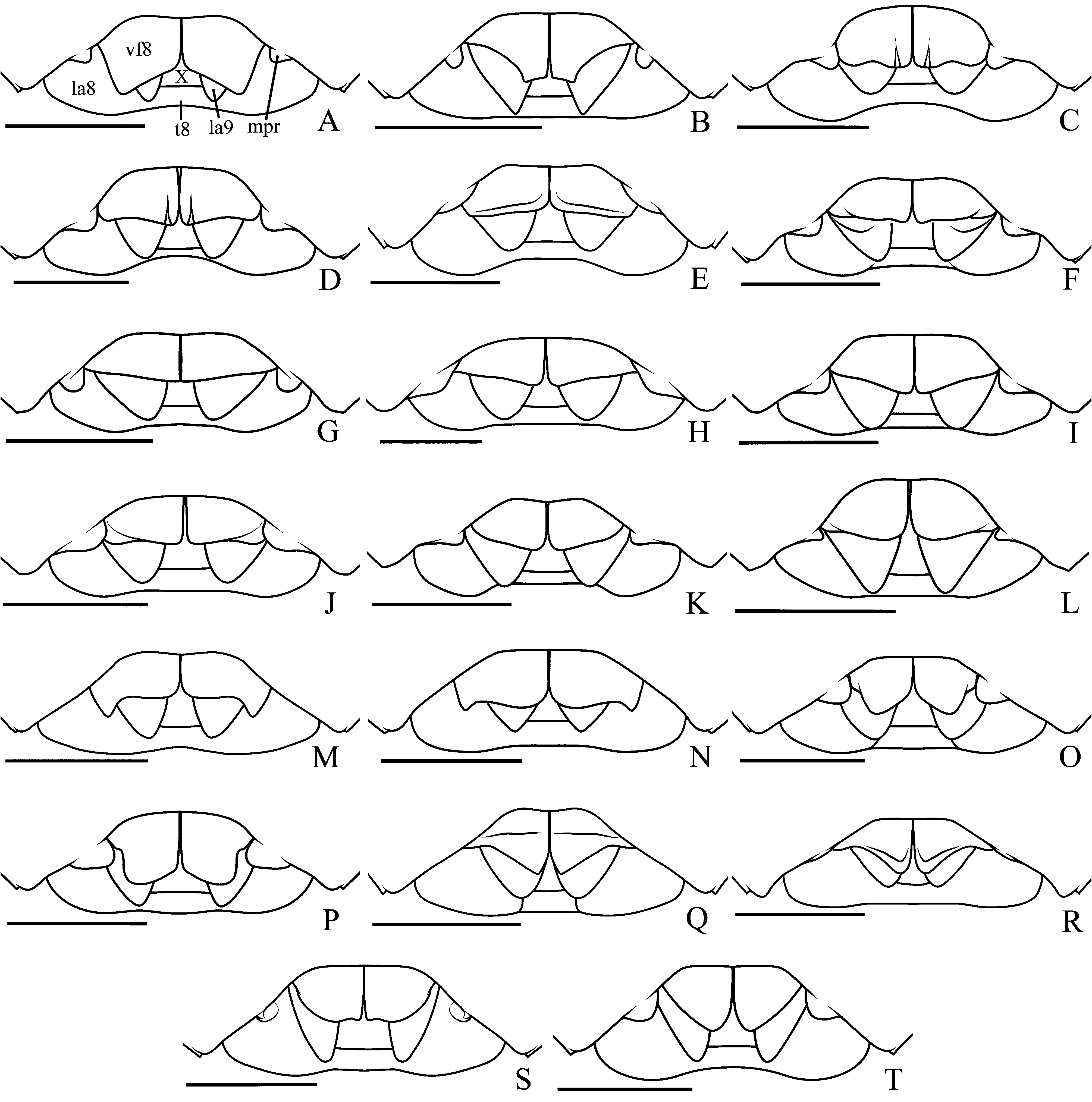
FIGURE 5. Female genital plates.A, Ischnopelta scutellata (Signoret, 1851); B, Ischnopelta bechyneorum Rosso & Campos, sp. n.; C, Ischnopelta confusa Rosso & Campos, sp. n.; D, Ischnopelta magna Rosso & Campos, sp. n.; E, Ischnopelta cylindrata Rosso & Campos, sp. n.; F, Ischnopelta impunctata Rosso & Campos, sp. n.; G, Ischnopelta luteicornis (Walker, 1867); H, Ischnopelta cristulata Rosso & Campos, sp. n.; I, Ischnopelta coralinae Rosso & Campos, sp. n.; J, Ischnopelta ruckesi Rosso & Campos, sp. n.; K, Ischnopelta pellucidula Rosso & Campos, sp. n.; L, Ischnopelta parvula Rosso & Campos, sp. n.; M, Ischnopelta vellozia Rosso & Campos, sp. n.; N, Ischnopelta wigodzinskyi Rosso & Campos, sp. n.; O, Ischnopelta alalonga Rosso & Campos, sp. n.; P, Ischnopelta crassula Rosso & Campos, sp. n.; Q, Ischnopelta cordiformis Rosso & Campos, sp. n.; R, Ischnopelta montana Rosso & Campos, sp. n.; S, Ischnopelta guarani Rosso & Campos, sp. n.; T, Ischnopelta marginella Rosso & Campos, sp. n.. Abbreviations: vf8, valvifers VIII; la8, laterotergites VIII; la9, laterotergites IX; mpr, projection on the lateral third of posterior margin of sternite VII; t8, mediotergite VIII; X, segment X. Scale bars = 1 mm.
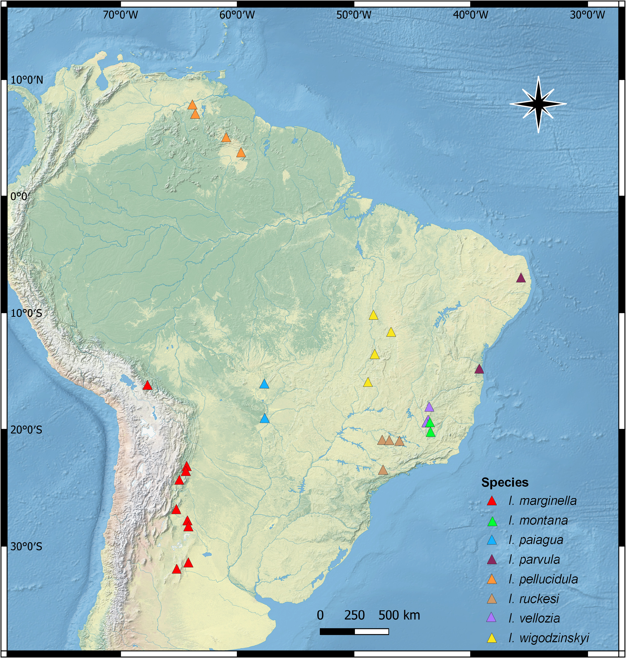
FIGURE 8. Distribution of Ischnopelta marginella Rosso & Campos, sp. n.; Ischnopelta montana Rosso & Campos, sp. n.; Ischnopelta paiagua Rosso & Campos, sp. n.; Ischnopelta parvula Rosso & Campos, sp. n.; Ischnopelta pellucidula Rosso & Campos, sp. n.; Ischnopelta ruckesi Rosso & Campos, sp. n.; Ischnopelta vellozia Rosso & Campos, sp. n.; Ischnopelta wigodzinskyi Rosso & Campos, sp. n..

FIGURE 34. Ischnopelta marginella Rosso & Campos, sp. n. Holotype Male. A, dorsal view; B, ventral view; C–E, pygophore: dorsal, ventral and posterior views respectively; F–I, parameters: dorsal, ventral, external and internal lateral views, respectively; J–K, phallus: lateral and dorsal views, respectively; L–M, segment X, dorsal and ventral views, respectively. Abbreviations: cls, cup like sclerites; dr, dorsal rim; pa, parameter; ph, phalloteca; pla, posterolateral angle; vr, ventral rim; vs, vesica; X, segment X.

FIGURE 35. Ischnopelta marginella Rosso & Campos, sp. n. Female. A, dorsal view; B, ventral view; C, genital plates ventroposterior view (45º); D, internal genitalia. Abbreviations: cl, collar; cs, seminalis capsule; dac, distal annular crest; drd, distal ductus receptaculi; drp, proximal ductus receptaculi; id, internal duct; idp, proximal internal duct; la8, laterotergite VIII; la9, laterotergite IX; md, medium duct; mdp, proximal medium duct; mpr, projection on the lateral third of posterior margin of sternite VII; od, external duct; odp, proximal external duct; pac, proximal annular crest; pi, pars intermedialis; pr, projection; t8, mediotergite VIII; va9, valvulae IX; vf8, valvifer VIII; vf9, valvifer IX; vi, thickening of vaginal intima; X, segment X.

FIGURE 36. Ischnopelta montana Rosso & Campos, sp. n. Holotype male.A, dorsal view; B, ventral view; C–E, pygophore: dorsal, ventral, and posterior views respectively; F–I, parameters: dorsal, ventral, external and internal lateral views, respectively; J–K, phallus: lateral and dorsal views, respectively; L–M, segment X, dorsal and ventral views, respectively. Abbreviations: dr, dorsal rim; pa, parameter; ph, phalloteca; pla, posterolateral angle; vr, ventral rim; vs, vesica; X, segment X.
No known copyright restrictions apply. See Agosti, D., Egloff, W., 2009. Taxonomic information exchange and copyright: the Plazi approach. BMC Research Notes 2009, 2:53 for further explanation.