
Stagmatoptera biocellata Saussure, 1869
|
publication ID |
https://doi.org/ 10.11646/zootaxa.4183.1.1 |
|
publication LSID |
lsid:zoobank.org:pub:0E576DCD-49EB-47DD-9CF2-14F3941BA0B5 |
|
DOI |
https://doi.org/10.5281/zenodo.6057584 |
|
persistent identifier |
https://treatment.plazi.org/id/03826A4A-FF93-FFB1-05B4-FC1E71751331 |
|
treatment provided by |
Plazi |
|
scientific name |
Stagmatoptera biocellata Saussure, 1869 |
| status |
|
Stagmatoptera biocellata Saussure, 1869 View in CoL
Male—Figures 3D, 5D, 7D, 9D, 11D, 13D–19D, 23A. Female—Figures 4C, 6C, 8C, 10C, 12C, 20C–22C, 23B, 24.
Stagmatoptera biocellata Saussure, 1869 View in CoL , p.67 (descr.); Saussure, 1870, p.231 (cit.); Saussure, 1871b, p.96, t.1, f.13 (redesc.); Saussure, 1872, p.257 (cit.); Westwood, 1889, p.16 (cit.); Kirby, 1904, p.299 (cit.); Rehn, 1911, p.12 (cit.); Giglio-Tos, 1914, p.32 (redesc.); Giglio-Tos, 1927, p.598 (redesc.); Terra, 1995, p.64 (cit.); Ehrmann, 2002, p.328 (cit.); Agudelo et al., 2007, p.125 (cit.); Ehrmann & Koçak, 2009, p.11 (cit.).
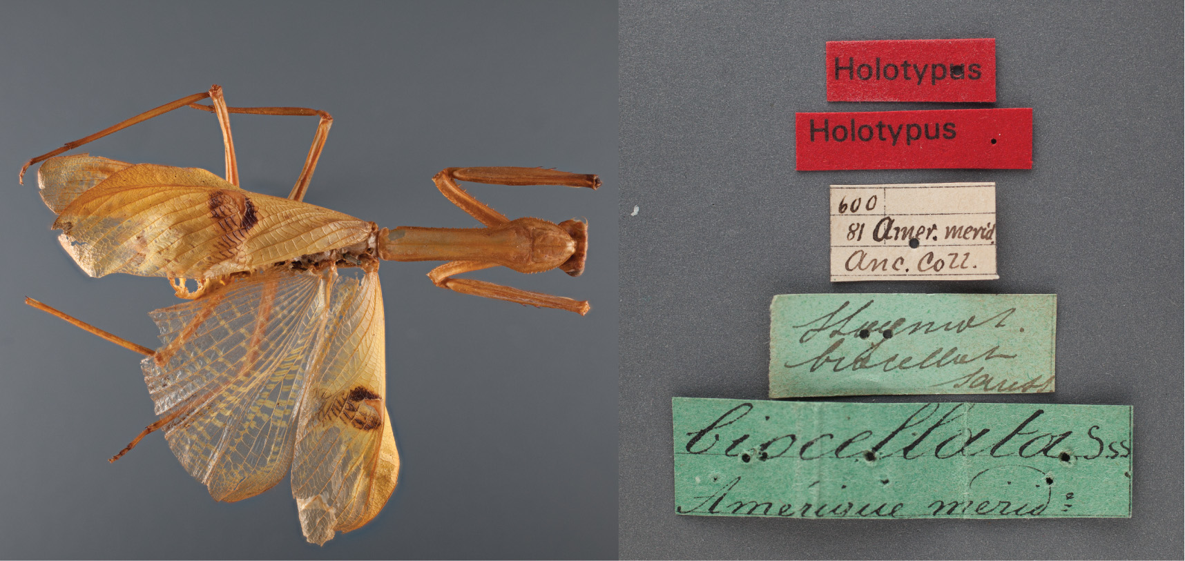
Holotype: 1♀ Brazil, Amer. merid. Anc. Cozz. ( MHNG) ( Fig. 24 View FIGURE 24 ; examined)
Type locality. Brazil.
Diagnosis. Frontal shield of females with four carinae. Prothorax relatively short and broad. Stigma spot oval, large and oblique, often reaching posterior margin of forewings.
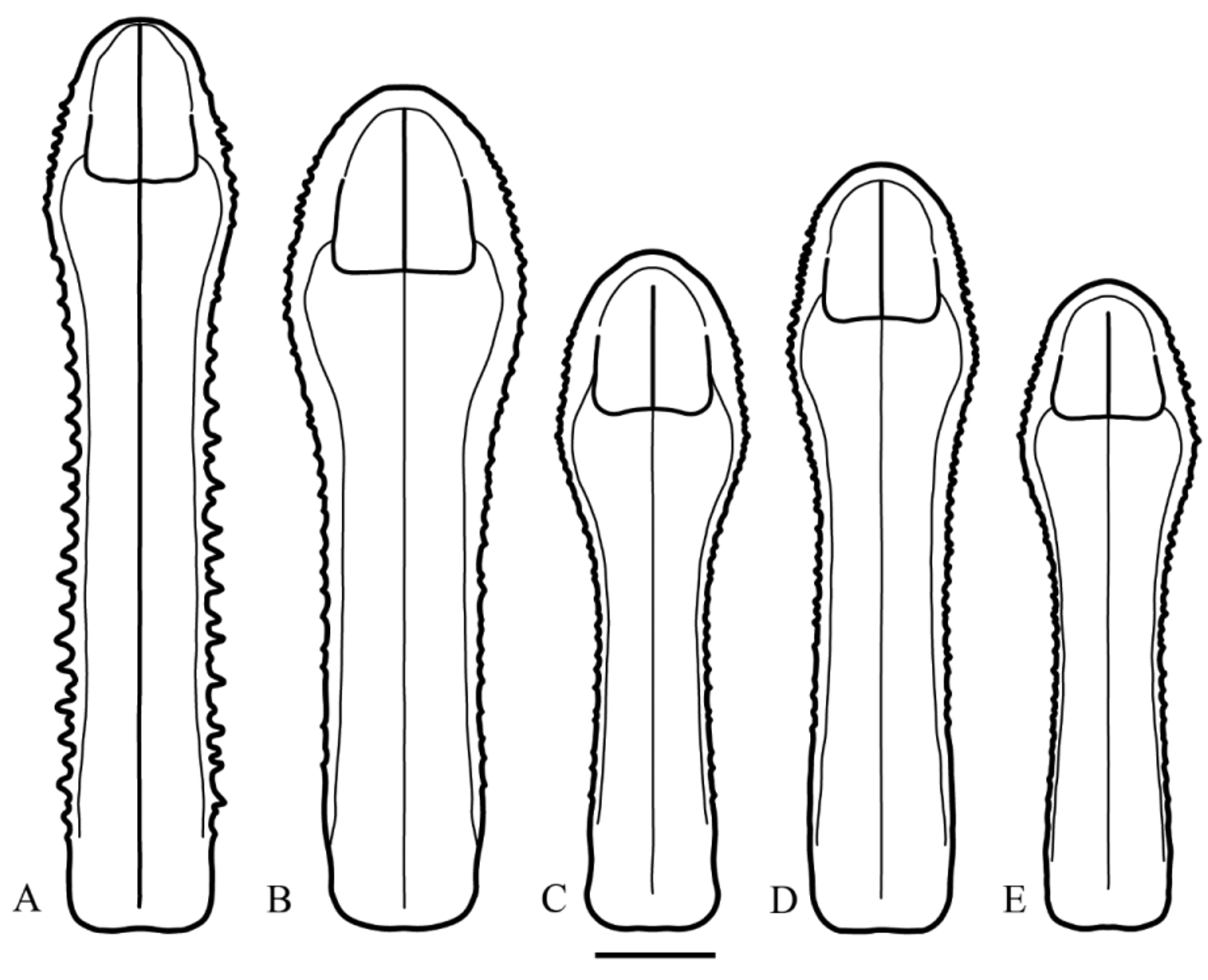
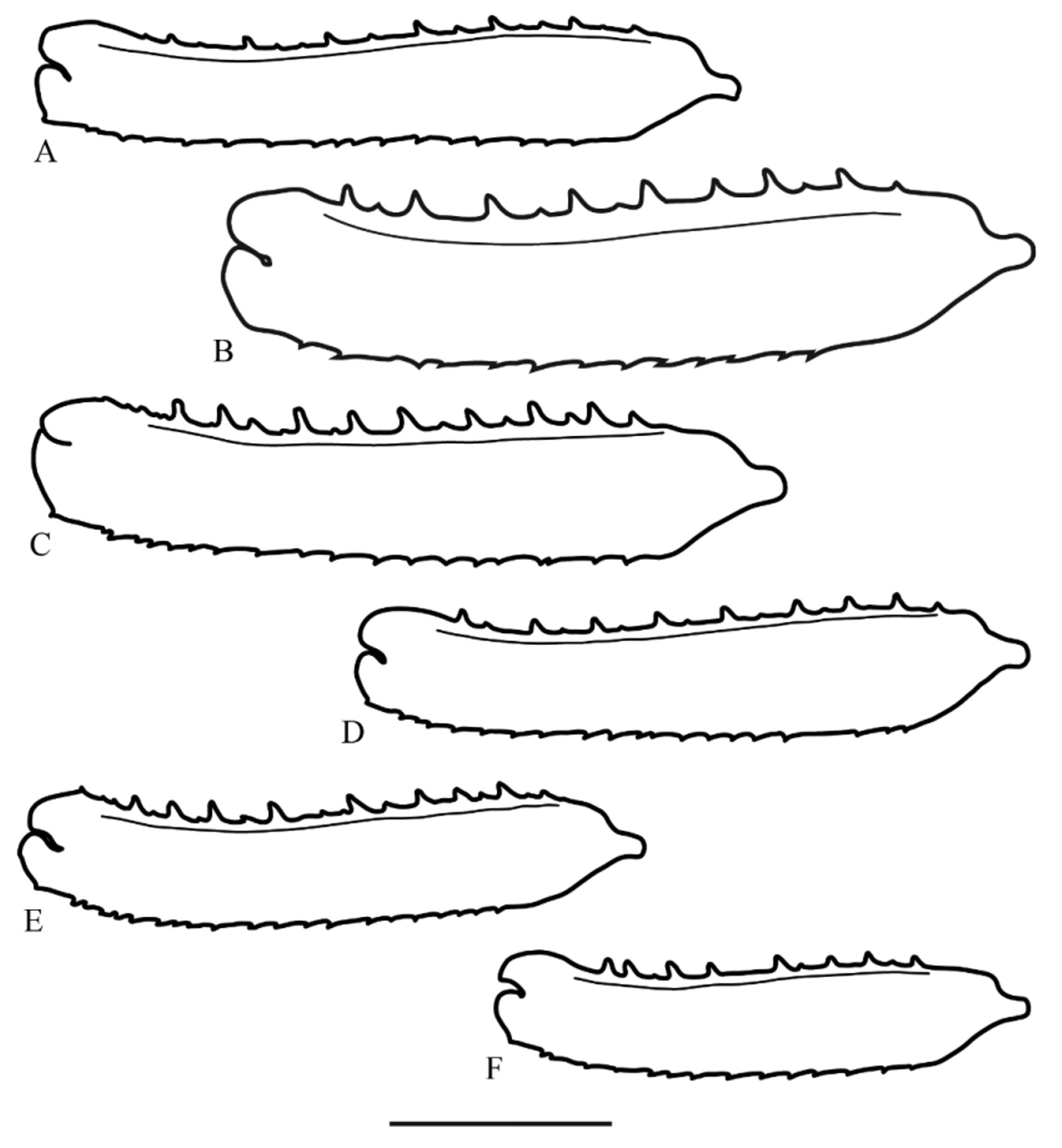
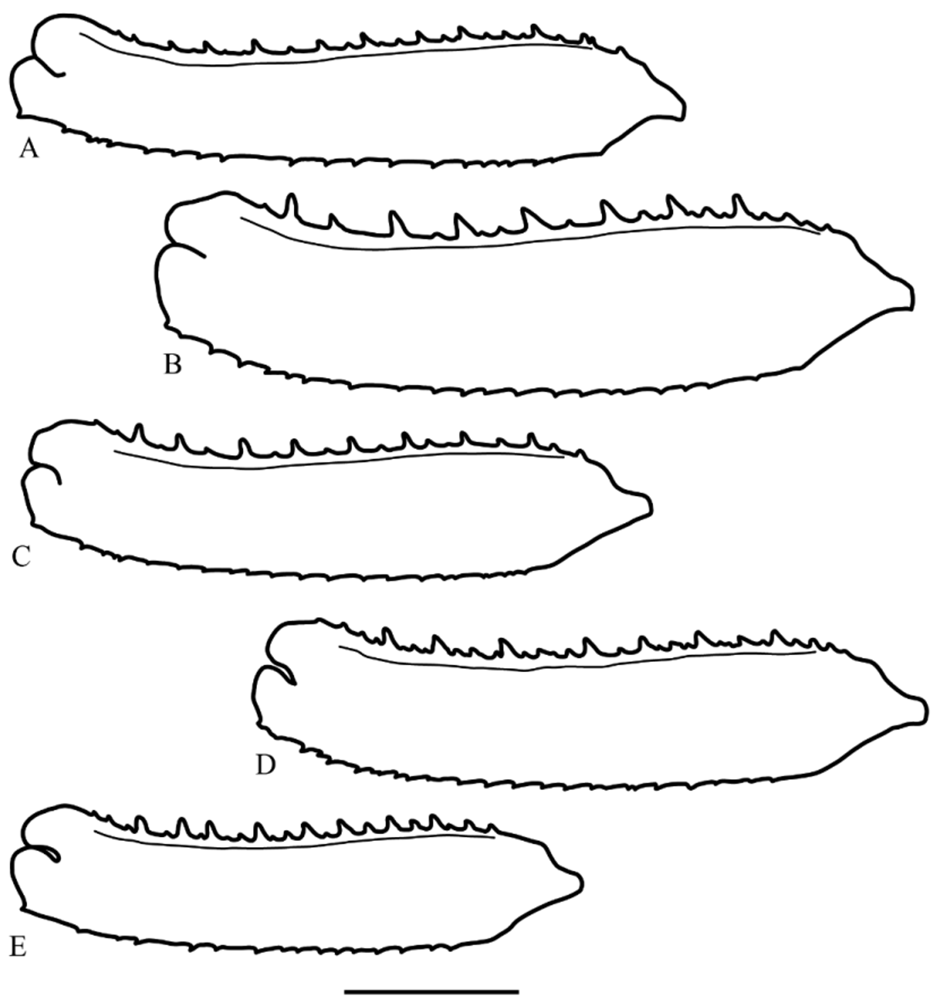
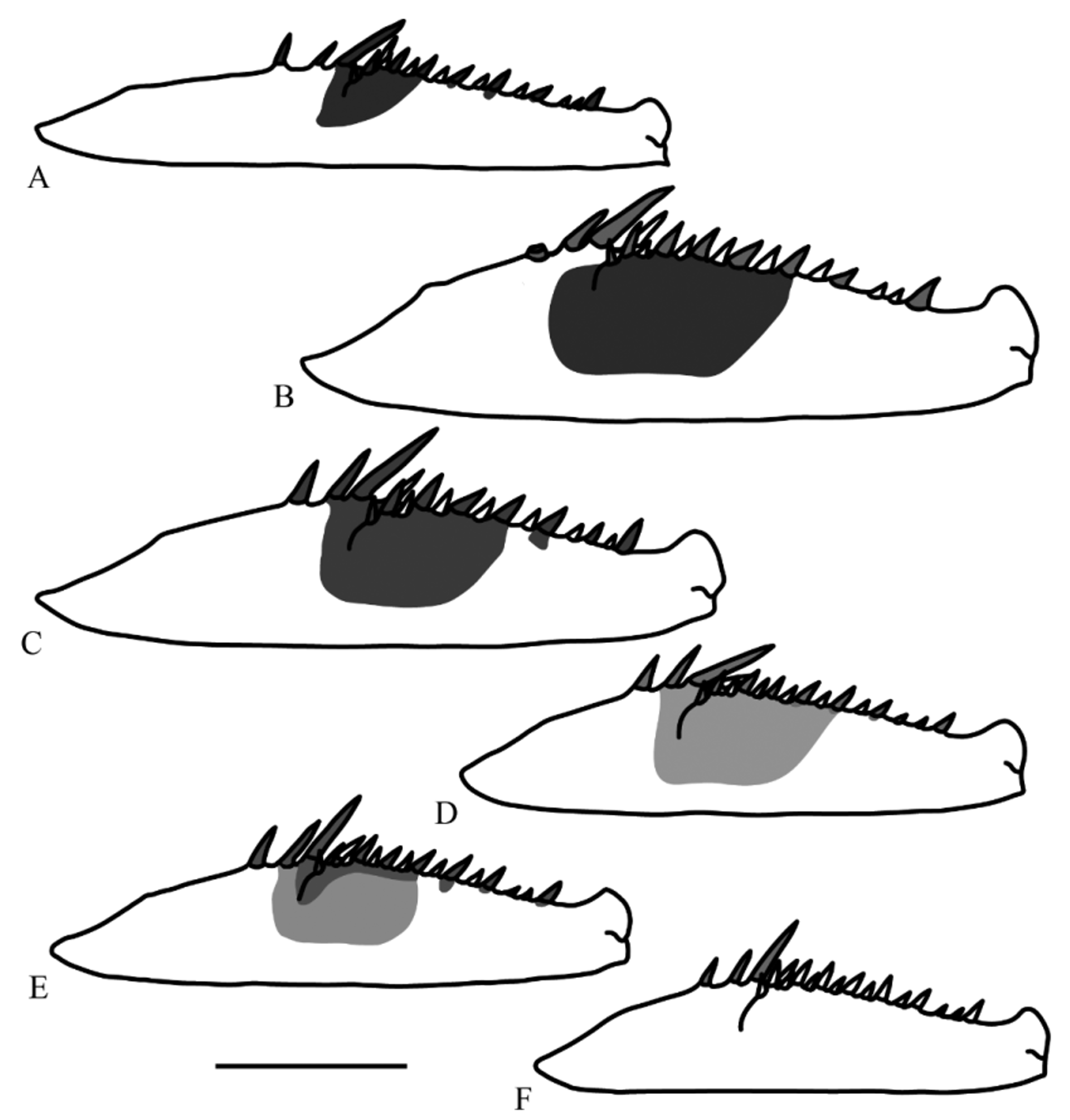
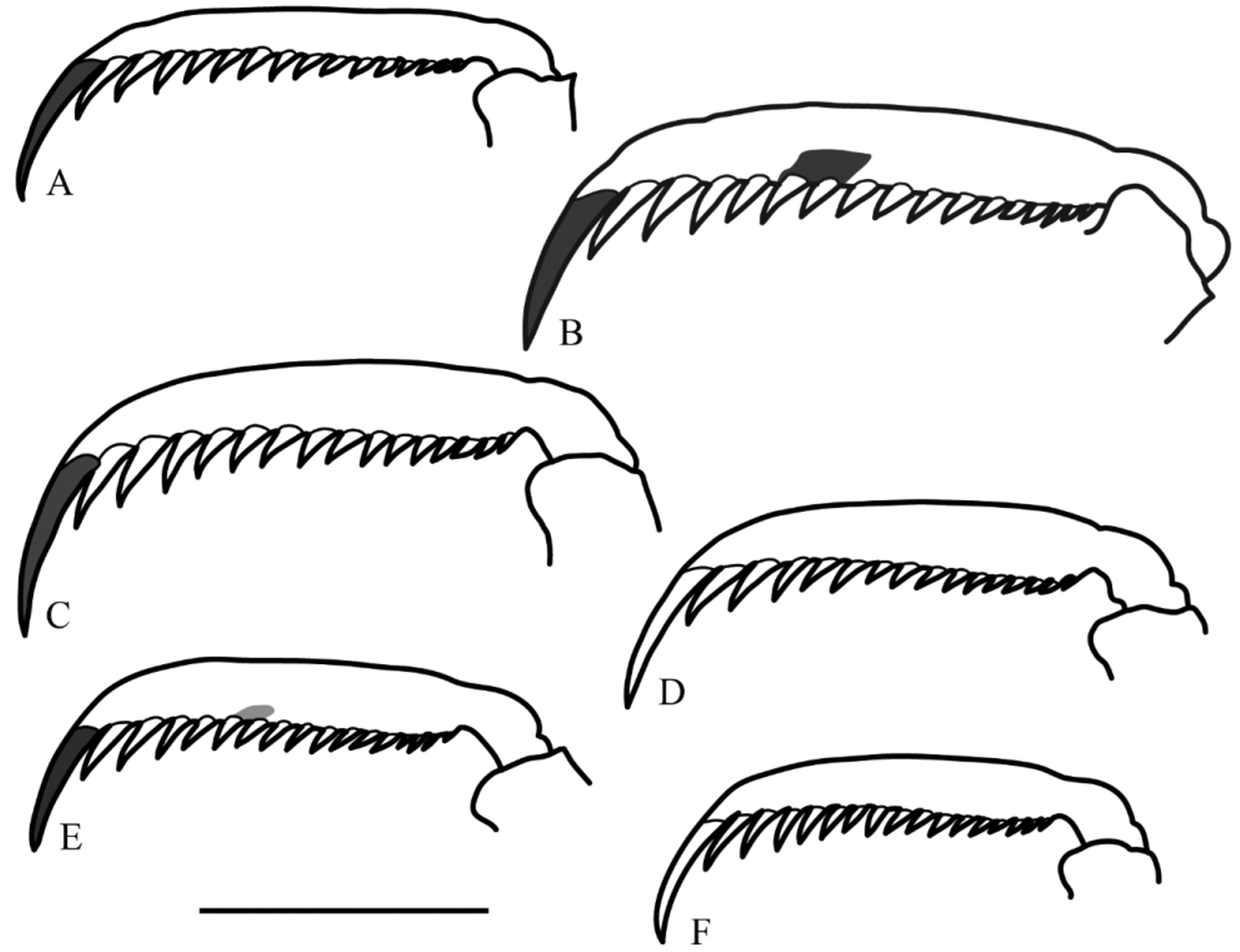
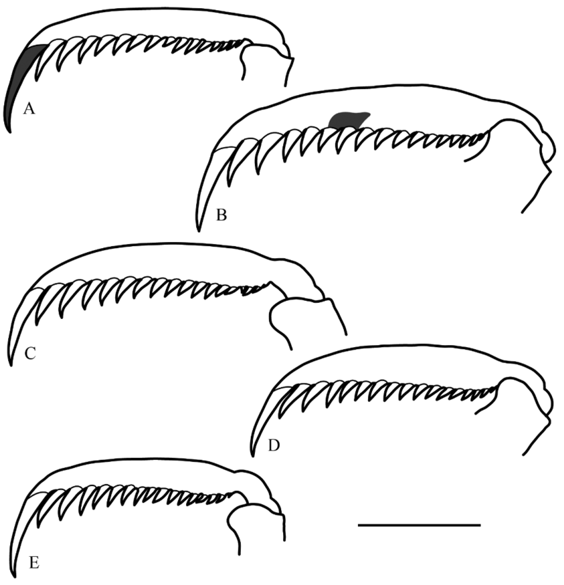
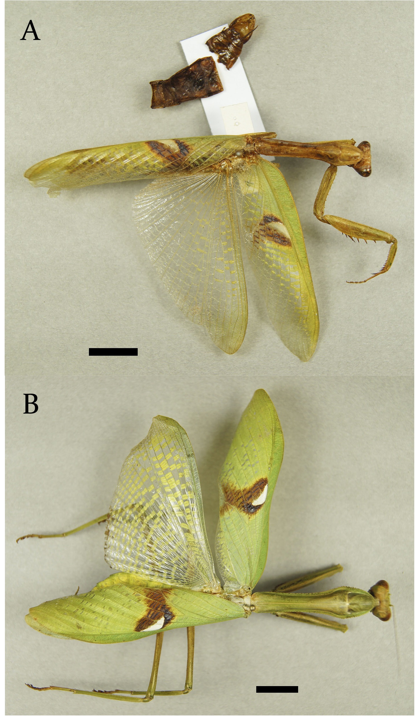
Redescription. Male —Frontal shield with two central carinae ( Fig. 3 View FIGURE 3 D). Prothorax short and broad, lateral margins of prozona slightly crenulated, of metazona smooth. Metazona with a weakly marked central keel ( Fig. 5 View FIGURE 5 D). Forecoxae with 6–9 large spines, which alternate with 2–8 smaller spines ( Fig. 7 View FIGURE 7 D). Forefemora elongated and broad, the first three discoidal spines dark on their anterior surface; 14–15 anteroventral spines, the first spine and all the large spines dark on the anterior surface, the 8th, 10th and 12th spines may feature a small spot of the same coloration on their insertion. Anterior femoral spot extending from the femoral groove to the 10th anteroventral spine, rectangular and effaced black color, which may be stronger near the insertion of the spines ( Fig. 9 View FIGURE 9 D). Foretibiae with 11–12 posteroventral spines and 15–16 anteroventral spines; tibial spur green on its anterior surface ( Fig. 11 View FIGURE 11 D). Foretarsi with apical black spots on the anterior surface of tarsomeres I to IV. Discoidal area of the forewings with an opaque-green stripe anteriorly, gradually becoming hyaline. Spot on the stigma oval, large, oblique, with a central, triangular white spot, a proximal, semicircular dark-brown spot, and a large, posterior and distal light-brown spot. Discoidal and anal area of the hindwings hyaline with yellow stripes on the crossveins at least on their anterior halves ( Fig. 23 View FIGURE 23 A).
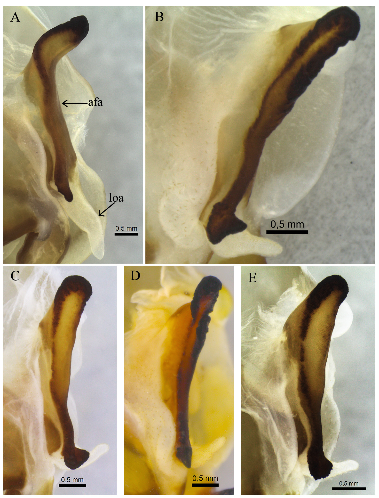
Genitalia. Left phallomere longer than wide; sclerite L4B longer than wide; paa elongated, slightly dilated on the left side of its base, angled approximately 30° from the body axis, afterwards bent to the left, apex simple, directed ventrally ( Fig. 13 View FIGURE 13 D); pda short, broad, flattened, sharply bent 90° to the right, strongly compressed before the apex, the apex also compressed at its base, strongly sclerotized, tapered ( Fig. 16 View FIGURE 16 D); afa elongated, oblique to the body axis ( Fig. 14 View FIGURE 14 D), right margin rugged and sinuous ( Fig. 15 View FIGURE 15 D), posterior apex simple, rugged and strongly sclerotized, anterior apex straight, margins strongly sclerotized, tumid, forming a groove ( Fig. 14 View FIGURE 14 D); loa short, bent to the right ( Fig. 14 View FIGURE 14 D); membrane adjacent to afa pilous ( Fig. 14 View FIGURE 14 D). Right phallomere with the posterior apex rounded; bm short, with a wide expansion ( Fig. 17 View FIGURE 17 D); pia short, strongly sclerotized ( Fig. 18 View FIGURE 18 D–19D); pva short, strongly sclerotized, angular apex ( Fig. 19 View FIGURE 19 D); an elongated, apex abruptly rounded ( Fig. 18 View FIGURE 18 D)
Measurements. Body length: 68.4–77.7; head width: 7.6–8.4; prozona length: 4.4–5.3; metazona length: 16.6–19.1; prothorax width: 4.7–6.2; forecoxae length: 11.8–14.5; forefemora length: 14.3–17.3; forefemora width: 3.1–3.5; foretibiae length: 7.8–9.3.
Ratios. Head/prothoracic-width: 1.35–1.62; metazona/prozona: 3.17–3.84; metazona length/prothoracic width: 3.05–3.6; metazona/forecoxae: 1.27–1.46; forefemora length/width: 4.33–4.97.
Female —Frontal shield with four carinae ( Fig. 4 View FIGURE 4 C). Prothorax short and broad, lateral margins slightly crenulated. Metazona with a weakly marked central keel ( Fig. 6 View FIGURE 6 C). Forecoxae with 7–9 large spines, which alternate with 5–8 smaller spines ( Fig. 8 View FIGURE 8 C). Forefemora elongated, broad, the first three discoidal spines dark on their anterior surface; 14–15 anteroventral spines, usually 15, the first spine and all the large spines dark on their anterior surface. Anterior femoral spot extending from the femoral groove to the 10th anteroventral spine, rectangular and effaced black color ( Fig. 10 View FIGURE 10 C). Foretibiae with 11–12 posteroventral spines, and 15–17 anteroventral spines; tibial spur green on its anterior surface ( Fig. 12 View FIGURE 12 C). Foretarsi with apical black spots on the anterior surface of tarsomeres I to IV. Spot on the stigma oval, large, oblique, with a central, triangular white spot, a proximal, semicircular dark-brown spot and a large, posterior and distal light-brown spot. Costal area of the hindwings hyaline, discoidal and anal areas hyaline with yellow stripes on the crossveins ( Fig. 23 View FIGURE 23 B).
Genitalia. Base of gonapophysis VIII projecting inwards ( Fig. 20 View FIGURE 20 C); bv roughly triangular ( Fig. 20 View FIGURE 20 C) the inner surface sculpted ( Fig. 21 View FIGURE 21 C); ib sclerotized and sculpted ( Fig. 21 View FIGURE 21 C); pe short, weakly sclerotized, with irregular margins ( Fig. 21 View FIGURE 21 C); gs sclerotized, smooth ( Fig. 22 View FIGURE 22 C); ls roughly rectangular, posterior process short, conical ( Fig. 22 View FIGURE 22 C).
Measurements. Body length: 71.8–76.7; head width: 9.3–9.6; prozona length: 5.9–7.3; metazona length: 21–24.7; prothorax width: 7.4–9.2; forecoxae length: 16.7–18.8; forefemora length: 20.4–23.8; forefemora width: 4.5–5.5; foretibiae length: 10.6–11.2.
Ratios. Head/prothoracic-width: 1.22–1.26; metazona/prozona: 3.23–3.78; metazona length/prothoracic width: 2.68–2.9; metazona/forecoxae: 1.18–1.31; forefemora length/width: 4.33–4.6.
Remarks. This species occurs in northern and northeastern Brazil. The vague original description resulted in various misidentifications of this species in the literature. For example, Werner (1925) described a few specimens as S. biocellata , but these actually are referable to S. hyaloptera . The senior author (HMR) identified this error while examining the same material reported by Werner (1925). Another problem was the common misidentification of S. diana sp. n. as S. biocellata ; in fact, all specimens identified as S. biocellata we studied were actually S. diana — except for the type species of the former. This situation makes it very difficult to verify the actual identity of specimens determined as S. biocellata in previous studies. Therefore, we here opted to cite all studies listing S. biocellata , even though their identity as such are reasonably dubious.
Distribution. Brazil (Maranhão, Pará).
Examined material. BRAZIL: Maranhão—Caxias , Faz. Gequiri, 13.x.1998, rede entom., col. F.S. Santos, 1♀ ( INPA) . Caxias, Fumo Verde , 24.x.1999, F. Limeira-de-Oliveira e J.T. Câmara, 1♀ ( INPA) . São P. Água Branca, Faz. Primavera, 03-10.iii.2002, Armadilha Malaise, F. Limeira-de-Oliveira e J.T. Câmara, 1♂ (INPA); Pará – S. Norte, Carajás , vii-viii.1985, Brandão & Benson col., 1♂ ( MZSP) ; without locality: 1♀ (NHMW).
Stagmatoptera cerdai Rodrigues , sp.n.
Male—Figures 3C, 5C, 7C, 9C, 11C, 13C–19C, 25.
Holotype: 1♂, “Au sud du barrage de Hillsborough, prés de Green Hill, Tobago, Trinidad & Tobago 30.x.2008, F. Langlois & Y. Bellanger, ♂ ( MNHN)”.
Paratype: 1♂, same data as holotype.
Type locality. South of Hillsborough , near Green Hill, Tobago, Trinidad & Tobago.
Diagnosis. Large size. Slender forefemora, internal femoral spot extending to the 8th spine, spot on the stigma circular and medium sized. This species is similar to Stagmatoptera diana sp. n., but the overall size is larger, and the internal femoral spot is smaller.
Description. Male —Prothorax slender, lateral margins slightly crenulated on the prozona, smooth on the metazona. Metazona with a weakly marked central keel ( Fig. 5 View FIGURE 5 C). Forecoxae with 9–12 large spines, which alternate with 3–6 smaller spines ( Fig. 7 View FIGURE 7 C). Forefemora slender, the first three discoidal spines dark on their anterior surface; 15–16 anteroventral spines, the first spine and all the larger spines dark on the anterior surface, the 10th and the 12th spines may present a small spot of the same coloration on their insertion. Anterior femoral spot extending from the femoral groove to the 8th anteroventral spine, rectangular and matte black color ( Fig. 9 View FIGURE 9 C). Foretibiae with 11–12 posteroventral spines and 15–17 anteroventral spines; tibial spur dark on its anterior surface, the tibiae have a dark spot on the middle of the anterior surface ( Fig. 11 View FIGURE 11 C). Foretarsi with apical black spots on the anterior and posterior surfaces of tarsomeres I to IV. Discoidal area of the forewings with an opaque-green anterior stripe, gradually becoming hyaline. Spot on the stigma circular, medium sized, without reaching the middle of the discoidal area, with an anterior white spot, a posterior brown spot, in the middle, between the other two spots, a small semi-hyaline spot. Discoidal and anal areas of the hindwings hyaline with yellow stripes on the crossveins ( Fig. 25 View FIGURE 25 ).
Genitalia. Left phallomere longer than wide; sclerite L4B as long as wide; paa elongated, slightly dilated on the left side of its base, angled approximately 30° from the body axis, afterwards curving to the left, apex simple, directed ventrally ( Fig. 13 View FIGURE 13 C). pda short, narrow, flattened, bent to the right, apex strongly sclerotized, tapered ( Fig. 16 View FIGURE 16 C); afa elongated, parallel to the body axis ( Fig. 14 View FIGURE 14 C), right margin smooth and sinuous ( Fig. 15 View FIGURE 15 C), posterior apex tumid, with a projection on the right margin, rugged and sclerotized, anterior apex straight, margins sclerotized and tumid, without forming a groove ( Fig. 14 View FIGURE 14 C); loa short, bent to the right ( Fig. 14 View FIGURE 14 C); membrane adjacent to afa glabrous ( Fig. 14 View FIGURE 14 C). Right phallomere with the posterior apex angular; bm short, with a large expansion near the apex ( Fig. 17 View FIGURE 17 C); pia short, strongly sclerotized ( Figs. 18 View FIGURE 18 C–19C); pva short, strongly sclerotized, with a rounded apex ( Fig. 19 View FIGURE 19 C); an elongated, apex abruptly rounded ( Fig. 18 View FIGURE 18 C).
Measurements. Body length: 85.1–91.7; head width: 8.4–8.5; prozona length: 5.5–5.6; metazona length: 21.7–22.4; prothorax width: 6–6.3; forecoxae length: 14.6–15.3; forefemora length: 16.9–18.4; forefemora width: 3.8–3.9; foretibiae length: 8.8–9.2.
Ratios. Head/prothoracic-width 1.33–1.42; metazona/prozona: 3.88–4.07; metazona length/prothoracic width: 3.56–3.62; metazona/forecoxae: 1.46–1.48; forefemora length/width: 4.33–4.84.
Female —Unknown.
Etymology. A noun in the genitive case, the specific epithet is a homage to Francisco Javier Cerdá, the first scientist to investigate the taxonomic value of male praying mantis genitalia in the Neotropical region; he also was the first person to suggest this species as undescribed.
Remarks. This species is similar to Stagmatoptera diana sp. n. but differs in having a narrower prothorax and overall larger males. Since it was not possible to find females of this new species, it remains unknown whether they are similar or not to those of S. diana sp. n. Female specimens collected in the distribution region should be carefully examined to avoid possible misidentifications. Cerdá reported a specimen of this species from Venezuela in his study of Mantodea male genitalia (1993).
Distribution. Trinidad & Tobago, Venezuela ( Cerdá, 1993).


No known copyright restrictions apply. See Agosti, D., Egloff, W., 2009. Taxonomic information exchange and copyright: the Plazi approach. BMC Research Notes 2009, 2:53 for further explanation.
|
Kingdom |
|
|
Phylum |
|
|
Class |
|
|
Order |
|
|
Family |
|
|
Genus |
Stagmatoptera biocellata Saussure, 1869
| Rodrigues, Henrique Miranda & Cancello, Eliana Marques 2016 |
Stagmatoptera biocellata
| Saussure 1869 |