
Leehelea Debenham
|
publication ID |
https://doi.org/10.11646/zootaxa.3879.1.1 |
|
publication LSID |
lsid:zoobank.org:pub:6423894B-97D9-4286-ABB9-D4AF072B57FD |
|
DOI |
https://doi.org/10.5281/zenodo.5593055 |
|
persistent identifier |
https://treatment.plazi.org/id/027587C9-BD71-3016-FD64-19514C2BE776 |
|
treatment provided by |
Felipe (2021-06-14 20:35:26, last updated 2024-11-28 17:36:46) |
|
scientific name |
Leehelea Debenham |
| status |
|
( Figs. 28A View FIGURE 28 , 31B View FIGURE 31 , 40C View FIGURE 40 , 46C–D View FIGURE 46 , 53A View FIGURE 53 , 68C View FIGURE 68 , 77B View FIGURE 77 )
DIAGNOSIS: Only pupa of Ceratopogonidae with the abdominal tubercles all apically pointed ( Fig. 68C View FIGURE 68 ), abdominal segment 4 with D-8-IV and D-9-IV on basally fused tubercles ( Fig. 68C View FIGURE 68 ) and abdominal segment 8 with the two ventral sensilla (V-5-VIII, V-6-VIII) on a single tubercle and V-5-VIII tiny and V-6-VIII elongate; not diagnosable as different from Sphaeromias .
DESCRIPTION: Total length = 6.50–6.97 mm. Without larval exuviae retained on abdomen. Exuviae with flagellum appressed against lateral margin of midleg, wing (as in Figs. 16B View FIGURE 16 , 33B View FIGURE 33 ). Ecdysial tear around base of antenna, along lateral margin of face to palpus (as in Figs. 17C View FIGURE 17 , 79H View FIGURE 79 ). Head: Dorsal apotome (as in Fig. 22A View FIGURE 22 ), with ventral line of weakness, without dorsomedial tubercle, without central dome; dorsolateral cephalic sclerite (as in Fig. 13H View FIGURE 13 ) fused to scutum, each side separated medially by dorsal apotome in whole pupa; mouthparts ( Fig. 28A View FIGURE 28 ) with mandible well-developed, lacinia absent; palpus extending equal to or just posterior to posterolateral margin of labium; labium entire (not divided medially); apex of antenna ( Fig. 40C View FIGURE 40 ) anterior to posterior extent of midlength portion of midleg (portion lateral to mesosternum), narrowed posteriorly; sensilla: dorsal apotomals (as in Fig. 22A View FIGURE 22 )—uncertain; dorsolateral cephalic sclerite sensilla—1 seta, 1 campaniform sensillum; clypeal-labrals ( Fig. 28A View FIGURE 28 )—2 slender setae; oculars ( Fig. 28A View FIGURE 28 )—2 setae, 1 campaniform sensillum. Thorax: Prothoracic extension ( Fig. 28A View FIGURE 28 ) wide, well-developed but narrow dorsolaterally, not extending to antenna; mesonotum with very short tubercles, not extending posteromedially, not dividing metathorax medially ( Fig. 53A View FIGURE 53 ); respiratory organ ( Figs. 46C–D View FIGURE 46 ) length/width = 3.39–4.08, elongate, moderately slender, somewhat flattened apically, with pores closely abutting at apex of respiratory organ, arranged in single row, outer surface with some wrinkles, with short, wide pedicel, base with elongate posteromedial apodeme, membranous base of respiratory organ moderately elongate, annulated, tracheal tube straight to slightly curved along length, with spirals restricted to base, wrinkles to half length; wing ( Fig. 40C View FIGURE 40 ) without apical tubercle or angle, separated medially by fore-, midlegs; halter apex and hind leg (similar to Fig. 33J View FIGURE 33 ) broadly abutting; halter apex extending posteriorly to 1/4 length of tergite 2; legs ( Fig. 40C View FIGURE 40 ) with lateral margin of foreleg near midlength of wing slightly angled; hind leg visible at lateral margin of wing (as in Fig. 33I View FIGURE 33 ); female with apex of foreleg ventral to apex of midleg; apex of hind leg abutting apex of midleg laterally; sensilla: anteromedials—2 elongate setae (as in Figs. 31L–M View FIGURE 31 ); anterolaterals—1 moderately long seta; dorsal setae ( Fig. 31B View FIGURE 31 )—D-1-T, D-2-T, D-4-T, D-5-T setae, D-3-T campaniform sensillum, D-3-T anterolateral to D-4-T; supraalar 2—campaniform sensillum; metathoracics ( Fig. 53A View FIGURE 53 )—1 campaniform sensillum; M-3-T distant from margin of metathorax (at least 1/3 length of metathorax). Abdomen: with tergite 1 with 1 medial spot, tergites 2–7 with medial area with stripe, 2 anterolateral spots, 2 pairs on tergite 8, sternites 3–7 with medial stripe, anterolateral spot, 2 spots on sternite 8, segment 2 as wide or slightly wider than segment 3, segments with undivided, thin to thick setae, with pointed, short to moderately elongate tubercles, tergites or sternites entire, each without membranous disc; segment 9 ( Fig. 77B View FIGURE 77 ) not strongly modified, terminal processes closely approximated basally, each projecting posterodorsolaterally, tapering to pointed apex; sensilla: tergite 1 ( Fig. 53A View FIGURE 53 ) with 8 setae, 2 campaniform sensilla, including 3 lateral sensilla, D-2-I, D-3-I closely approximated, D-7-I situated anteriorly near D-3-I; segment 4 ( Fig. 68C View FIGURE 68 )—D-2-IV, D-3-IV short to moderately elongate setae on pointed tubercles; D-5-IV, D-8-IV, D-9-IV short to moderately elongate setae; D-5-IV on single tubercle, D-8-IV, D-9-IV on basally fused, closely approximated tubercles, posterior dorsal sensilla in transverse row, arranged medially to laterally: D-5-IV, D-4-IV, D-8-IV, D-9-IV; D-7-IV near D-3-IV; L-1-IV elongate seta from surface, moderately anterior of posterior lateral setae; L-2-IV, L-3-IV, L-4-IV short on pointed tubercles, V-5-IV, V-6-IV, V-7-IV short setae on short tubercles, all closely approximated, V-5-IV, V-6-IV with tubercles fused basally; segment 8 without D-3-VIII, without L-1-VIII; with V-5-VIII, V-6-VIII on single tubercle, V-5-VIII tiny, V-6-VIII elongate; segment 9 ( Fig. 77B View FIGURE 77 )—with D-5-IX, D-6-IX campaniform sensilla.
DISTRIBUTION AND HABITAT: The genus Leehelea is known from nine species in the Oriental and Australasian Regions ( Borkent 2014 ). Pupae have been found in sand from a river margin and from a lily pond.
TAXONOMIC DISCUSSION: The pupae of two species of Leehelea are known ( Tables 2–3 View TABLE 2 View TABLE 3 ). Male and female pupae of Homohelea and Sphaeromias , two genera closely related to Leehelea , are sexually dimorphic in the arrangement of their forelegs. In males, the foreleg is relatively short ( Figs. 40A, D View FIGURE 40 ) while in females the foreleg overlaps the midleg entirely ( Figs. 40B, E View FIGURE 40 ). Only female pupae of Leehelea are known ( Fig. 40C View FIGURE 40 ) and it is likely that the males, once discovered, will share this dimorphism (see character 44).
The dorsal apotome of one specimen was present but at too great an angle to illustrate. Seta DA-A-1 could not be seen but was likely broken off (considering that all Ceratopogonidae other than some Forcipomyia have a seta present). Overall, the shape of the dorsal apotome appeared similar to that of Sphaeromias longipennis .
Debenham (1974) noted that the pupae of Leehelea were similar to those of Sphaeromias and neither could I find any differences between the two of any significance. Considering these strong similarities, the validity of recognizing the two genera should be reconsidered. Regarding the differences given by Debenham (1974), Sphaeromias may be paraphyletic in relation to Leehelea . They are separated primarily on the basis of a single difference in the shape of the female claw (presence or absence of an inner tooth and their length) and degree of fusion of the male parameres. Further differences seem to me to be minor (size of leg setae and their bases) or invalid (the brush on the ventral surface of Ta 4 is present in at least S. longipennis ). Nandi et al. (2012) lists other differences but at least most apply to both genera.
Elson-Harris (1987) keyed the pupae of the two Australian species, L. hispida and L. wasselli .
MATERIAL EXAMINED: L. hispida : 1 pupal exuviae, Nepean River, Menangle, New South Wales, Australia, 9-XII-1968 (ANIC). L. wasselli : 1 pupal exuviae, Roper River Mission, Northern Territory, Australia, 8- XI-1956 (ANIC).
Borkent, A. (2014) World Species of Biting Midges (Diptera: Ceratopogonidae). Available from: http: // www. inhs. illinois. edu / research / FLYTREE / Borkent. html (accessed 20 May 2014)
Debenham, M. L. (1974) A revision of the Australian and New Guinea predatory Ceratopogonidae (Diptera: Nematocera) of the tribes Heteromyiini and Sphaeromiini. Australian Journal of Zoology, Supplementary Series, 28, 1 - 92.
Elson-Harris, M. M. (1987) The immature stages of some Australian Sphaeromiini (Diptera: Ceratopogonidae). Journal of the Australian Entomological Society, 26, 45 - 61. http: // dx. doi. org / 10.1111 / j. 1440 - 6055.1987. tb 00258. x
Nandi, M., Mazumdar, A. & Chaudhuri, P. K. (2012) Biting midges of the genus Leehelea Debenham (Diptera: Ceratopogonidae) in India. Zootaxa, 3399, 53 - 60.

FIGURE 13. Anterior portion of heads and thoraces, in anterior or anterodorsal view. A. Leptoconops kerteszi, ecdysial sutures of head enhanced to show position. B. Culicoides denticulatus. C. Ceratopogon nr. abstrusus. D. Brachypogon sp. (from 6 km E. Falkland, BC, Canada). E. Alluaudomyia bella, dorsal apotome absent. F. Clinohelea curriei. G. Pellucidomyia leei. H. Mallochohelea caudellii. I. Bezzia sp. (from Bolean Lake, BC, Canada).

FIGURE 16. Anterior portion of heads. A. Parabezzia huberti, in ventral view (distorted slide specimen). B. Parabezzia sp. (5 km. E. Danby, Vermont, USA), ventrolateral view. C. Clinohelea curriei, in ventral view. D. Heteromyia clavata, in ventral view, dorsal apotome not present. E. Heteromyia wokei, in ventral, slightly lateral view. F. Pellucidomyia leei, in ventral view, dorsal apotome not present.

FIGURE 17. Anterior portion of heads. A. Jenkinshelea sp., in ventral view (from Ottawa, ON, Canada). B. Mallochohelea caudellii., in ventral view. C. Sphaeromias longipennis, in ventral view. D. Bezzia sp., in ventral view (6 km E Salmon Arm, BC, Canada). E. Palpomyia sp. (Black Lake, Quebec, Canada), in ventral view. F. Phaenobezzia sp., in ventral view (from Skulsuza, South Africa).
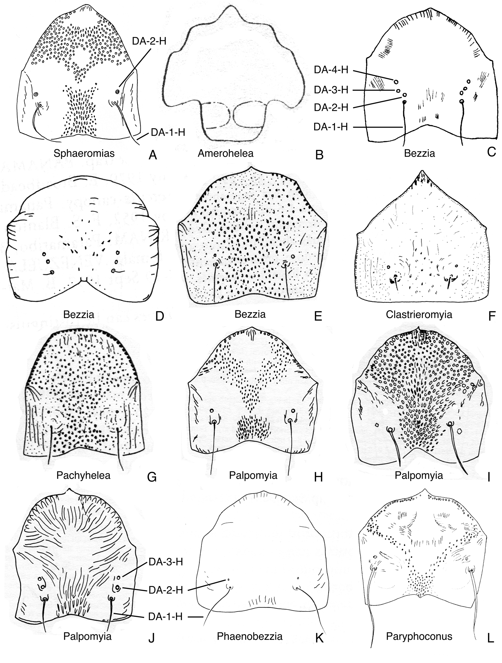
FIGURE 22. Dorsal apotomes, anterior view. A. Sphaeromias longipennis (from Wirth & Grogan 1979). B. Amerohelea sordidipes (from Lane et al. 1955). C. Bezzia mollis (from Mayer 1934c). D. Bezzia roldani (from Spinelli & Wirth 1989). E. Bezzia brevicornis (from Spinelli 1983a). F. Clastrieromyia dycei (from Spinelli & Grogan 1986). G. Pachyhelea pachymera (from Spinelli 1983a). H. Palpomyia lineata (from Grogan & Wirth 1979). I. Palpomyia belkini (from Grogan & Wirth 1979, modified). J. Palpomyia flaviceps (from Grogan & Wirth 1979). K. Phaenobezzia opaca (from Wirth & Grogan 1982). L. Paryphoconus oliveirai (from Ronderos et al. 2007).

FIGURE 28. Posterior portion of heads, in ventral view. A. Leehelea hispida. B. Sphaeromias longipennis. C. Xenohelea galatea. D. Bezzia nobilis. E. Clastrieromyia dycei. F. Pachyhelea pachymera. G. Palpomyia subasper. H. Phaenobezzia opaca. I. Paryphoconus grandis.
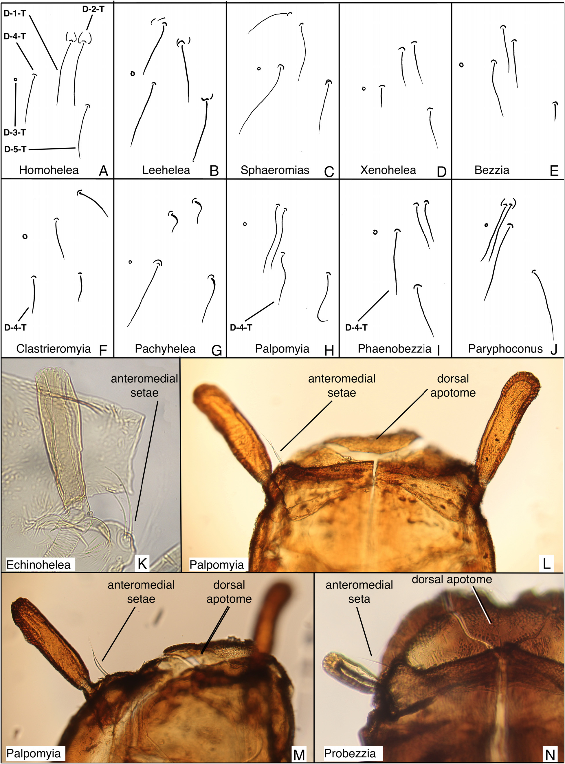
FIGURE 31. Dorsal setae from left side of thoraces; anteromedial setae. A. Homohelea delanoe. B. Leehelea hispida. C. Sphaeromias longipennis. D. Xenohelea galatea. E. Bezzia nobilis. F. Clastrieromyia dycei. G. Pachyhelea pachymera. H. Palpomyia jonesi. I. Phaenobezzia mashonensis. J. Paryphoconus oliveirai. K. Echinohelea lanei, partial lateral view (distorted slide specimen). L. Palpomyia (JAD685), dorsal view. M. Palpomyia (JAD685), dorsolateral view. N. Probezzia seminigra, dorsal view of left side.

FIGURE 33. Cephalothoraces and abdominal segments 1–3, in lateral view. A. Parabezzia sp. (5 km. E. Danby, Vermont, USA). B. Clinohelea curriei. C. Pellucidomyia leei. D. Hebetula tonnoiri. E. Jenkinshelea sp. (from Ottawa, ON, Canada). F. Mallochohelea caudellii. G. Probezzia seminigra. H. Nilobezzia sp. (from Kruger NP, South Africa). I. Sphaeromias longipennis. J. Bezzia sp. (6 km E. Salmon Arm, BC, Canada) K. Palpomyia sp. (Black Lake, Quebec, Canada). L. Phaenobezzia sp. (from Skulsuza, South Africa).
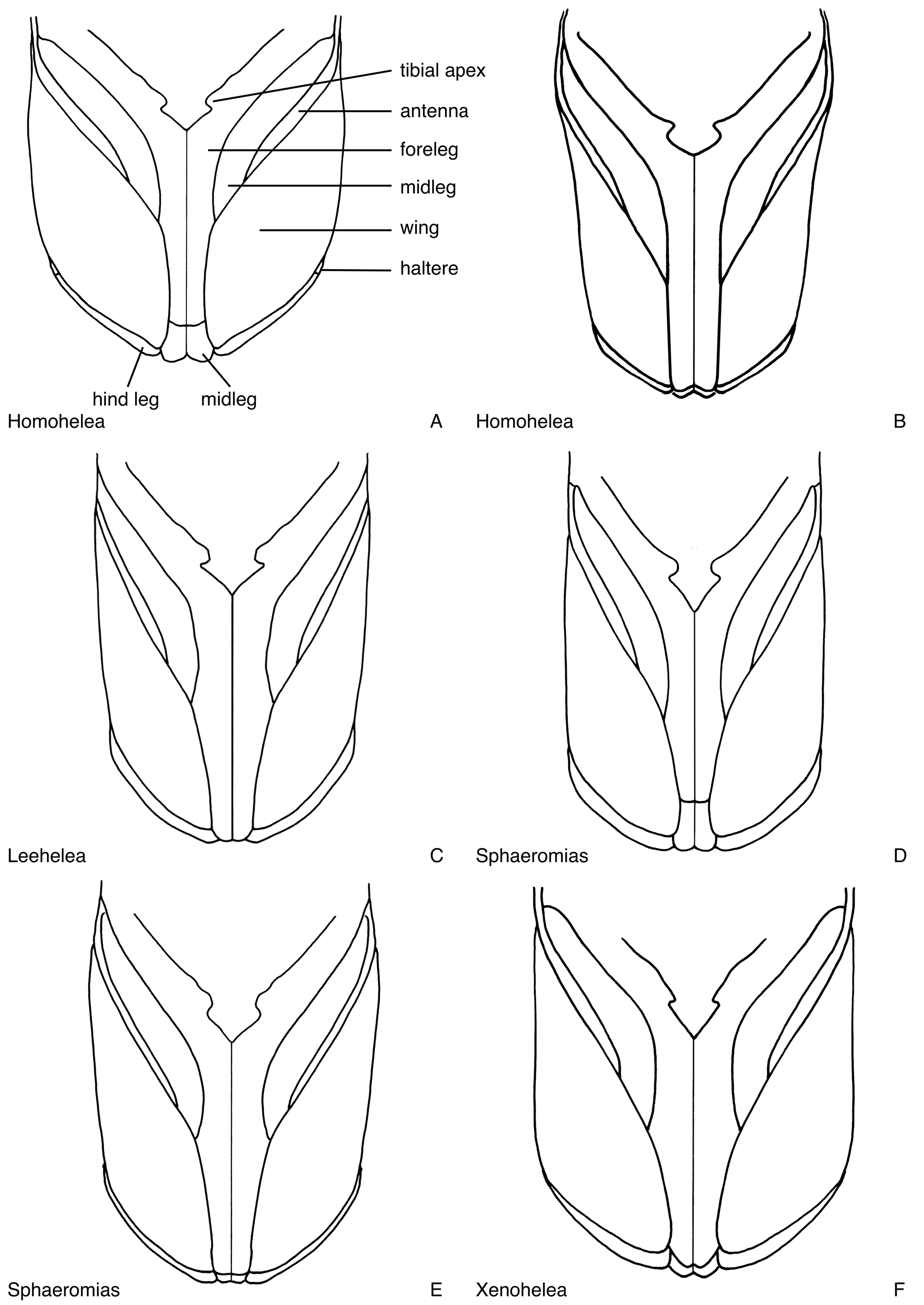
FIGURE 40. Legs, wings and apices of the antennae, in ventral view. A. Homohelea delanoe, male (reconstructed from distorted specimen). B. Homohelea albitudinis, female (drawn from photograph). C. Leehelea hispida, female. D. Sphaeromias longipennis, male. E. Sphaeromias longipennis, female. F. Xenohelea galatea, female (reconstructed from distorted specimen).

FIGURE 46. Respiratory organs, most in dorsal view, Figs. 46G. in lateral view, pattern on tracheal tubes shown only in some. A. Homohelea albitudinis (from de Meillon & Wirth 1981). B. Homohelea delanoe (from de Meillon & Wirth 1981). C. Leehelea wasselli (from Elson-Harris 1987). D. Leehelea hispida (from Elson-Harris 1987). E. Sphaeromias longipennis (from Wirth & Grogan 1979). F. Xenohelea galatea. G. Amerohelea sordidipes (from Lane et al. 1955). H. Bezzia glabra (from Wirth 1983b). I. Bezzia obelisca (from Wirth 1983a). J. Bezzia dorsasetula (from Wirth 1983a). K. Bezzia turkmenica (from Glukhova 1979b). L. Clastrieromyia dycei (from Spinelli & Grogan 1986). M. Pachyhelea pachymera (from Spinelli 1983a). N. Palpomyia lineata (from Grogan & Wirth 1979). O. Palpomyia jonesi (from Grogan & Wirth 1979). P. Palpomyia belkini (from Grogan & Wirth 1979). Q. Palpomyia rufa (from Grogan & Wirth 1979). R. Palpomyia flaviceps (from Grogan & Wirth 1979). S. Phaenobezzia opaca (from Wirth & Grogan 1982). T. Paryphoconus oliveirai (from Ronderos et al. 2007).
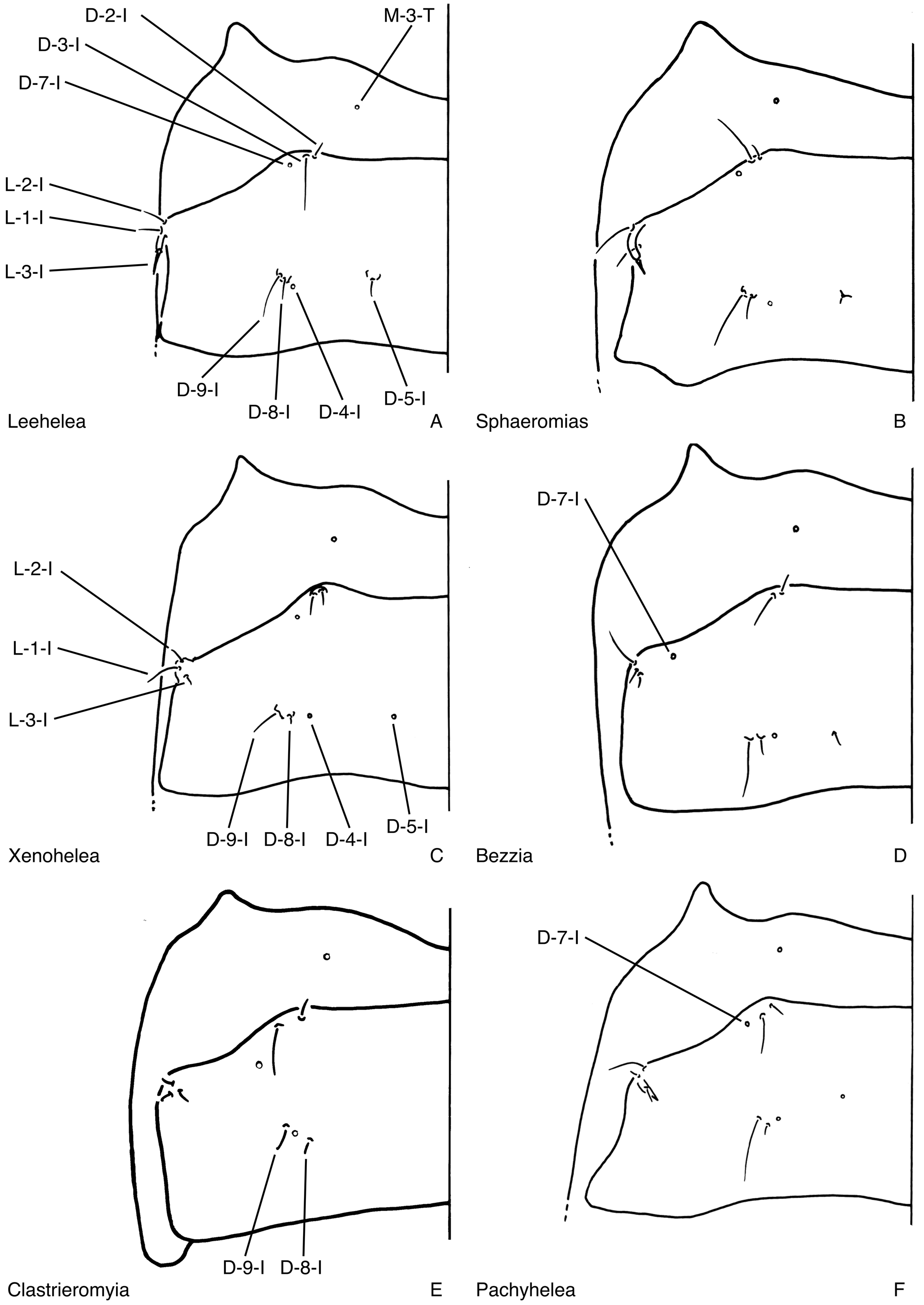
FIGURE 53. Metathoraces and tergites 1, in dorsal view. A. Leehelea hispida. B. Sphaeromias longipennis. C. Xenohelea galatea. D. Bezzia nobilis. E. Clastrieromyia dycei. F. Pachyhelea pachymera.

FIGURE 68. Abdominal segments four, in dorsal and ventral view (shagreen not shown). A. Anebomyia atripes. B. Homohelea delanoe. C. Leehelea hispida (reconstructed from distorted specimen).
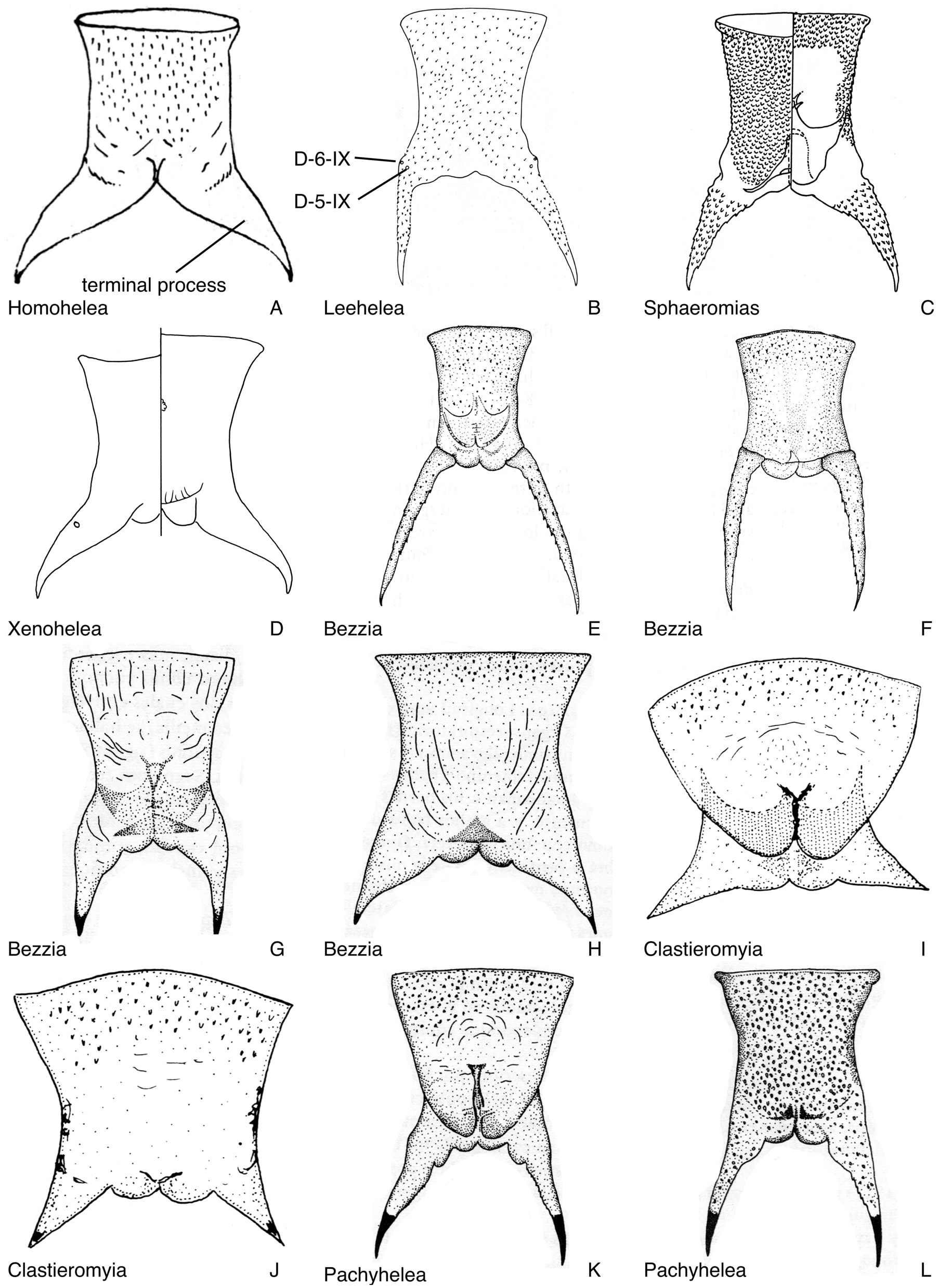
FIGURE 77. Abdominal segments 9. A. Homohelea delanoe, female, in dorsal view (from de Meillon & Wirth 1981). B. Leehelea wasselli, female, in dorsal view (from Elson-Harris 1987). C. Sphaeromias longipennis, male, in dorsal and ventral view (from Wirth & Grogan 1979). D. Xenohelea galatea, female, in dorsal and ventral view (shagreen not shown). E. Bezzia roldani, male, in dorsal view (from Spinelli & Wirth 1989). F. Bezzia roldani, female, in dorsal view (from Spinelli & Wirth 1989). G. Bezzia brevicornis, male, in dorsal view (from Spinelli 1983a). H. Bezzia acanthodes, female, in dorsal view (from Spinelli 1983a). I. Clastrieromyia dycei, male, in ventral view (from Spinelli & Grogan 1986). J. Clastrieromyia dycei, female, in ventral view (from Spinelli & Grogan 1986). K. Pachyhelea pachymera, male, in ventral view (from Spinelli 1983a). L. Pachyhelea pachymera, female, in dorsal view (from Spinelli 1983a).

FIGURE 79. Evolution of ecdysial sutures and splits in pupal exuviae of Ceratopogonidae, head and anterior portion of thorax in ventral view. Numbers refer to character states of characters 2 and 8 in text. Red indicates ecdysial splits. A. Leptoconopinae. B. Forcipomyiinae. C. Dasyheleinae. D. Most Ceratopogonini. E. Most Ceratopogonini, Clinohelea. F. Allohelea, Atyphohelea, Parabezzia. G. Heteromyia, Pellucidomyia, Hebetula. H. most Sphaeromiini s. lat., most Palpomyiini, Paryphoconus; some Bezzia are nearly completed fused (as in Fig. 79I), with only the dorsal apotome separate, along with an ecdysial split along the base of the antenna and the dorsal thoracic split. I. Jenkinshelea, Macropeza, Mallochohelea, Probezzia, Neobezzia.
No known copyright restrictions apply. See Agosti, D., Egloff, W., 2009. Taxonomic information exchange and copyright: the Plazi approach. BMC Research Notes 2009, 2:53 for further explanation.
|
Kingdom |
|
|
Phylum |
|
|
Class |
|
|
Order |
|
|
Family |
|
|
SubFamily |
Ceratopogoninae |
|
Tribe |
Sphaeromiini |
1 (by felipe, 2021-06-14 20:35:26)
2 (by ExternalLinkService, 2021-10-20 03:23:19)
3 (by diego, 2021-10-22 14:59:52)
4 (by ExternalLinkService, 2021-10-22 15:18:37)
5 (by ExternalLinkService, 2021-10-22 15:46:02)
6 (by ExternalLinkService, 2022-01-30 07:28:53)
7 (by diego, 2022-05-23 12:43:12)
8 (by admin, 2022-07-14 20:19:55)
9 (by ExternalLinkService, 2023-11-02 23:08:14)