
Sphaeromias Curtis
|
publication ID |
https://doi.org/10.11646/zootaxa.3879.1.1 |
|
publication LSID |
lsid:zoobank.org:pub:6423894B-97D9-4286-ABB9-D4AF072B57FD |
|
DOI |
https://doi.org/10.5281/zenodo.5593057 |
|
persistent identifier |
https://treatment.plazi.org/id/027587C9-BD4E-3014-FD67-1FD049E9E2FC |
|
treatment provided by |
Felipe (2021-06-14 20:35:26, last updated 2024-11-28 17:36:46) |
|
scientific name |
Sphaeromias Curtis |
| status |
|
Sphaeromias Curtis View in CoL View at ENA
( Figs. 2F View FIGURE 2 , 12E View FIGURE 12 , 17C View FIGURE 17 , 22A View FIGURE 22 , 28B View FIGURE 28 , 31C View FIGURE 31 , 33I View FIGURE 33 , 40D–E View FIGURE 40 , 46E View FIGURE 46 , 53B View FIGURE 53 , 69A View FIGURE 69 , 77C View FIGURE 77 )
DIAGNOSIS: Only pupa of Ceratopogonidae with the abdominal tubercles all apically pointed ( Fig. 68C View FIGURE 68 ), abdominal segment 4 with D-8-IV and D-8-IV on only basally fused tubercles ( Fig. 68C View FIGURE 68 ) and abdominal segment 8 with the two ventral sensilla (V-5-VIII, V-6-VIII) on a single tubercle and V-5-VIII tiny and V-6-VIII elongate; not diagnosable as different from Leehelea , a genus known only from the Oriental and Australasian Regions.
DESCRIPTION: Habitus as in Fig. 12E View FIGURE 12 . Total length = 6.63–8.53 mm. Without larval exuviae retained on abdomen. Exuviae with flagellum appressed against lateral margin of midleg, wing ( Figs. 17C View FIGURE 17 , 33I View FIGURE 33 ). Ecdysial tear around base of antenna, along lateral margin of face to palpus (as in Figs. 17C View FIGURE 17 , 79H View FIGURE 79 ). Head: Dorsal apotome ( Fig. 22A View FIGURE 22 ), with ventral line of weakness, without dorsomedial tubercle, without central dome; dorsolateral cephalic sclerite (as in Fig. 13H View FIGURE 13 ) fused to scutum, each side separated medially by dorsal apotome in whole pupa; mouthparts ( Fig. 28B View FIGURE 28 ) with mandible well-developed, lacinia absent; palpus extending just posterior to well posterior to posterolateral margin of labium; labium entire (not divided medially); apex of antenna ( Figs. 40D–E View FIGURE 40 ) anterior to posterior extent of midlength portion of midleg (portion lateral to mesosternum), narrowed posteriorly; sensilla: dorsal apotomals ( Fig. 22A View FIGURE 22 )—1 elongate seta, 1 campaniform sensillum; dorsolateral cephalic sclerite sensilla—1 seta, 1 campaniform sensillum; clypeal-labrals ( Fig. 28B View FIGURE 28 )—2 slender setae; oculars ( Fig. 28B View FIGURE 28 )—2 setae, 1 campaniform sensillum. Thorax: Prothoracic extension ( Fig. 28B View FIGURE 28 ) wide, well-developed but narrow dorsolaterally, not extending to antenna; mesonotum without tubercles, not extending posteromedially, not dividing metathorax medially ( Fig. 53B View FIGURE 53 ); respiratory organ ( Fig. 46E View FIGURE 46 ) length/width = 3.20–4.06, elongate, moderately slender, somewhat flattened apically, with pores closely abutting at apex of respiratory organ, arranged in single row, outer surface with a few wrinkles, with moderately elongate pedicel, base with moderate elongate posteromedial apodeme, membranous base of respiratory organ short to moderately elongate and annulated, tracheal tube straight to slightly curved along length, with spirals restricted to base, wrinkles for most of length; wing ( Figs. 40D–E View FIGURE 40 ) without apical tubercle or angle, separated medially by fore-, midlegs; halter apex and hind leg ( Fig. 33I View FIGURE 33 ) broadly abutting; halter apex extending posteriorly to 1/6 length of tergite 2; legs ( Figs. 40D–E View FIGURE 40 ) with lateral margin of foreleg near midlength of wing evenly curved; hind leg visible at lateral margin of wing ( Fig. 33I View FIGURE 33 ); male with apex of foreleg moderately anterior to apex of midleg, female with apex of foreleg ventral to apex of midleg; apex of hind leg abutting apex of midleg laterally; sensilla: anteromedials—2 elongate setae (as in Figs. 31L–M View FIGURE 31 ); anterolaterals—1 moderately long seta; dorsal setae ( Fig. 31C View FIGURE 31 )—D-1-T, D-2-T, D-4-T, D-5-T setae, D-3- T campaniform sensillum, D-3-T lateral to D-4-T; supraalar 2—campaniform sensillum; metathoracics ( Fig. 53B View FIGURE 53 )—1 campaniform sensillum; M-3-T distant from margin of metathorax (at least 1/3 length of metathorax). Abdomen: with tergite 1 with 3 medial spots, tergites 2–7 with medial area with stripe, 2 anterolateral spots, sternites 3–7 with medial stripe, anterolateral spot, 2 spots on sternite 8; sternite 8 with dark posteromedial apodeme, segment 2 as wide or slightly wider than segment 3, segments with undivided, thin to thick setae, with pointed, short to moderately elongate tubercles, tergites or sternites entire, each without membranous disc; segment 9 ( Fig. 77C View FIGURE 77 ) not strongly modified, terminal processes closely approximated basally, each projecting posterodorsolaterally, tapering to pointed apex; sensilla: tergite 1 ( Fig. 53B View FIGURE 53 ) with 8 setae, 2 campaniform sensilla, including 3 lateral sensilla, D-2-I, D-3-I closely approximated, D-7-I situated anteriorly near D-3-I; segment 4 ( Fig. 69A View FIGURE 69 )—D-2-IV, D-3-IV short to moderately elongate setae on short tubercles; D-5-IV, D-8-IV, D-9-IV short to moderately elongate setae, D-7-IV present or absent; D-5-IV on single tubercle, D-8-IV, D-9-IV on basally fused, closely approximated tubercles, posterior dorsal sensilla in transverse row, arranged medially to laterally: D-5-IV, D-4-IV, D-8-IV, D-9-IV; D-7-IV, if present, near D-3-IV; L-1-IV elongate seta on short tubercle, just anterior of base of tubercle with L-2-IV, L-3-IV; L-2-IV, L-3-IV, L-4-IV moderately elongate setae, L-2-IV, L-3-IV on single pointed tubercle, L-4-IV on elongate tubercle, V-5-IV, V-6-IV, V-7-IV short setae on short tubercles, all closely approximated, V-5-IV, V-6-IV with tubercles fused basally; segment 8 without D-3-VIII, without L-1-VIII; with V- 5-VIII, V-6-VIII on single tubercle, V-5-VIII tiny, V-6-VIII elongate; segment 9 ( Fig. 77C View FIGURE 77 )—with D-5-IX, D-6-IX campaniform sensilla.
DISTRIBUTION AND HABITAT: The genus Sphaeromias is known from 30 species from every Region worldwide other than the Neotropical Region ( Borkent 2014 ; minus one species, see below). Immatures are known from water hyacinth, Pistia stratiotes , from peat soil in Rhizophora mangroves, shady swamps, a grassy wetland, rivers, and lakes (sometimes in blanket algae). Larvae of Holarctic species are reported from the benthos of lakes. Knausenberger (1987) provides further details of various microhabitats within the genus.
TAXONOMIC DISCUSSION: The pupae of five species of Sphaeromias are known ( Tables 2–3 View TABLE 2 View TABLE 3 ). Sphaeromias theileri de Meillon & Wirth (1981) is now considered a species of Nilobezzia (see taxonomic discussion under that genus).
Male and female pupae of Sphaeromias and Homohelea are sexually dimorphic in the arrangement of their forelegs. In males, the foreleg is relatively short ( Figs. 40A, D View FIGURE 40 ) while in females the foreleg overlaps the midleg entirely ( Figs. 40B, E View FIGURE 40 ) (see character 44). The feature is likely present in two related genera, Xenohelea and Leehelea .
Some pupae of Sphaeromias are the largest of all Ceratopogonidae pupae ( Fig. 2F View FIGURE 2 ).
MATERIAL EXAMINED: S. bifidus : 1 pupal exuviae, Black Lake, North Burgess Township, Ontario, Canada, 21-VI-1971 (CNCI). S. fasciatus : 2 pupae, Tegeler See, Germany, 20-V-1931 (ZSMC); 7 pupal exuviae, Strelna, Leningrad Province, Russia, 30-V-1998 (ZIN). S. longipennis : 1 pupal exuviae, Black Lake, Stanleyville, Ontario, Canada, 24-VI-1975 (USNM); 2 pupal exuviae, as previous locality, 25-VI-1975 (USNM); 5 pupal exuviae, Rideau River, Ottawa, Ontario, Canada, 29-V-1960 (USNM); 1 pupal exuviae (in glycerin), Black Lake, Quebec, Canada, 21-VI-1971 (CNCI); 1 pupal exuviae, Bemus Point, Chautauqua Lake, New York, USA, 31-V- 1963 (USNM); 1 pupal exuviae, Wanakena, St. Lawrence County, New York, USA, 25-VI-1963 (USNM); 1 pupal exuviae, Cranberry Lake, St. Lawrence County, New York, USA, 25-VI-1963 (USNM). S. pictus : 4 pupal exuviae, Raigorodok, Donetsk Province, Ukraine, 30-V-1970 (ZIN). S. sp.: 2 pupal exuviae, White Lake, Ontario, Canada, 29-V-1967 (CNCI); 1 pupal exuviae, Rutka Tartak nr Suwałki, Poland, 2-VII-1993 ( IZUG).
Borkent, A. (2014) World Species of Biting Midges (Diptera: Ceratopogonidae). Available from: http: // www. inhs. illinois. edu / research / FLYTREE / Borkent. html (accessed 20 May 2014)
De Meillon, B. & Wirth, W. W. (1981) Subsaharan Ceratopogonidae (Diptera) VI. New species and records of South African biting midges collected by A. L. Dyce. Annals of the Natal Museum, 24, 525 - 561.
Knausenberger, W. I. (1987) Contributions to the autecology and ecosystematics of immature Ceratopogonidae (Diptera), with emphasis on the tribes Heteromyiini and Sphaeromiini in the Middle Atlantic United States. Ph. D. Dissertation, Virginia Polytechnic Institute and State University, Blacksburg, Virginia, xxix + 729 pp.

FIGURE 2. Pupae in unusual habitats, preservation and relative sizes. A. Forcipomyia pupae, Rancho Grande, Venezuela; photo by Steve Marshall. B. Forcipomyia pupae under bark of log of Pinus sylvestris, Poland; photo by Marek W. Kozlowski. C. Pupae of Mallochohelea sp. on emergent plants at edge of Bolean Lk., 6 km NE Falkland, British Columbia. D. Palpomyia sp. showing mounting technique on pin, 8 km E. Sicamous, British Columbia. E. Permanent slide mount of dissected adult and pupal exuviae of Mallochohelea albibasis. F. Pupal exuviae, from left to right, of Baeodasymyia michaeli, Culicoides denticulatus and Sphaeromias longipennis showing size differences within the family. Total length of Baeodasymyia michaeli = 1.2 mm, of Sphaeromias = 7.3 mm.

FIGURE 12. Habitus. A. Pellucidomyia leei, male, in dorsal view (from Debenham 1970). B. Dibezzia prominens, sex unknown, in dorsal view (form Mayer 1934c). C. Macropeza albitarsis, male, in dorsal view (from Szadziewski & Dominiak, 2007, modified). D. Mallochohelea munda, male, in ventral view (from Szadziewski et al. 1997). E. Sphaeromias fasciata, sex unknown, in dorsal view (from Mayer 1934a). F. Bezzia glabra, female, in ventral view (from Thomsen 1937). G. Palpomyia sp., male, in dorsal view (from Grogan & Wirth 1979). H. Paryphoconus oliveirai, female, in ventral view (modified from Ronderos et al. 2007).

FIGURE 13. Anterior portion of heads and thoraces, in anterior or anterodorsal view. A. Leptoconops kerteszi, ecdysial sutures of head enhanced to show position. B. Culicoides denticulatus. C. Ceratopogon nr. abstrusus. D. Brachypogon sp. (from 6 km E. Falkland, BC, Canada). E. Alluaudomyia bella, dorsal apotome absent. F. Clinohelea curriei. G. Pellucidomyia leei. H. Mallochohelea caudellii. I. Bezzia sp. (from Bolean Lake, BC, Canada).

FIGURE 17. Anterior portion of heads. A. Jenkinshelea sp., in ventral view (from Ottawa, ON, Canada). B. Mallochohelea caudellii., in ventral view. C. Sphaeromias longipennis, in ventral view. D. Bezzia sp., in ventral view (6 km E Salmon Arm, BC, Canada). E. Palpomyia sp. (Black Lake, Quebec, Canada), in ventral view. F. Phaenobezzia sp., in ventral view (from Skulsuza, South Africa).
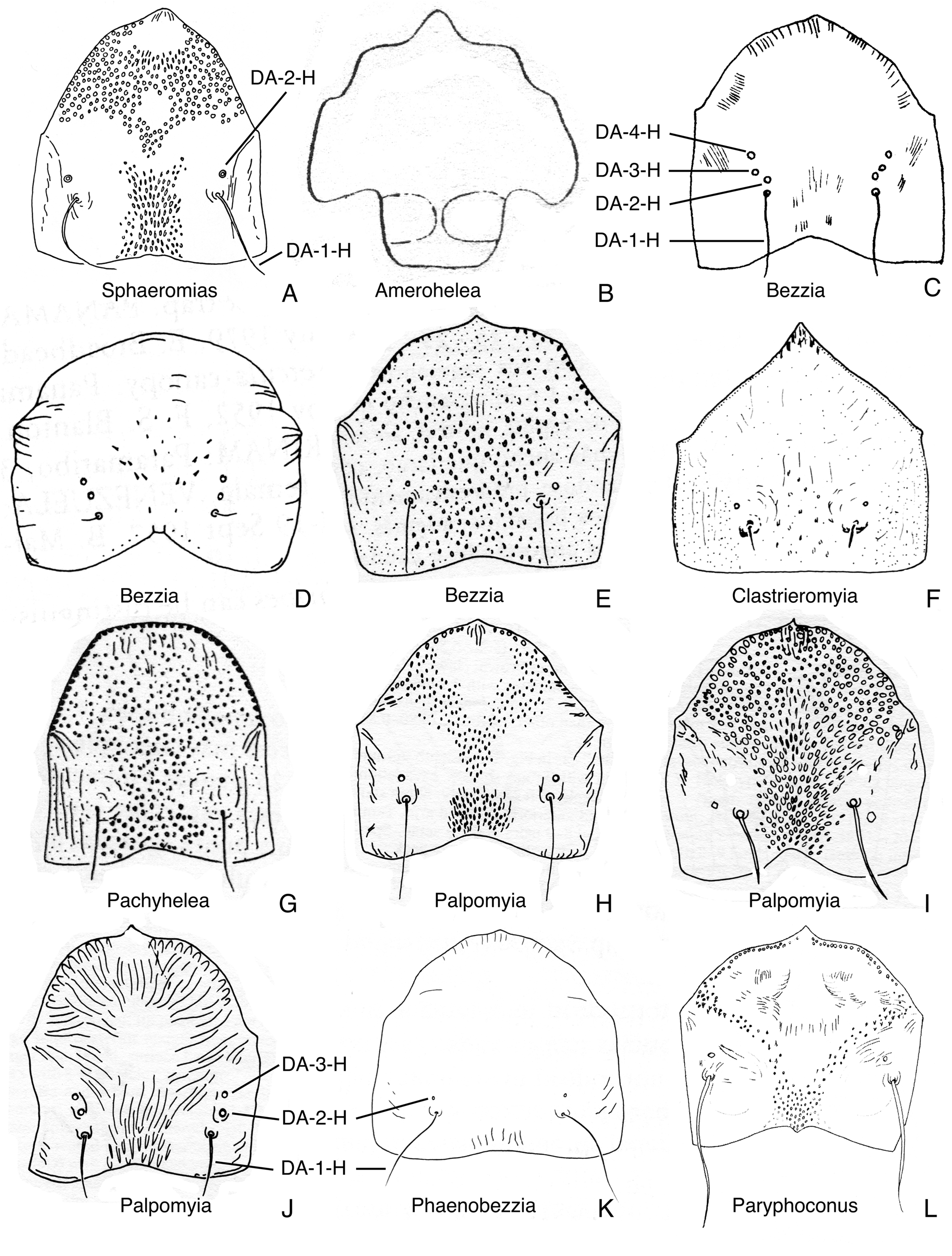
FIGURE 22. Dorsal apotomes, anterior view. A. Sphaeromias longipennis (from Wirth & Grogan 1979). B. Amerohelea sordidipes (from Lane et al. 1955). C. Bezzia mollis (from Mayer 1934c). D. Bezzia roldani (from Spinelli & Wirth 1989). E. Bezzia brevicornis (from Spinelli 1983a). F. Clastrieromyia dycei (from Spinelli & Grogan 1986). G. Pachyhelea pachymera (from Spinelli 1983a). H. Palpomyia lineata (from Grogan & Wirth 1979). I. Palpomyia belkini (from Grogan & Wirth 1979, modified). J. Palpomyia flaviceps (from Grogan & Wirth 1979). K. Phaenobezzia opaca (from Wirth & Grogan 1982). L. Paryphoconus oliveirai (from Ronderos et al. 2007).

FIGURE 28. Posterior portion of heads, in ventral view. A. Leehelea hispida. B. Sphaeromias longipennis. C. Xenohelea galatea. D. Bezzia nobilis. E. Clastrieromyia dycei. F. Pachyhelea pachymera. G. Palpomyia subasper. H. Phaenobezzia opaca. I. Paryphoconus grandis.
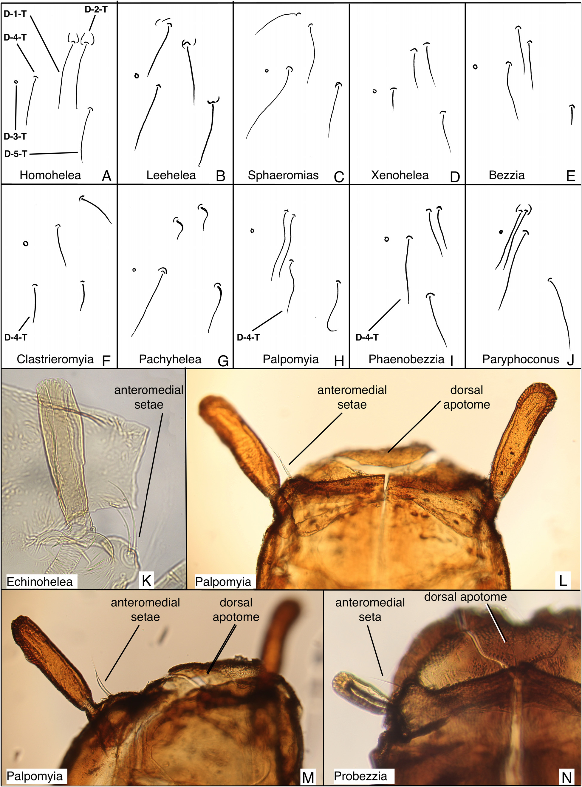
FIGURE 31. Dorsal setae from left side of thoraces; anteromedial setae. A. Homohelea delanoe. B. Leehelea hispida. C. Sphaeromias longipennis. D. Xenohelea galatea. E. Bezzia nobilis. F. Clastrieromyia dycei. G. Pachyhelea pachymera. H. Palpomyia jonesi. I. Phaenobezzia mashonensis. J. Paryphoconus oliveirai. K. Echinohelea lanei, partial lateral view (distorted slide specimen). L. Palpomyia (JAD685), dorsal view. M. Palpomyia (JAD685), dorsolateral view. N. Probezzia seminigra, dorsal view of left side.

FIGURE 33. Cephalothoraces and abdominal segments 1–3, in lateral view. A. Parabezzia sp. (5 km. E. Danby, Vermont, USA). B. Clinohelea curriei. C. Pellucidomyia leei. D. Hebetula tonnoiri. E. Jenkinshelea sp. (from Ottawa, ON, Canada). F. Mallochohelea caudellii. G. Probezzia seminigra. H. Nilobezzia sp. (from Kruger NP, South Africa). I. Sphaeromias longipennis. J. Bezzia sp. (6 km E. Salmon Arm, BC, Canada) K. Palpomyia sp. (Black Lake, Quebec, Canada). L. Phaenobezzia sp. (from Skulsuza, South Africa).
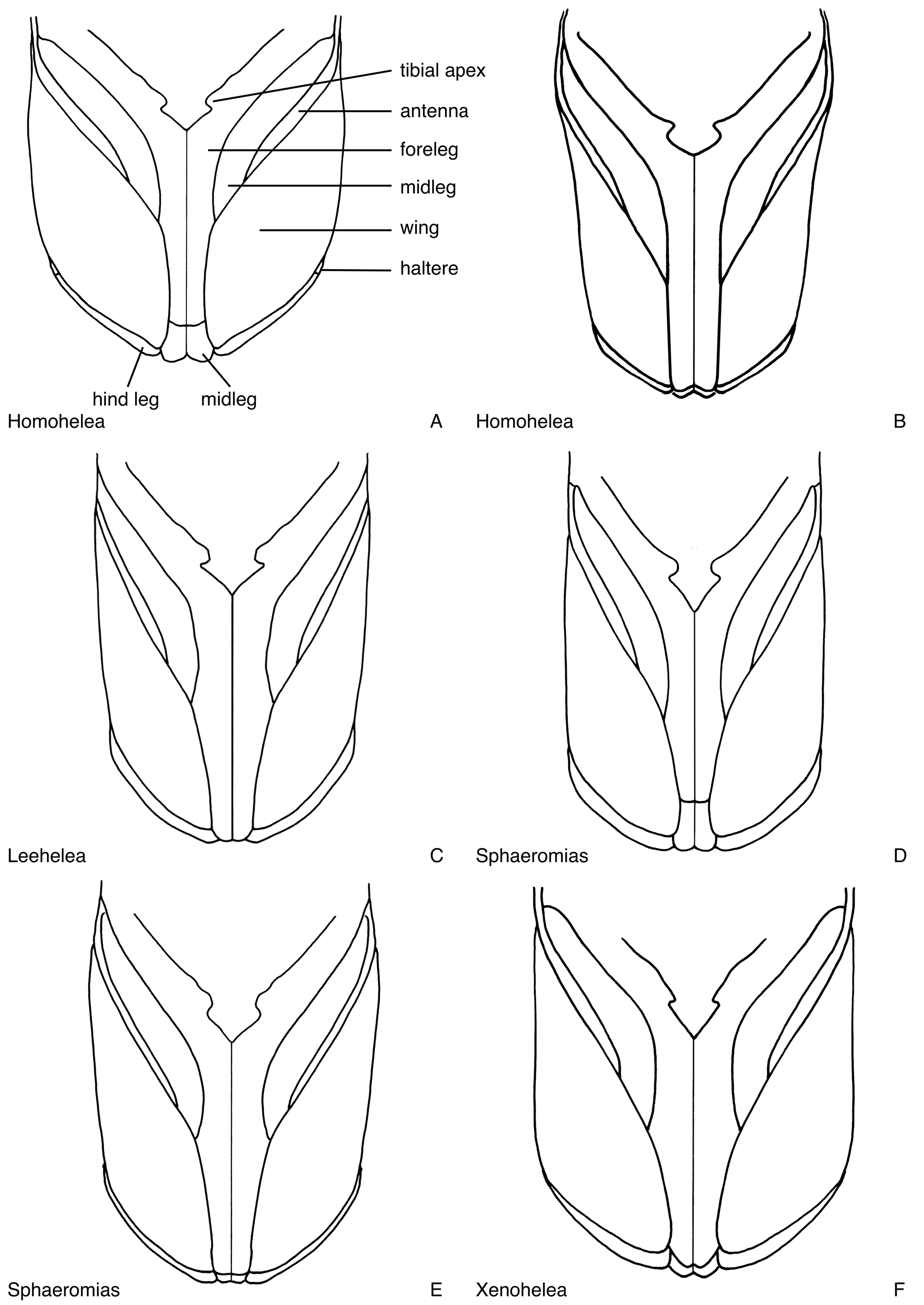
FIGURE 40. Legs, wings and apices of the antennae, in ventral view. A. Homohelea delanoe, male (reconstructed from distorted specimen). B. Homohelea albitudinis, female (drawn from photograph). C. Leehelea hispida, female. D. Sphaeromias longipennis, male. E. Sphaeromias longipennis, female. F. Xenohelea galatea, female (reconstructed from distorted specimen).

FIGURE 46. Respiratory organs, most in dorsal view, Figs. 46G. in lateral view, pattern on tracheal tubes shown only in some. A. Homohelea albitudinis (from de Meillon & Wirth 1981). B. Homohelea delanoe (from de Meillon & Wirth 1981). C. Leehelea wasselli (from Elson-Harris 1987). D. Leehelea hispida (from Elson-Harris 1987). E. Sphaeromias longipennis (from Wirth & Grogan 1979). F. Xenohelea galatea. G. Amerohelea sordidipes (from Lane et al. 1955). H. Bezzia glabra (from Wirth 1983b). I. Bezzia obelisca (from Wirth 1983a). J. Bezzia dorsasetula (from Wirth 1983a). K. Bezzia turkmenica (from Glukhova 1979b). L. Clastrieromyia dycei (from Spinelli & Grogan 1986). M. Pachyhelea pachymera (from Spinelli 1983a). N. Palpomyia lineata (from Grogan & Wirth 1979). O. Palpomyia jonesi (from Grogan & Wirth 1979). P. Palpomyia belkini (from Grogan & Wirth 1979). Q. Palpomyia rufa (from Grogan & Wirth 1979). R. Palpomyia flaviceps (from Grogan & Wirth 1979). S. Phaenobezzia opaca (from Wirth & Grogan 1982). T. Paryphoconus oliveirai (from Ronderos et al. 2007).
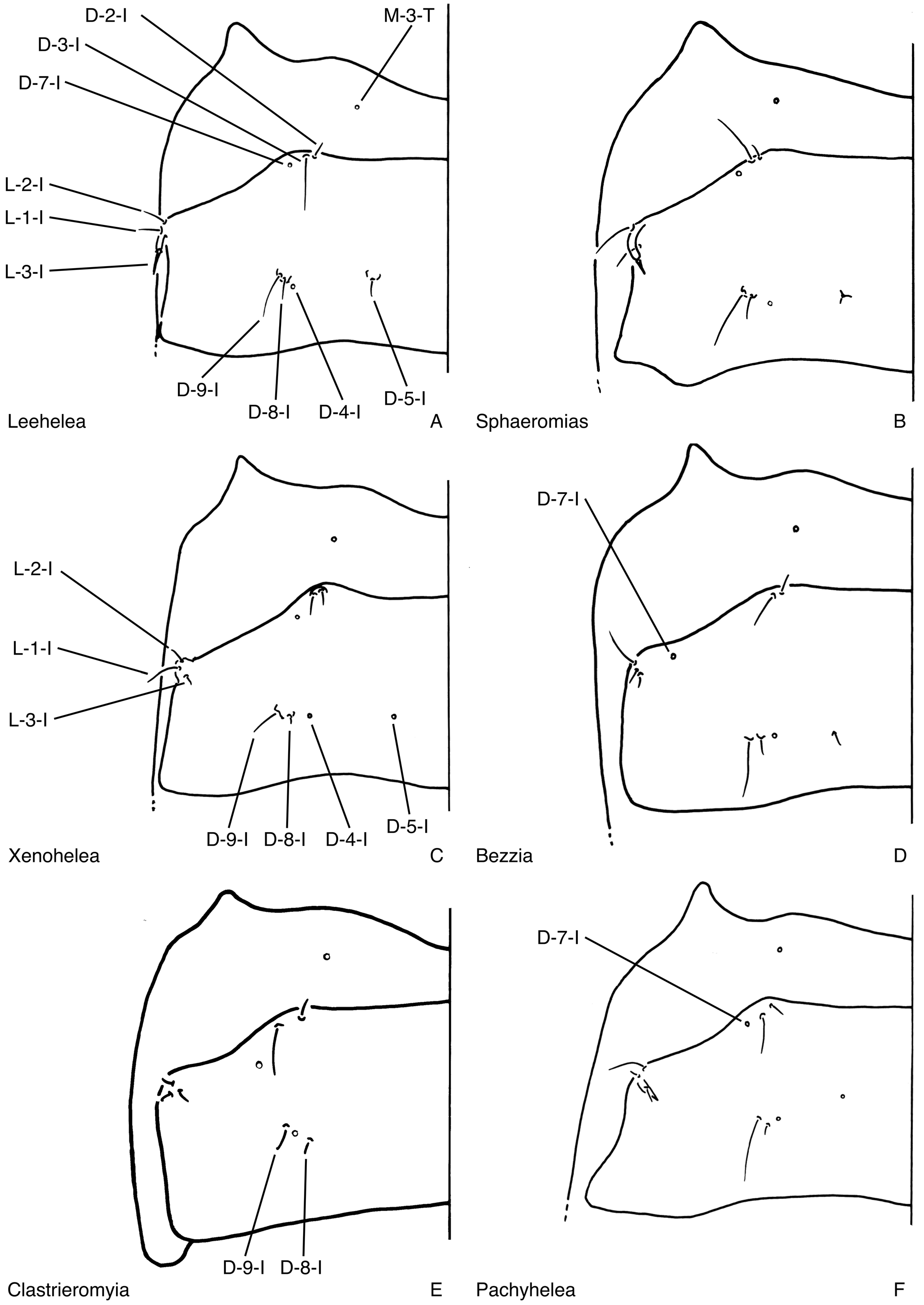
FIGURE 53. Metathoraces and tergites 1, in dorsal view. A. Leehelea hispida. B. Sphaeromias longipennis. C. Xenohelea galatea. D. Bezzia nobilis. E. Clastrieromyia dycei. F. Pachyhelea pachymera.

FIGURE 68. Abdominal segments four, in dorsal and ventral view (shagreen not shown). A. Anebomyia atripes. B. Homohelea delanoe. C. Leehelea hispida (reconstructed from distorted specimen).

FIGURE 69. Abdominal segments four, in dorsal and ventral view (shagreen not shown). A. Sphaeromias longipennis. B. Xenohelea galatea. C. Bezzia nobilis.
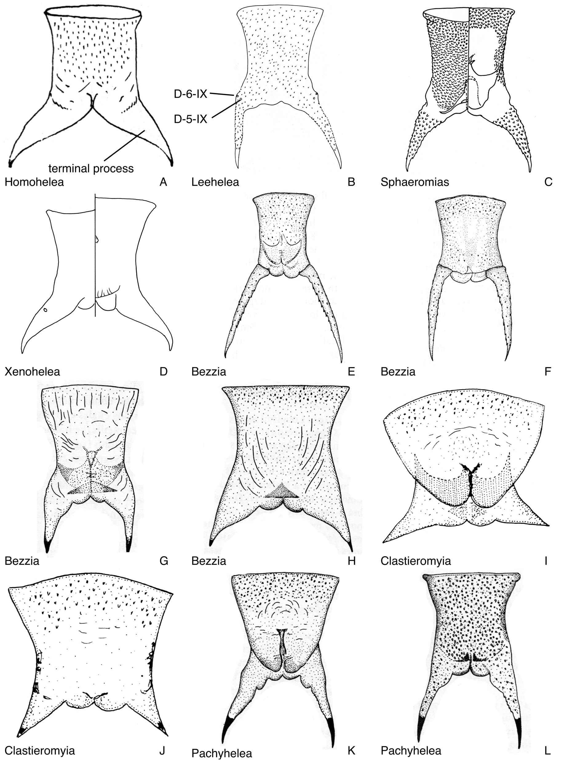
FIGURE 77. Abdominal segments 9. A. Homohelea delanoe, female, in dorsal view (from de Meillon & Wirth 1981). B. Leehelea wasselli, female, in dorsal view (from Elson-Harris 1987). C. Sphaeromias longipennis, male, in dorsal and ventral view (from Wirth & Grogan 1979). D. Xenohelea galatea, female, in dorsal and ventral view (shagreen not shown). E. Bezzia roldani, male, in dorsal view (from Spinelli & Wirth 1989). F. Bezzia roldani, female, in dorsal view (from Spinelli & Wirth 1989). G. Bezzia brevicornis, male, in dorsal view (from Spinelli 1983a). H. Bezzia acanthodes, female, in dorsal view (from Spinelli 1983a). I. Clastrieromyia dycei, male, in ventral view (from Spinelli & Grogan 1986). J. Clastrieromyia dycei, female, in ventral view (from Spinelli & Grogan 1986). K. Pachyhelea pachymera, male, in ventral view (from Spinelli 1983a). L. Pachyhelea pachymera, female, in dorsal view (from Spinelli 1983a).

FIGURE 79. Evolution of ecdysial sutures and splits in pupal exuviae of Ceratopogonidae, head and anterior portion of thorax in ventral view. Numbers refer to character states of characters 2 and 8 in text. Red indicates ecdysial splits. A. Leptoconopinae. B. Forcipomyiinae. C. Dasyheleinae. D. Most Ceratopogonini. E. Most Ceratopogonini, Clinohelea. F. Allohelea, Atyphohelea, Parabezzia. G. Heteromyia, Pellucidomyia, Hebetula. H. most Sphaeromiini s. lat., most Palpomyiini, Paryphoconus; some Bezzia are nearly completed fused (as in Fig. 79I), with only the dorsal apotome separate, along with an ecdysial split along the base of the antenna and the dorsal thoracic split. I. Jenkinshelea, Macropeza, Mallochohelea, Probezzia, Neobezzia.
| IZUG |
Istituto di Zoologia dell'Universita |
No known copyright restrictions apply. See Agosti, D., Egloff, W., 2009. Taxonomic information exchange and copyright: the Plazi approach. BMC Research Notes 2009, 2:53 for further explanation.
|
Kingdom |
|
|
Phylum |
|
|
Class |
|
|
Order |
|
|
Family |
|
|
SubFamily |
Ceratopogoninae |
|
Tribe |
Sphaeromiini |
1 (by felipe, 2021-06-14 20:35:26)
2 (by ExternalLinkService, 2021-10-20 03:23:19)
3 (by diego, 2021-10-22 14:59:52)
4 (by ExternalLinkService, 2021-10-22 15:18:37)
5 (by ExternalLinkService, 2021-10-22 15:46:02)
6 (by ExternalLinkService, 2022-01-30 07:28:53)
7 (by diego, 2022-05-23 12:43:12)
8 (by admin, 2022-07-14 20:19:55)
9 (by ExternalLinkService, 2023-11-02 23:08:14)