
Borkent, Borkent, 2014Borkent, Borkent, 2014Borkent, Borkent, 2014
|
publication ID |
https://dx.doi.org/10.11646/zootaxa.3879.1.1 |
|
publication LSID |
lsid:zoobank.org:pub:6423894B-97D9-4286-ABB9-D4AF072B57FD |
|
persistent identifier |
https://treatment.plazi.org/id/027587C9-BD47-301B-FD5F-1CA94C9AE13C |
|
treatment provided by |
Felipe |
|
scientific name |
Borkent Borkent Borkent |
| status |
|
( Figs. 2D View FIGURE 2 12G View FIGURE 12 , 17E View FIGURE 17 , 22H–J View FIGURE 22 , 28G View FIGURE 28 , 31H, 31L–M View FIGURE 31 , 33K View FIGURE 33 , 41D View FIGURE 41 , 46N–R View FIGURE 46 , 54A View FIGURE 54 , 70C View FIGURE 70 , 71A View FIGURE 71 , 78A–H View FIGURE 78 )
DIAGNOSIS: Only pupa of Ceratopogonidae with the metathorax with only one campaniform sensillum (M-3-T) situated at least ⅓ the length of the metathorax from its anterior margin ( Fig. 54A View FIGURE 54 ), apex of the halter extending posteriorly to about 1/6 length of tergite 2 (as in Fig. 33L View FIGURE 33 ), abdominal segment 4 with V-5-IV, V-6-IV and V-7-IV closely approximated ( Fig. 70C View FIGURE 70 ) or, if V-7-IV is closer to L-4-IV then L-3-IV is closer to L-2-IV than to an elongate L-1-IV (as in Fig. 69C View FIGURE 69 ) (not as in Fig. 70B View FIGURE 70 ), abdominal segment 8 has V-5-VIII and V-6-VIII on separate tubercles or if on partially to completely fused tubercles, then V-5-VIII is well-developed (not minute), and segment 8 is without L-1-VIII (not diagnosable as different from Bezzia and Phaenobezzia ); however, most species of Bezzia have two or more campaniform sensilla on the dorsal apotome ( Figs. 22C–D View FIGURE 22 ), a nearly unique condition found otherwise only in P. flavipes and P. jonesi (the latter distinctively with two setae ( Fig. 22J View FIGURE 22 ).
DESCRIPTION: Habitus as in Fig. 12G View FIGURE 12 . Total length = 2.25–7.03 mm. Without larval exuviae retained on abdomen. Exuviae with flagellum appressed against lateral margin of midleg, wing ( Figs. 17E View FIGURE 17 , 33K View FIGURE 33 ). Ecdysial tear around base of antenna, along lateral margin of face to palpus ( Figs. 17E View FIGURE 17 , 79H View FIGURE 79 ). Head: Dorsal apotome ( Figs. 22H–J View FIGURE 22 ), with partial ventral line of weakness, without dorsomedial tubercle, without central dome; dorsolateral cephalic sclerite (as in Fig. 13H View FIGURE 13 ) fused to scutum, each side separated medially by dorsal apotome in whole pupa; mouthparts ( Fig. 28G View FIGURE 28 ) with mandible well-developed, lacinia absent; palpus extending posterior to posterolateral margin of labium; labium entire or separated medially by labrum, hypopharynx; apex of antenna ( Fig. 41D View FIGURE 41 ) well anterior to posterior to, posterior extent of midlength portion of midleg (portion lateral to mesosternum), narrowed posteriorly; sensilla: dorsal apotomals ( Figs. 22H–J View FIGURE 22 )—1 elongate seta, 1 campaniform sensillum or 2 elongate setae, 2 campaniform sensilla; dorsolateral cephalic sclerite sensilla—1 seta, 1 campaniform sensillum; clypeallabrals ( Fig. 28G View FIGURE 28 )—2 slender setae; oculars ( Fig. 28G View FIGURE 28 )—1–2 setae, 1 campaniform sensillum or 1 seta, 2 campaniform sensilla. Thorax: Prothoracic extension ( Fig. 28G View FIGURE 28 ) wide, well-developed but in some narrow dorsolaterally, extending from palpus to antenna; mesonotum without tubercles, not extending posteromedially, not dividing metathorax medially ( Fig. 54A View FIGURE 54 ); respiratory organ ( Figs. 46N–R View FIGURE 46 ) length/width = 2.46–4.73, moderately elongate to elongate, somewhat flattened apically, with pores closely abutting at apex of respiratory organ, arranged in single straight to curved row, outer surface smooth or with some wrinkles, with short, wide pedicel, base with short posteromedial apodeme, membranous base of respiratory organ short, annulated, tracheal tube straight to slightly curved along length, with spirals restricted to base, wrinkles to half length or more; wing ( Fig. 41D View FIGURE 41 ) without apical tubercle or angle, separated medially by fore-, midlegs; halter apex and hind leg ( Fig. 33K View FIGURE 33 ) broadly abutting; halter apex extending posteriorly to 1/6 length of tergite 2; legs ( Fig. 41D View FIGURE 41 ) with lateral margin of foreleg near midlength of wing evenly curved; hind leg visible at lateral margin of wing ( Fig. 33K View FIGURE 33 ); with apex of foreleg moderately anterior to apex of midleg; apex of hind leg abutting apex of midleg laterally; sensilla: anteromedials—2 elongate setae, 1 campaniform sensillum ( Figs. 31L–M View FIGURE 31 ); anterolaterals—1 moderately long seta; dorsal setae ( Fig. 31H View FIGURE 31 )—D-1-T, D-2-T, D-5-T, D-4-T setae, D-3-T campaniform sensillum, D-3-T lateral to anterolateral to D-4-T; supraalar 2—campaniform sensillum; metathoracics ( Fig. 54A View FIGURE 54 )—1 campaniform sensillum; M-3-T distant from margin of metathorax (at least 1/3 length of metathorax). Abdomen: pigmentation light to dark brown, tergites 1–7 with medial area with stripe, 2 spots, sternites 3–7 with medial stripe, anterolateral spot, segment 2 as wide or slightly wider than segment 3, segments with undivided, peg-like or thin to thick setae, with rounded to pointed, short to moderately elongate tubercles, tergites or sternites entire, each without membranous disc; segment 9 ( Figs. 78A–H View FIGURE 78 ) not strongly modified, terminal processes closely approximated to separated basally, each projecting posterodorsolaterally, tapering to pointed apex; sensilla: tergite 1 ( Fig. 54A View FIGURE 54 ) with 8 setae, 2 campaniform sensilla, including 4 lateral sensilla, D-2-I, D-3-I closely approximated, D-7-I situated anterolaterally near L-1-I; segment 4 ( Figs. 70C View FIGURE 70 , 71A View FIGURE 71 )—D-2-IV, D-3-IV short to moderately elongate setae on short to pointed tubercles; D-5-IV peg-like or slender seta, D-8-IV, D-9-IV short to moderately elongate setae, D-7-IV present or absent; D-5-IV without or on short tubercle, D-8-IV, D-9-IV on basally fused or separate but closely approximated tubercles, posterior dorsal sensilla in transverse row, arranged medially to laterally: D-5-IV, D-4-IV, D-8-IV, D-9- IV; D-7-IV, if present, near D-3-IV; L-1-IV short to elongate seta on rounded tubercle, just anterior of base of tubercle with L-2-IV, L-3-IV; L-2-IV, L-3-IV, L-4-IV elongate setae on pointed tubercles, L-2-IV, L-3-IV on single tubercle in some, V-5-IV, V-6-IV, V-7-IV short to moderately elongate setae on short tubercles, all closely approximated or with V-7-IV closer to L-4-IV; segment 8 without D-3-VIII, without L-1-VIII; segment 9 ( Figs. 78 View FIGURE 78 A-H)—with D-5-IX, D-6-IX campaniform sensilla.
DISTRIBUTION AND HABITAT: The genus Palpomyia is known from 270 species from every Region worldwide ( Borkent 2014, additional species below). Immatures have been collected from streams, hot springs, river margins, marshes, bogs, fens, ponds, lakes and reservoirs.
TAXONOMIC DISCUSSION: There are 41 species of Palpomyia known as pupae ( Tables 2–3). Thienemann (1928), Mayer (1934a) and Lenz (1934) provided similar keys to a few European species groups known at that time. Grogan & Wirth (1979), in their masterful revision of Nearctic Palpomyia , provide a key to the pupae of 16 species known from the Nearctic. However, it is unfortunate that Palpomyia pupae cannot be diagnosed at the generic level so that we cannot yet identify a Palpomyia pupa as such. The key therefore requires foreknowledge that the specimen is a member of the genus (e.g. reared to adult).
Liu & Yu (1991) described the monotypic genus Nemoromyia Liu & Yu based on their newly described N. nemorosa Liu & Yu. They stated that the genus was a member of the Heteromyiini . Borkent (1998) indicated that the species was actually a species of Palpomyia based on various described features but that the type should be reexamined before nomenclatural changes are made. Yu et al. (2005) continued to place the genus and species in the Heteromyiini , although the evidence for this was unclear (it is not noted in the English abstract, but I have not translated the longer Chinese text). I have reexamined the female holotype and associated pupal exuviae and it is clearly a member of the Palpomyia distincta species group and, within this group, is very similar to P. rufa Loew ( Grogan & Wirth 1979) . The female has the characteristic synapomorphy of posteromedially directed lobes on sternite 8 of the P. distincta group. Furthermore, tergite 8 is very short (much shorter than tergite 7), a feature which is a synapomorphy of the Palpomyiini (the feature is unique in the Ceratopogonidae ), further supporting its position as a species of Palpomyia . The pupa of N. nemorosa is very similar to a number of other species of Palpomyia (as indicated in the key). Liu & Yu (1991) noted that the adult female lacked the abdominal tergal apodemes which is a synapomorphy of the Palpomyiini . However, as shown by Borkent & Craig (1994), newly emerged female adults of this group do not show the apodemes which sclerotize and darken after emergence. It is not surprising, therefore, that this reared specimen was reported as lacking the apodemes. I therefore recognize Nemoromyia as a new synonym of Palpomyia and the species as a new combination as follows:
Palpomyia nemorosa (Liu & Yu) , 1991: 26 ( Nemoromyia ). Raohe, Heilongjiang Province, China. new combination.
The pupa of P. lineata has been described more than once by the same authors ( Table 2) but with different names (now synonyms), suggesting either misidentifications or the possibility of more than one species actually being present in Europe.
Ronderos et al. (2004) gave a detailed description of the pupa of P. guarani . Their figure of the cephalothorax (their fig. 14) shows the apex of the halter extending only barely past the anterior margin of tergite 2. This would be unique within the Palpomyia + Bezzia + Phaenobezzia + Clastrieromyia + Stenoxenini clade, where the halter extends to about 1/6 the length of tergite 2. Paul et al. (2014) recently thoroughly described the pupa of a species from India, the first known from the Oriental Region, but misidentified some of the sensilla (e.g. SA-2-T, L-3-IV, L-4-IV).
MATERIAL EXAMINED: P. aldrichi : 2 pupal exuviae, Agoura, Los Angeles County, California, USA, 7- IV-1954 ( USNM) ; 2 pupal exuviae, Hopland, Mendocino County, California, USA, 19-V-1964 ( USNM) . P. altispina : 9 pupal exuviae (of paratypes), Taughannock Falls , Tompkins County, New York, USA, 15-VI-1963 ( USNM) . P. armatipes : 1 pupal exuviae, Victoria , British Columbia, Canada, VII-1965 ( USNM) ; 7 pupal exuviae, Beaver Creek , Rio Grande County, Colorado, USA, 21-VI-1972 ( USNM) ; 2 pupal exuviae, Cross Creek margin, Rio Grande County, Colorado, USA, 24-VI-1972 ( USNM) ; 3 pupal exuviae, South Fork , Rio Grande County, Colorado, USA, 21-VI-1972 ( USNM) . P. basilis : 1 pupal exuviae, Allegany State Park , New York, USA, 28-V- 1963 ( USNM) ; 1 pupal exuviae, Blue Ridge, New York, USA, 25-V-1959 ( USNM) ; 1 pupal exuviae, as previous locality, 26-V-1959 ( USNM) ; 2 pupal exuviae, Lake Ravenel , Highlands, Macon County, North Carolina, USA, 10-VI-1986 ( USNM) ; 1 pupal exuviae, as previous locality, 11-VI-1986 ( USNM) . P. belkini : 1 pupal exuviae (of paratype): Los Angeles River , Reseda, Los Angeles County, California, USA, 6-IV-1955 ( USNM) ; 1 pupal exuviae (of paratype), Agoura, Los Angeles County, California, USA, 7-IV-1954 ( USNM) . P. distincta : 1 pupa, 2 pupal exuviae, springs at Die , Schleswig-Holstein, Malente, Germany, ( ZSMC) . P. flaviceps : 1 pupal exuviae, Mud Creek , Freeville, Tompkins County, New York, USA, 19-VI-1963 ( USNM) ; 2 pupal exuviae, Lunzer Untersee, lower Austria, Austria, 1940 ( ZSMC) . P. flavipes : 1 pupal exuviae, Suputinka river , Ussuri Nature Reserve, Primorskii Territory, Russia, 21-VI-1973 ( ZIN) . P. hastata : 1 pupal exuviae (of paratype), Blue Mountain Lake , Hamilton County, New York, USA, 10-VI-1960 ( USNM) ; 1 pupal exuviae, Mud Creek , Freeville, Tompkins County, New York, USA, 19-VI-1963 ( USNM) ; 1 pupal exuviae, Letchworth State Park , New York, USA, 13-VI- 1963 ( USNM) ; 1 pupal exuviae, Mount Solon , Virginia, USA, 4-VII-1951 ( USNM) . P. jamnbacki : 1 pupal exuviae (of holotype), Mud Pond outlet, Blue Mountain Lake, New York, USA, 14-V-1958 ( USNM) ; 1 pupal exuviae (of paratype), Blue Ridge, Essex County, New York, USA, 19-V-1959 ( USNM) . P. jonesi : 1 pupal exuviae, Black Lake , North Burgess Township, Ontario, Canada, 4-VI-1967 ( CNCI) ; 1 pupal exuviae (of paratype), Washburn County, Wisconsin, USA, 18-V-1953 ( USNM) ; 1 pupal exuviae (of paratype), as previous locality, V-1953 ( USNM) ; 2 pupal exuviae (of paratypes), as previous locality, 22-V-1953 ( USNM) ; 4 pupal exuviae, Fishing Creek Pond, Newcomb, New York, USA, 28-V-1958 (3 NYSM, 1 USNM) ; 1 pupal exuviae, Blue Mountain Lake , New York, USA, 14-V-1959 ( NYSM) ; 4 pupal exuviae (of paratypes), no locality, 20-V-1953 ( USNM) ; 2 pupal exuviae, no locality, 30-V-1953 ( USNM) ; 1 pupal exuviae (of paratype), no locality, 16-V-1953 ( USNM) ; 4 pupal exuviae (of paratypes), no locality, 22-V-1953 ( USNM) . P. lineata : 1 pupal exuviae, Algonquin Park , Ontario, Canada, 8- VI-1960 ( USNM) ; 1 pupal exuviae, Morgan Arboretum, St. Anne de Bellevue , Quebec, Canada, 1964 ( USNM) ; 1 pupal exuviae, Snow Hill, Maryland, USA, 19-V-1968 ( USNM) ; 1 pupal exuviae, Lakeland Pond, College Park, Prince George’s County, Maryland, USA, 23-V-1975 ( USNM) ; 1 pupal exuviae, as previous locality, 27-IV-1977 ( VPIC) ; 1 pupal exuviae, Greensport, Long Island , New York, USA, 24-V-1963 ( USNM) ; 1 pupal exuviae, Montauk, Long Island , New York, USA, 24-V-1963 ( USNM) ; 1 pupal exuviae, Fishing Creek , Newcomb, New York, USA, 28-V-1958 ( NYSM) ; 1 pupal exuviae, Beaver Lake Reservoir, Pocahontas State Park, Virginia, USA, 9-V-1977 ( VPIC) ; 1 pupa, Eppendorfer Moor near Hamburg, Germany ( ZSMC) ; 1 pupal exuviae, Schöhsee, Plön , Slesvig-Holstein, Germany ( ZSMC) ; 2 pupae, locality uncertain ( ZSMC) ; 1 pupal exuviae, no locality, 16-V-1953 ( USNM) ; 6 pupal exuviae, Shushary, Leningrad Province, Russia, 19-V-1997 ( ZIN) . P. magali : 1 pupal exuviae (of holotype), Magaliesberg Agricultural School , Transvaal, South Africa, 13-XI-1973 ( NMSA) . P. melacheira : 1 pupal exuviae, Suputinka river , Ussuri Nature Reserve, Primorskii Territory, Russia, 30-V-1973 ( ZIN) . P. nemorosa : 1 pupal exuviae (of holotype), Raohe , Heilongjiang Province, China, VI-1983 ( IMBC) ; P. novitibilialis : 1 pupal exuviae (of paratype), Allegany State Park , New York, USA, 28-V–3-VI-1963 ( USNM) ; 1 pupal exuviae (of paratype), as previous locality, 28-V-1963 ( USNM) ; 2 pupal exuviae (of paratypes), Ivory, Chautauqua County, New York, USA, 31-V-1963 ( USNM) ; 1 pupal exuviae (of paratype), Sinclairville, Chautauqua County, New York, USA, 31-V-1963 ( USNM) . P. occidentalis : 1 pupal exuviae, 17 km N. Sedona, Arizona, USA, 11-V-1987 ( CNCI) ; 18 pupal exuviae, Rio Penasco approx. 2 mi W of Dunken, New Mexico, USA, 22-V-1973 ( WLGC) ; 1 pupal exuviae, Spanish Queen Mine, Mt. Jemez Springs , Sandoval, New Mexico, USA, 19-VIII-1972 ( WLGC) ; 9 pupal exuviae (of paratypes), Beaver Creek , Lawrence County, South Dakota, USA, 15-VI-1969 ( USNM) ; 1 pupal exuviae (of paratype), Spearfish Creek , Lawrence County, South Dakota, USA, 14-VI-1969 ( USNM) ; 1 pupal exuviae (of paratype), Little White River , Mellette County, South Dakota, USA, 4-VI-1969 ( USNM) . P. plebeja : 4 pupal exuviae, Holmes Run, Falls Church, Fairfax County, Virginia, USA, 22-VII-1951 ( USNM) . P. reversa : 1 pupal exuviae, Issyk-Kul lake , Kyrgyzstan, 16-VI-1971 ( ZIN) . P. rubiginosa : 9 pupal exuviae (of paratypes), Algonquin Park , Ontario, Canada, 7-VI-1960 ( USNM) . P. rufa : 1 pupal exuviae (of paratype), Whetstone Gulf, Lewis County, New York, USA, 20-VI-1963 ( USNM) ; 1 pupal exuviae (of paratype), 2 pupal exuviae, Mud Creek , Freeville, Tompkins County, New York, USA, 19-VI-1963 ( USNM) ; 1 pupal exuviae (of paratype), Blue Mountain Lake , Hamilton County, New York, USA, 10-VI-1960 ( USNM) ; 1 pupal exuviae, Patuxent Rescue Center, Prince George’s County, Maryland, USA, 6-V-1976 ( USNM) ; 3 pupal exuviae, as previous locality, 13-V-1976 ( USNM) . P. rufipes : 1 pupal exuviae, Priluki , Belarus, V-1967 ( ZIN) . P. scalpellifera : 3 pupal exuviae, Letchworth State Park , New York, USA, 13-VI-1963 ( USNM) . P. serripes : 1 pupal exuviae, Kellersee , Malente, Slesvig-Holstein, Germany ( ZSMC) . P. spinipes : 1 pupal exuviae, Zaklin'ye, Luga District , Leningrad Province, Russia, 30-VI-1972 ( ZIN) . P. stonei : 2 pupal exuviae, Allegany State Park , New York, USA, 28-V-1963 ( USNM) . P. subaspera : 2 pupal exuviae, Shafter Water Reservoir, Kern County, California, USA, 5-VI-1946 ( USNM) ; 2 pupal exuviae, Shafter, Kern County, California, USA, 5-VI-1946 ( USNM) ; 1 pupal exuviae, Fellsmere, Florida, USA, 7-V-1973 ( USNM) . P. tibialis : 2 pupal exuviae, B. Brod, Luga District , Leningrad Province, Russia, 29-VI-1969 ( ZIN) . P. tuvae : 1 pupal exuviae, Cholpon-Atinka river , Tien-Shan, Kyrgyzstan, 7-VI-1971 ( ZIN) . P. nr. lineata : 1 pupal exuviae, Bolean Lake , 6 km NE of Falkland, British Columbia, Canada, 1-VII-2009 ( CNCI) ; 1 pupal exuviae, 11 km S of Patagonia , Arizona, USA, 1-V-1987 ( CNCI) . P. nr. novitibialis : 2 pupal exuviae, Beaver Lake , Pocahontas State Forest, Chesterfield County, Virginia, USA, 30-IV-1977 ( VPIC) ; 1 pupal exuviae, Hoot Owl Barn, VPI & SU campus, Montgomery County, Virginia, USA, 15-IV-1976 ( VPIC) . P. nr. plebeia: 1 pupal exuviae, Huntington Road , Newcomb, New York, USA, 14-V-1959 ( WLGC) . P. nr. rubiginosa : 1 pupal exuviae, Lake Norman , North Carolina, USA, 28-VII-1978 ( VPIC) . P. sp.: 1 pupal exuviae, 6 km E of Salmon Arm , British Columbia, Canada, 6- VI-1990 ( CNCI) ; 1 pupal exuviae, 9 km S of Salmon Arm , British Columbia, Canada, 19-VII-1988 ( CNCI) ; 4 pupal exuviae, 8 km E of Sicamous , British Columbia, Canada, 1-VI-1992 ( CNCI) ; 1 pupal exuviae, 24 km E of Enderby , British Columbia, Canada, 8-VI-1991 ( CNCI) ; 1 pupal exuviae, Gibson Lake , Kokanee Glacier Provincial Park, British Columbia, Canada, 9-VII-2008 ( CNCI) ; 2 pupal exuviae, 20 km E of Anola , Manitoba, Canada, 16-VI-1990 ( CNCI) ; 1 pupal exuviae, Trail Bay , Manitoba, Canada ( CNCI) ; 1 pupal exuviae (in glycerin), Black Lake , Gatineau, Quebec, Canada, 1-VI-1972, ( CNCI) ; 5 pupal exuviae, 17 km N of Sedona , Arizona, USA, 11-V-1987 ( CNCI) ; 1 pupal exuviae, Quoque, Long Island , New York, USA, 9-V-1957 ( WLGC) ; 1 pupal exuviae, Hoot Owl Barn, VPI & SU campus, Montgomery County, Virginia, USA, 15-IV-1976 ( VPIC) ; P. sp.: 1 pupal exuviae, 5 km NE of Tarcoles , Costa Rica, 26-VII-1993 ( CNCI) ; 2 pupal exuviae, Iquitos, Loreto, Peru, 28-III- 2003 ( CNCI) ; 1 pupal exuviae, as previous locality, 26-IV-2003 ( CNCI) ; 3 pupal exuviae, as previous locality, 28- IV-2003 ( CNCI) ; 1 pupal exuviae, as previous locality, 15-I-2003 ( CNCI) ; 3 pupal exuviae (identified by Mayer as Bezzia annulipes ), Rotmoos on Mittersee , Lower Austria, Austria, 10-VI-1942 ( ZSMC) ; 1 pupal exuviae, Krzeszna, Poland, 19-V-1993 ( IZUG) ; 1 pupal exuviae, Gdańsk Osowa, Poland, 18-V-1993 ( IZUG) ; 10 pupal exuviae, Yanchep Ponds, Western Australia, Australia ( ANIC) ; 5 pupal exuviae, as previous locality, 25-X-1985 ( ANIC) ; 1 pupal exuviae, Yeerongpilly, Brisbane, Queensland, Australia ( ANIC) ; 5 pupal exuviae, Griffith, New South Wales, Australia, 16-XI-1956 ( ANIC) ; 2 pupal exuviae, Hornsby, New South Wales, Australia, 25-X-1956 ( ANIC) ; 1 pupal exuviae, as previous locality, 18-XI-1969 ( ANIC) ; 1 pupal exuviae, Middle Creek , Narrabeen, New South Wales, Australia, 4-X-1986 ( ANIC) ; 3 pupal exuviae, Deniliquin, New South Wales, Australia, 19-XI- 1956 ( ANIC) ; 1 pupal exuviae, Breakfast Creek , Spencer, New South Wales, Australia, 3-III-1969 ( ANIC) ; 1 pupal exuviae, Oxford Falls , New South Wales, Australia, 18-I-1967 ( ANIC) ; 5 pupal exuviae, Gap Creek , The Crags, Mittagong, New South Wales, Australia, 10-II-1966 ( ANIC) ; 5 pupal exuviae, Nattai River , Mittagong, New South Wales, Australia, 18-X-1968 ( ANIC) ; 7 pupal exuviae, as previous locality, 6-I-1969 ( ANIC) ; 1 pupal exuviae, as previous locality, 25-X-1968 ( ANIC) ; 2 pupal exuviae, as previous locality, 4-XI-1964 ( ANIC) ; 1 pupal exuviae, as previous locality, 5-II-1969 ( ANIC) ; 3 pupal exuviae, as previous locality, 25-X-1968 ( ANIC) ; 1 pupal exuviae, no location, 30-V-1953 ( USNM) .
( Figs. 17F View FIGURE 17 , 22K View FIGURE 22 , 28H View FIGURE 28 , 31I View FIGURE 31 , 33L View FIGURE 33 , 41E View FIGURE 41 , 46S View FIGURE 46 , 54B View FIGURE 54 , 71B View FIGURE 71 , 78I–K View FIGURE 78 )
DIAGNOSIS: Only pupa of Ceratopogonidae with the male with the metathorax with only M-3-T present and situated at least 1/3 length of metathorax and genital lobes very short, extending no more than about 0.8 length of segment 9 (measured medially) ( Figs. 78I–J View FIGURE 78 ); female (without genital lobes) with the metathorax with only one campaniform sensillum (M-3-T) situated at least ⅓ the length of the metathorax from its anterior margin ( Fig. 54B View FIGURE 54 ), apex of the halter extending posteriorly to about 1/6 length of tergite 2 ( Fig. 33L View FIGURE 33 ), abdominal segment 4 with V-5-IV, V-6-IV and V-7-IV closely approximated ( Fig. 71B View FIGURE 71 ), abdominal segment 8 with V-5-VIII and V-6-VIII on separate tubercles or if on partially to completely fused tubercles, then V-5-VIII well-developed (not minute), without L-1-VIII (not diagnosable as different from Bezzia and Palpomyia ); however, most species of Bezzia have two or more campaniform sensilla on the dorsal apotome ( Figs. 22C–D View FIGURE 22 ), a nearly unique condition found otherwise only in P. flavipes and P. jonesi ( Fig. 22J View FIGURE 22 ).
DESCRIPTION: Total length = 2.25–4.78 mm. Without larval exuviae retained on abdomen. Exuviae with flagellum appressed against lateral margin of midleg, wing ( Figs. 17F View FIGURE 17 , 33L View FIGURE 33 ). Ecdysial tear around base of antenna, along lateral margin of face to palpus ( Figs. 17F View FIGURE 17 , 79H View FIGURE 79 ). Head: Dorsal apotome ( Fig. 22K View FIGURE 22 ), uncertain ventral line of weakness, without dorsomedial tubercle, without central dome; dorsolateral cephalic sclerite (as in Fig. 13H View FIGURE 13 ) fused to scutum, each side separated medially by dorsal apotome in whole pupa; mouthparts ( Fig. 28H View FIGURE 28 ) with mandible well-developed, lacinia absent; palpus extending posterior to posterolateral margin of labium; labium separated medially by labrum, hypopharynx; apex of antenna ( Fig. 41E View FIGURE 41 ) posterior to posterior extent of midlength portion of midleg (portion lateral to mesosternum), narrowed posteriorly; sensilla: dorsal apotomals ( Fig. 22K View FIGURE 22 )—1 elongate seta, 1 campaniform sensillum; dorsolateral cephalic sclerite sensilla—1 seta, 1 campaniform sensillum; clypeallabrals ( Fig. 28H View FIGURE 28 )—2 slender setae; oculars ( Fig. 28H View FIGURE 28 )—1 seta, 1 campaniform sensillum. Thorax: Prothoracic extension ( Fig. 28H View FIGURE 28 ) wide, well-developed, extending from palpus to antenna; mesonotum with short tubercles, not extending posteromedially, not dividing metathorax medially ( Fig. 54B View FIGURE 54 ); respiratory organ ( Fig. 46S View FIGURE 46 ) length/width = 3.86–4.93, elongate, moderately slender, somewhat flattened apically, with pores closely abutting at apex of respiratory organ, arranged in single curved row, outer surface with some wrinkles, with short, wide pedicel, base with moderate elongate posteromedial apodeme, membranous base of respiratory organ short, annulated, tracheal tube straight to slightly curved along length, with spirals restricted to base, wrinkles to half length; wing ( Fig. 41E View FIGURE 41 ) without apical tubercle or angle, separated medially by fore-, midlegs; halter apex and hind leg ( Fig. 33L View FIGURE 33 ) broadly abutting; halter apex extending posteriorly to 1/6 length of tergite 2; legs ( Fig. 41E View FIGURE 41 ) with lateral margin of foreleg near midlength of wing evenly curved; hind leg visible at lateral margin of wing ( Fig. 33L View FIGURE 33 ); with apex of foreleg moderately anterior to apex of midleg; apex of hind leg abutting apex of midleg laterally; sensilla: anteromedials—2 elongate setae, 1 campaniform sensillum (as in Figs. 31L–M View FIGURE 31 ); anterolaterals—1 moderately long seta; dorsal setae ( Fig. 31I View FIGURE 31 )—D-1-T, D-2-T, D-4-T, D-5-T setae, D-3-T campaniform sensillum, D-3-T lateral to D- 4-T; supraalar 2—campaniform sensillum; metathoracics ( Fig. 54B View FIGURE 54 )—1 campaniform sensillum; M-3-T distant from margin of metathorax (at least 1/3 length of metathorax). Abdomen: pigmentation light to dark brown, with tergites 1–7 with medial area with stripe, 2 spots or 1 with single medial spot and tergites 2–7 with stripe and 2 spots, medial stripe barely discernable in some, sternites 3–7 with medial stripe, anterolateral spot, segment 2 as wide or slightly wider than segment 3, segments with undivided, thin to thick setae, with rounded to pointed, short to moderately elongate tubercles, tergites or sternites entire, each without membranous disc; segment 9 ( Figs. 78I–K View FIGURE 78 ) not strongly modified, terminal processes closely approximated to separated basally, each projecting posterodorsolaterally, tapering to pointed apex; apex of male genital lobe anterior or to level of base of terminal process ( Figs. 78I–J View FIGURE 78 ); sensilla: tergite 1 ( Fig. 54B View FIGURE 54 ) with 8 setae, 2 campaniform sensilla, including 4 lateral sensilla, D-2-I, D-3-I closely approximated, D-7-I situated anterolaterally near L-1-I; segment 4 ( Fig. 71B View FIGURE 71 )—D-2-IV, D-3- IV moderately elongate setae on moderately developed tubercles; D-5-IV, D-8-IV short setae, D-9-IV elongate seta, D-7-IV present or absent; D-5-IV on short tubercle, D-8-IV, D-9-IV on basally fused, closely approximated tubercles, posterior dorsal sensilla in transverse row, arranged medially to laterally: D-5-IV, D-4-IV, D-8-IV, D-9- IV; D-7-IV, if present, near D-3-IV or L-2-IV; L-1-IV elongate seta on rounded tubercle, just anterior of base of tubercle with L-2-IV, L-3-IV; L-2-IV, L-3-IV, L-4-IV elongate setae on pointed tubercles, L-2-IV, L-3-IV on single tubercle, V-5-IV, V-7-IV moderately elongate setae, V-6-IV elongate seta, on short tubercles, all closely approximated, with tubercles of V-5-IV, V-6-IV fused basally; segment 8 without D-3-VIII, without L-1-VIII; with V-5-VIII, V-6-VIII on single tubercle, V-5-VIII tiny, V-6-VIII elongate. Segment 9 ( Figs. 78 View FIGURE 78 I-K)—with D-5-IX, D- 6-IX campaniform sensilla.
DISTRIBUTION AND HABITAT: The genus Phaenobezzia is known from 34 species from every Region worldwide other than the Australasian Region ( Borkent 2014). Immatures have been collected from stream and river margins, seepage pools, rice fields, back waters of rivers, marshes (usually among vegetation), among Pistia stratiotes , lake margins and reservoirs. Knausenberger (1987) provides some further microhabitat information for P. opaca .
TAXONOMIC DISCUSSION: The pupae of nine species of Phaenobezzia are known ( Tables 2–3).
Haeselbarth (1965) used the shape of the terminal processes to distinguish the pupae of the four African species he studied and indicated some further character states which may separate some species. Thomsen (1937) drew the dorsal apotome of P. opaca with two setae (and no campaniform sensillum); she likely mistook the campaniform sensillum as the broken base of a seta. Wirth & Grogan (1982) drew abdominal segment 4 of P. opaca with V-7-IV situated laterally, close to L-4-IV and this is significantly different than that observed here for this species and all other Phaenobezzia ( Fig. 71B View FIGURE 71 ).
MATERIAL EXAMINED: P. cinnae : 2 pupal exuviae, Shaw’s Drift, Eshowe, Zululand, South Africa, 9-IX- 1936 (1 USNM, 1 SAIM); 2 pupal exuviae, no data ( ZSMC) . P. fulvithorax : 3 pupal exuviae, Bainville, Roosevelt County, Montana, USA, 9-VI-1969 ( USNM) ; 1 pupal exuviae, Cranberry Lake , St. Lawrence County, New York, USA, 25-VI-1963 ( USNM) ; 2 pupal exuviae, Rio Frio , 5 mi NW of Leakey, Real County, Texas, USA, 23-V-1972 ( USNM) ; 1 pupal exuviae, Pedernales River , Gillespie County, Texas, USA, 19-III-1972 ( USNM) . P. mashonensis : 1 pupal exuviae, Tugela River , Middledrift, South Africa, 6-IX-1935 ( USNM) ; 2 pupal exuviae, Burgershall, Hazyview, East Transvaal , South Africa, 3-XII-1973 ( NMSA) ; 1 pupal exuviae, Sand River between White River and Hazyview, East Transvaal, South Africa, 2-XII-1973 ( NMSA) ; 1 pupal exuviae, Mseleni River , North Zululand, South Africa, 7-II-1936 (1 ZSMC, 1 SAIM) ; 1 pupal exuviae, N'Kwaleni, Zululand, South Africa, 10- IX-1935 ( SAIM) . P. opaca : 1 pupal exuviae, Mississippi River , Carleton County, Ontario, Canada, 24-VI-1966 ( VPIC) ; 4 pupal exuviae, Turin, Lewis County, New York, USA, 21-VI-1963 ( USNM) ; 1 pupal exuviae, Independence River , Glenfield, Lewis County, New York, USA, 22-VI-1963 ( USNM) ; 1 pupal exuviae, Meachum Lake , New York, USA, 27-VI-1986 ( CNCI) ; 1 pupal exuviae, Pillsburg State Park , New Hampshire, USA, 26-VI- 1986 ( CNCI) ; 1 pupal exuviae, Dunmore Springs, Pocahontas County, Virginia, USA, 24-VI-1976 ( VPIC) ; 2 pupal exuviae, Bear Creek Lake , Cumberland County, Virginia, USA, 30-IV-1977 ( VPIC) ; 1 pupal exuviae, Jackson River , Allegheny County, Virginia, USA, 4-VI-1977 ( VPIC) ; 1 pupal exuviae, Hamilton County, Florida , USA, 3- VI-1993 ( JHEC) . P. pistiae : 1 pupal exuviae, Chipinga , Zimbabwe, VIII-1941 ( SAIM) ; 1 pupal exuviae, no location/date ( SAIM). P. rubiginosa : 8 pupal exuviae, Anninskoe lake , Pskov Province, Russia, 15-VII-1996 ( ZIN) . P. vacunae : 4 pupal exuviae, stream nr. Umlalazi River , Eshowe, Zululand, South Africa, 19-IX-1935 ( SAIM) . P. sp.: 1 pupal exuviae, Canoe, British Columbia, Canada, 7-VII-1990 ( CNCI) ; 1 pupa, 7 pupal exuviae, Dark Canyon Creek , Eddy County, New Mexico, USA, 13-IV-1991 ( CNCI) ; 1 pupal exuviae, 10 mi SE of Middleburg, Lake Dunmore , Vermont, USA, 23-VI-1986 ( CNCI) ; 1 pupal exuviae, Meachum Lake , New York, USA, 21-VI-1986 ( CNCI) ; 1 pupal exuviae, Dunmore Springs, Pocahontas County, West Virginia, USA, 24-VI- 1976 ( VPIC) ; 1 pupal exuviae, Rheocrene, Rio Bento Gomes , Mato Grosso, Brazil, 29-VIII-1994 ( CNCI) ; 1 pupal exuviae (in glycerin), Skulsuza, South Africa, 11-IV-1993 ( ANIC) ; 1 pupal exuviae, Nam Houng river , Moung Sayaboury, Sayaboury Province, Laos, 17-XII-1967 ( BPBM) ; 1 pupal exuviae, as previous locality, 6-I-1968 ( BPBM) ; 2 pupal exuviae, Bamboo Creek , Kimberley, Western Australia, Australia ( ANIC) ; 1 pupal exuviae, Lenard River gorge, Kimberley, Western Australia, Australia, 12-X-1995 ( ANIC) ; 1 pupal exuviae, Mitchell Falls , Kimberley, Western Australia, Australia (ANIC ; 1 pupal exuviae, Lower Durack River , Kimberley, Western Australia, Australia ( ANIC) ; 1 pupal exuviae, Crossing Pool, Fortescue River , Pilbara, Western Australia, Australia ( ANIC) ; 2 pupal exuviae, Nattai River , Mittagong, New South Wales, Australia, 15-XI-1968 ( ANIC) .
( Figs. 12H View FIGURE 12 , 22L View FIGURE 22 , 28I View FIGURE 28 , 31J View FIGURE 31 , 41F View FIGURE 41 , 46T View FIGURE 46 , 54C View FIGURE 54 , 71C View FIGURE 71 , 78L View FIGURE 78 )
DIAGNOSIS: Only pupa of Ceratopogonidae with abdominal segment 8 with D-3-VIII ( Fig. 12H View FIGURE 12 ); also, the female is the only Ceratopogonidae with wings abutting ventromedially ( Figs. 12H View FIGURE 12 , 41F View FIGURE 41 ).
DESCRIPTION: Habitus (female) as in Fig. 12H View FIGURE 12 . Total length = 4.50–6.16 mm. Without larval exuviae retained on abdomen. Exuviae with flagellum appressed against lateral margin of midleg, wing (as in Figs. 16B View FIGURE 16 , 33B View FIGURE 33 ). Ecdysial tear around base of antenna, along lateral margin of face to palpus (as in Figs. 17C View FIGURE 17 , 79H View FIGURE 79 ). Head: Dorsal apotome ( Fig. 22L View FIGURE 22 ), uncertain ventral line of weakness, without dorsomedial tubercle, without central dome; dorsolateral cephalic sclerite (as in Fig. 13H View FIGURE 13 ) fused to scutum, each side separated medially by dorsal apotome in whole pupa; mouthparts ( Fig. 28I View FIGURE 28 ) with mandible well-developed, lacinia absent; palpus extending posterior to posterolateral margin of labium; labium entire (not divided medially); apex of antenna ( Fig. 41F View FIGURE 41 ) posterior to posterior extent of midlength portion of midleg (portion lateral to mesosternum), not narrowed sharply posteriorly; sensilla: dorsal apotomals ( Fig. 22L View FIGURE 22 )—1 elongate seta, 1 campaniform sensillum; dorsolateral cephalic sclerite sensilla—1 seta, 1 campaniform sensillum; clypeal-labrals ( Fig. 28I View FIGURE 28 )—2 thick setae; oculars ( Fig. 28I View FIGURE 28 )—1 seta, 1 campaniform sensillum? or possibly merely an indentation? Thorax: Prothoracic extension ( Fig. 28I View FIGURE 28 ) wide, well-developed but narrow dorsolaterally, extending from palpus to antenna; mesonotum yes tubercles, not extending posteromedially, not dividing metathorax medially ( Fig. 54C View FIGURE 54 ); respiratory organ ( Fig. 46T View FIGURE 46 ) length/width = 3.06–5.06, elongate, moderately slender, somewhat flattened apically, with pores closely abutting at apex of respiratory organ, arranged in single curved row, outer surface with some wrinkles, with short, wide pedicel, base with elongate posteromedial apodeme, membranous base of respiratory organ short, annulated, tracheal tube straight to slightly curved along length, with spirals restricted to base, plates to half length; wing ( Fig. 41F View FIGURE 41 ) without apical tubercle or angle, with males wings separated medially by fore-, midlegs, female wings abutting medially; halter apex and hind leg (as in Fig. 33J View FIGURE 33 ) broadly abutting; halter apex extending posteriorly to 1/6 length of tergite 2; legs ( Fig. 41F View FIGURE 41 ) with lateral margin of foreleg near midlength of wing evenly curved; hind leg visible at lateral margin of wing (as in Fig. 33K View FIGURE 33 ); male with apex of foreleg moderately anterior to apex of midleg (as in Fig. 41E View FIGURE 41 ), female with apices of fore-, mid leg not externally visible, dorsal to fused wings; apex of hind leg abutting apex of midleg laterally (dorsal to wing in female); sensilla: anteromedials—2 elongate setae, 1 campaniform sensillum (as in Figs. 31L–M View FIGURE 31 ); anterolaterals—1 elongate seta; dorsal setae ( Fig. 31J View FIGURE 31 )—D-1-T, D-2-T, D-4-T, D-5-T setae, D-3- T campaniform sensillum; D-1-T, D-2-T separate or on single tubercle, D-3-T lateral to D-4-T; supraalar 2—campaniform sensillum; metathoracics ( Fig. 54C View FIGURE 54 )—1 campaniform sensillum; M-3-T distant from margin of metathorax (at least 1/3 length of metathorax). Abdomen: with tergite 1 with 3 medial spots, tergites 2–7 with medial area with stripe and 2 anterolateral spots or with bare patches in 3 medial and anterolateral areas, sternites 3–7 with medial stripe, anterolateral spot; sternite 8 with dark posteromedial apodeme, segment 2 as wide or slightly wider than segment 3, segments with undivided, thin to thick setae, with rounded to pointed, short to moderately elongate tubercles, tergites or sternites entire, each without membranous disc; segment 9 ( Fig. 78L View FIGURE 78 ) not strongly modified, terminal processes closely approximated basally, each projecting posterodorsolaterally, tapering to pointed apex; sensilla: tergite 1 ( Fig. 54C View FIGURE 54 ) with 7 setae, 1 campaniform sensillum, including 3 lateral sensilla, D- 2-I, D-3-I closely approximated, D-7-I absent; segment 4 ( Fig. 71C View FIGURE 71 )—D-2-IV, D-3-IV elongate setae on elongate tubercles; D-5-IV, D-8-IV, D-9-IV elongate setae; D-5-IV on single elongate tubercle, D-8-IV, D-9-IV on separate but closely approximated elongate tubercles, posterior dorsal sensilla in transverse row, arranged medially to laterally: D-5-IV, D-4-IV, D-8-IV, D-9-IV; D-7-IV near D-3-IV; L-1-IV elongate seta on elongate tubercle, just anterior of base of tubercle with L-3-IV; L-2-IV, L-3-IV, L-4-IV elongate setae on pointed tubercles, V-5-IV, V-6- IV, V-7-IV elongate setae, on short or moderately elongate tubercles, V-6-IV, V-7-IV somewhat closely approximated; segment 8 with D-3-VIII, with or without L-1-VIII; segment 9 ( Fig. 78L View FIGURE 78 )—with D-5-IX, D-6-IX campaniform sensilla.
DISTRIBUTION AND HABITAT: The genus Paryphoconus is known from 41 species in the Nearctic (one species) and Neotropical Regions ( Borkent 2014). Pupae have been collected among vegetation along creeks or on the sandy bottom of a shallow stream.
TAXONOMIC DISCUSSION: The pupae of four species of Paryphoconus are known ( Tables 2–3). A female pupa of a Mallochohelea in the ZSMC was labeled " Paryphoconus lanei " but on a modern label and with recent handwriting. The specimen was among material studied by Mayer but does not correspond to his description of Paryphoconus flavidus (as lanei ) ( Mayer 1959b). The Paryphoconus pupae studied by Mayer (1959b) were not located.
MATERIAL EXAMINED: P. grandis : 2 pupal exuviae, Ruta Nacional 12, Corrientes, Argentina, 18-IX- 2010 ( MLPA). P. oliveirai : 1 pupal exuviae, Parque das Garcas, Amazonas Igarape, Brazil, 14-X-2005 ( INPA) .
Borkent, A. & Craig, D. A. (1994) The structure and function of the abdominal eversible sacs of female Bezzia varicolor (Coquillett) (Ceratopogonidae: Diptera). Canadian Entomologist, 126, 533 - 541. http: // dx. doi. org / 10.4039 / Ent 126533 - 3
Borkent, A. (1998) A revision of Neurobezzia Wirth & Ratanaworabhan and Neurohelea Kieffer, with a description of a new genus and discussion of their phylogenetic relationships (Diptera: Ceratopogonidae). Entomologica Scandinavica, 29, 137 - 160. http: // dx. doi. org / 10.1163 / 187631298 X 00258
Borkent, A. (2014) World Species of Biting Midges (Diptera: Ceratopogonidae). Available from: http: // www. inhs. illinois. edu / research / FLYTREE / Borkent. html (accessed 20 May 2014)
Grogan, W. L. & Wirth, W. W. (1979) The North American predaceous midges of the genus Palpomyia Meigen (Diptera: Ceratopogonidae). Memoirs of the Entomological Society of Washington, 8, 1 - 125.
Haeselbarth, E. (1965) Phaenobezzia, a new genus of biting midges (Diptera: Ceratopogonidae), with a review of the African species. Zeitschrift fur Angewandte Zoologie, 52, 297 - 324.
Knausenberger, W. I. (1987) Contributions to the autecology and ecosystematics of immature Ceratopogonidae (Diptera), with emphasis on the tribes Heteromyiini and Sphaeromiini in the Middle Atlantic United States. Ph. D. Dissertation, Virginia Polytechnic Institute and State University, Blacksburg, Virginia, xxix + 729 pp.
Lenz, F. (1934) B. Die Metamorphose der Heleidae. In: Lindner, E. (Ed.), Die Fliegen der palaearktischen Region. Vol. 3. Stuttgart, pp. 95 - 128.
Liu, G. - P. & Yu, Y. - X. (1991) Nemoromyia a new Palaeactical (sic) genus of Heteromyiini (Diptera: Ceratopogonidae). Contributions to Blood-sucking Dipteran Insects (Beijing) 3, 25 - 29 [in Chinese, English summary]
Mayer, K. (1934 a) Die Metamorphose der Ceratopogonidae (Dipt.). Ein Beitrag zur Morphologie, Systematik. Okologie und Biologie der Jugendstadien dieser Dipterenfamilie. Archiv fur Naturgeschicthe, 3, 205 - 288.
Mayer, K. (1959 b) Die Puppen brasilianischer Heleiden (Diptera). Deutsche Entomologische Zeitschrift, 6, 230 - 233. http: // dx. doi. org / 10.1002 / mmnd. 19590060122
Paul, N., Harsha, R. & Mazumdar, A. (2014) A new species of Palpomyia Meigen (Diptera: Ceratopogonidae) described in all life stages from Shillong plateau, India. Zootaxa, 3755 (4), 368 - 378. http: // dx. doi. org / 10.11646 / zootaxa. 3755.4.3
Thienemann, A. (1928) Chironomiden - Metamorphosen I. Archiv fur Hydrobiologie, 19, 585 - 623.
Thomsen, L. C. (1937) Aquatic Diptera Part V. Ceratopogonidae. Cornell University Agricultural Experiment Station Memoir, 210, 57 - 80, pls. 10 - 18.
Wirth, W. W. & Grogan, W. L. (1982) The predaceous midges of the genus Phaenobezzia in North America (Diptera: Ceratopogonidae). Memoirs of the Entomological Society of Washington, 10, 179 - 192.
Yu, Y. - X., Liu, J. - H., Liu, G. - P., Liu, Z. - J., Hao, B. - S., Yan, G. & Zhao, T. - S. (2005) Ceratopogonidae of China, Insecta, Diptera. Volumes 1 - 2. Military Medical Science Press, Beijing, 1699 pp. [in Chinese]

FIGURE 2. Pupae in unusual habitats, preservation and relative sizes. A. Forcipomyia pupae, Rancho Grande, Venezuela; photo by Steve Marshall. B. Forcipomyia pupae under bark of log of Pinus sylvestris, Poland; photo by Marek W. Kozlowski. C. Pupae of Mallochohelea sp. on emergent plants at edge of Bolean Lk., 6 km NE Falkland, British Columbia. D. Palpomyia sp. showing mounting technique on pin, 8 km E. Sicamous, British Columbia. E. Permanent slide mount of dissected adult and pupal exuviae of Mallochohelea albibasis. F. Pupal exuviae, from left to right, of Baeodasymyia michaeli, Culicoides denticulatus and Sphaeromias longipennis showing size differences within the family. Total length of Baeodasymyia michaeli = 1.2 mm, of Sphaeromias = 7.3 mm.

FIGURE 12. Habitus. A. Pellucidomyia leei, male, in dorsal view (from Debenham 1970). B. Dibezzia prominens, sex unknown, in dorsal view (form Mayer 1934c). C. Macropeza albitarsis, male, in dorsal view (from Szadziewski & Dominiak, 2007, modified). D. Mallochohelea munda, male, in ventral view (from Szadziewski et al. 1997). E. Sphaeromias fasciata, sex unknown, in dorsal view (from Mayer 1934a). F. Bezzia glabra, female, in ventral view (from Thomsen 1937). G. Palpomyia sp., male, in dorsal view (from Grogan & Wirth 1979). H. Paryphoconus oliveirai, female, in ventral view (modified from Ronderos et al. 2007).

FIGURE 13. Anterior portion of heads and thoraces, in anterior or anterodorsal view. A. Leptoconops kerteszi, ecdysial sutures of head enhanced to show position. B. Culicoides denticulatus. C. Ceratopogon nr. abstrusus. D. Brachypogon sp. (from 6 km E. Falkland, BC, Canada). E. Alluaudomyia bella, dorsal apotome absent. F. Clinohelea curriei. G. Pellucidomyia leei. H. Mallochohelea caudellii. I. Bezzia sp. (from Bolean Lake, BC, Canada).

FIGURE 16. Anterior portion of heads. A. Parabezzia huberti, in ventral view (distorted slide specimen). B. Parabezzia sp. (5 km. E. Danby, Vermont, USA), ventrolateral view. C. Clinohelea curriei, in ventral view. D. Heteromyia clavata, in ventral view, dorsal apotome not present. E. Heteromyia wokei, in ventral, slightly lateral view. F. Pellucidomyia leei, in ventral view, dorsal apotome not present.

FIGURE 17. Anterior portion of heads. A. Jenkinshelea sp., in ventral view (from Ottawa, ON, Canada). B. Mallochohelea caudellii., in ventral view. C. Sphaeromias longipennis, in ventral view. D. Bezzia sp., in ventral view (6 km E Salmon Arm, BC, Canada). E. Palpomyia sp. (Black Lake, Quebec, Canada), in ventral view. F. Phaenobezzia sp., in ventral view (from Skulsuza, South Africa).
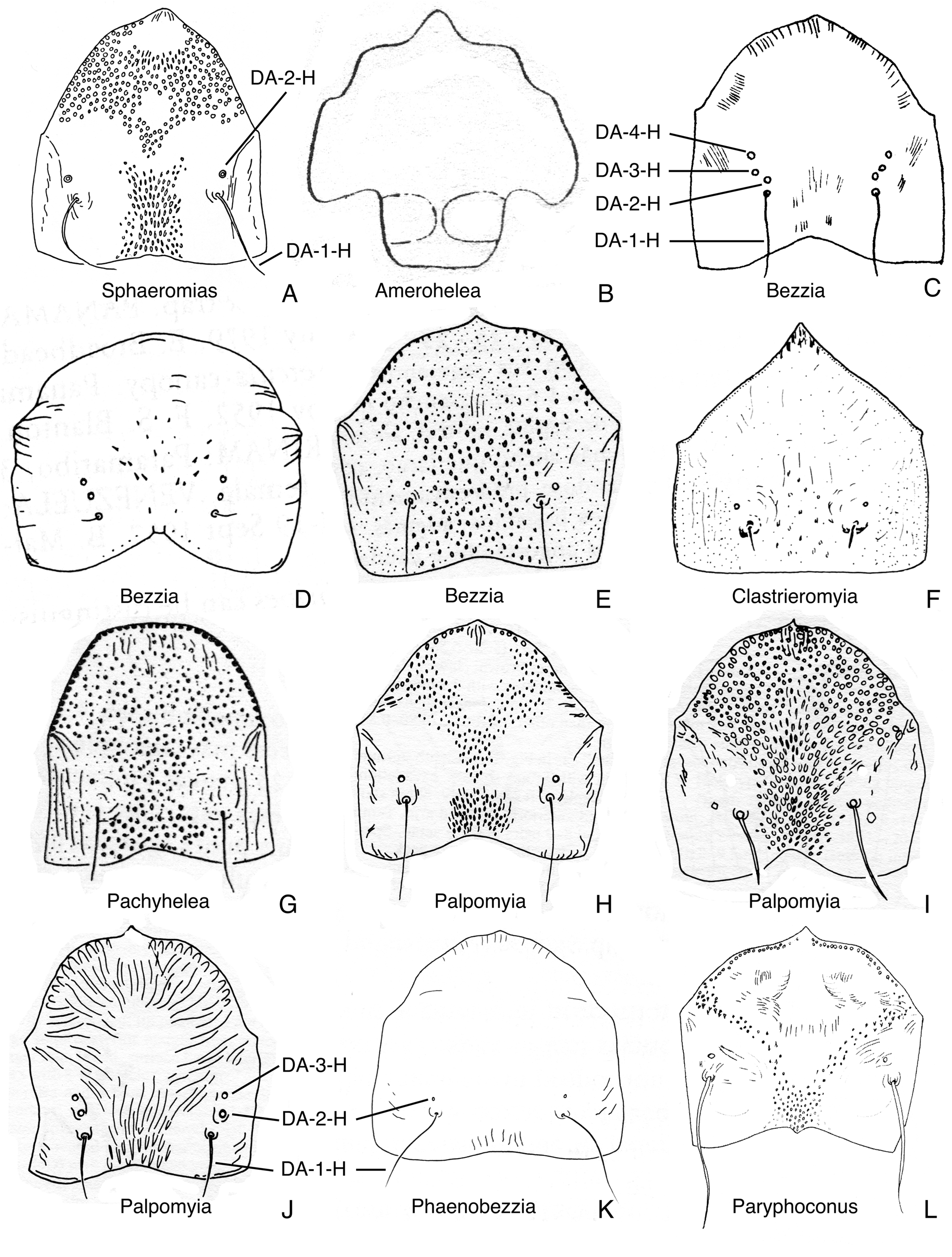
FIGURE 22. Dorsal apotomes, anterior view. A. Sphaeromias longipennis (from Wirth & Grogan 1979). B. Amerohelea sordidipes (from Lane et al. 1955). C. Bezzia mollis (from Mayer 1934c). D. Bezzia roldani (from Spinelli & Wirth 1989). E. Bezzia brevicornis (from Spinelli 1983a). F. Clastrieromyia dycei (from Spinelli & Grogan 1986). G. Pachyhelea pachymera (from Spinelli 1983a). H. Palpomyia lineata (from Grogan & Wirth 1979). I. Palpomyia belkini (from Grogan & Wirth 1979, modified). J. Palpomyia flaviceps (from Grogan & Wirth 1979). K. Phaenobezzia opaca (from Wirth & Grogan 1982). L. Paryphoconus oliveirai (from Ronderos et al. 2007).

FIGURE 28. Posterior portion of heads, in ventral view. A. Leehelea hispida. B. Sphaeromias longipennis. C. Xenohelea galatea. D. Bezzia nobilis. E. Clastrieromyia dycei. F. Pachyhelea pachymera. G. Palpomyia subasper. H. Phaenobezzia opaca. I. Paryphoconus grandis.
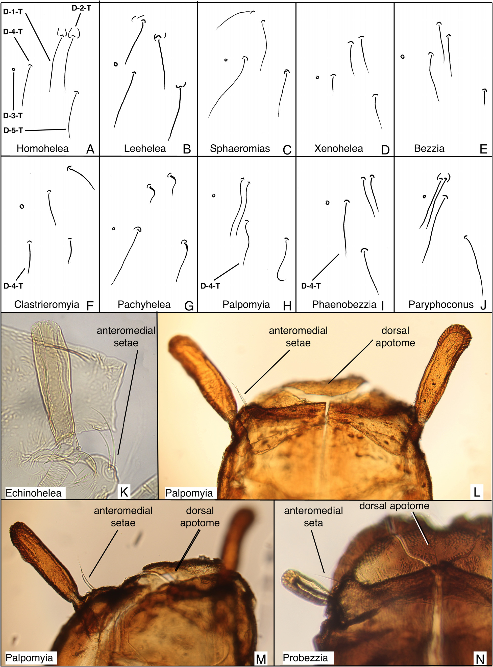
FIGURE 31. Dorsal setae from left side of thoraces; anteromedial setae. A. Homohelea delanoe. B. Leehelea hispida. C. Sphaeromias longipennis. D. Xenohelea galatea. E. Bezzia nobilis. F. Clastrieromyia dycei. G. Pachyhelea pachymera. H. Palpomyia jonesi. I. Phaenobezzia mashonensis. J. Paryphoconus oliveirai. K. Echinohelea lanei, partial lateral view (distorted slide specimen). L. Palpomyia (JAD685), dorsal view. M. Palpomyia (JAD685), dorsolateral view. N. Probezzia seminigra, dorsal view of left side.

FIGURE 33. Cephalothoraces and abdominal segments 1–3, in lateral view. A. Parabezzia sp. (5 km. E. Danby, Vermont, USA). B. Clinohelea curriei. C. Pellucidomyia leei. D. Hebetula tonnoiri. E. Jenkinshelea sp. (from Ottawa, ON, Canada). F. Mallochohelea caudellii. G. Probezzia seminigra. H. Nilobezzia sp. (from Kruger NP, South Africa). I. Sphaeromias longipennis. J. Bezzia sp. (6 km E. Salmon Arm, BC, Canada) K. Palpomyia sp. (Black Lake, Quebec, Canada). L. Phaenobezzia sp. (from Skulsuza, South Africa).
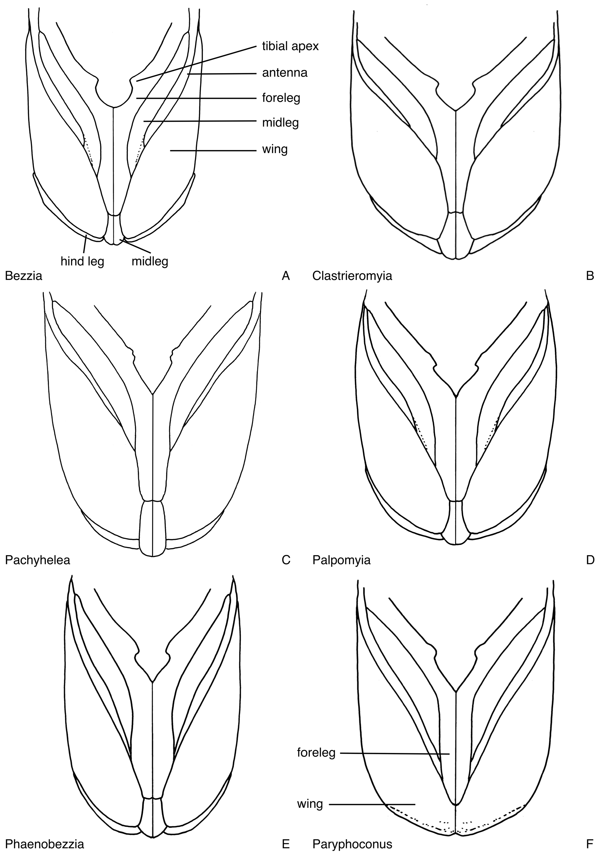
FIGURE 41. Legs, wings and apices of the antennae, in ventral view. A. Bezzia nobilis. B. Clastrieromyia dycei (reconstructed from distorted specimen). C. Pachyhelea pachymera. D. Palpomyia occidentalis. E. Phaenobezzia opaca. F. Paryphoconus oliveirai (reconstructed from distorted specimen).

FIGURE 46. Respiratory organs, most in dorsal view, Figs. 46G. in lateral view, pattern on tracheal tubes shown only in some. A. Homohelea albitudinis (from de Meillon & Wirth 1981). B. Homohelea delanoe (from de Meillon & Wirth 1981). C. Leehelea wasselli (from Elson-Harris 1987). D. Leehelea hispida (from Elson-Harris 1987). E. Sphaeromias longipennis (from Wirth & Grogan 1979). F. Xenohelea galatea. G. Amerohelea sordidipes (from Lane et al. 1955). H. Bezzia glabra (from Wirth 1983b). I. Bezzia obelisca (from Wirth 1983a). J. Bezzia dorsasetula (from Wirth 1983a). K. Bezzia turkmenica (from Glukhova 1979b). L. Clastrieromyia dycei (from Spinelli & Grogan 1986). M. Pachyhelea pachymera (from Spinelli 1983a). N. Palpomyia lineata (from Grogan & Wirth 1979). O. Palpomyia jonesi (from Grogan & Wirth 1979). P. Palpomyia belkini (from Grogan & Wirth 1979). Q. Palpomyia rufa (from Grogan & Wirth 1979). R. Palpomyia flaviceps (from Grogan & Wirth 1979). S. Phaenobezzia opaca (from Wirth & Grogan 1982). T. Paryphoconus oliveirai (from Ronderos et al. 2007).

FIGURE 54. Metathoraces and tergites 1, in dorsal view. A. Palpomyia jonesi. B. Phaenobezzia opaca. C. Paryphoconus oliveirai.

FIGURE 69. Abdominal segments four, in dorsal and ventral view (shagreen not shown). A. Sphaeromias longipennis. B. Xenohelea galatea. C. Bezzia nobilis.
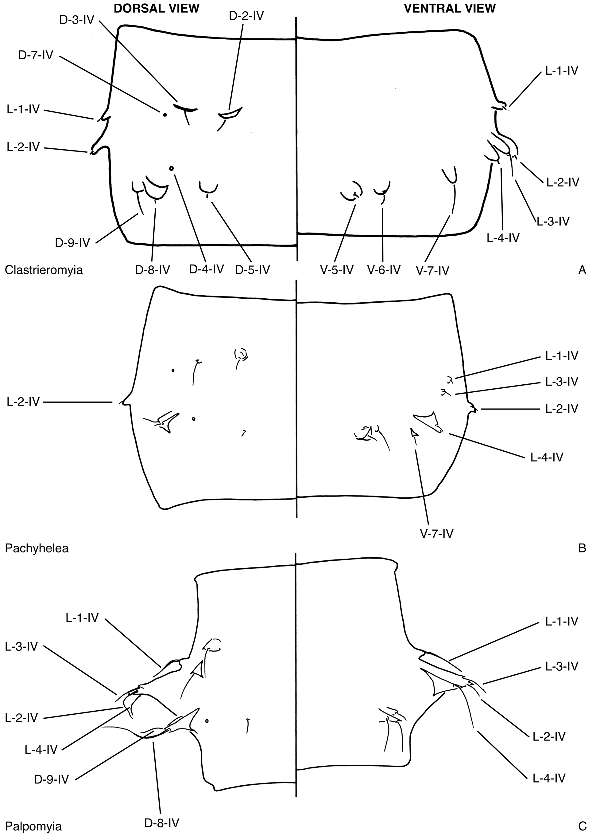
FIGURE 70. Abdominal segments four, in dorsal and ventral view (shagreen not shown). A. Clastrieromyia dycei. B. Pachyhelea pachymera. C. Palpomyia jonesi.
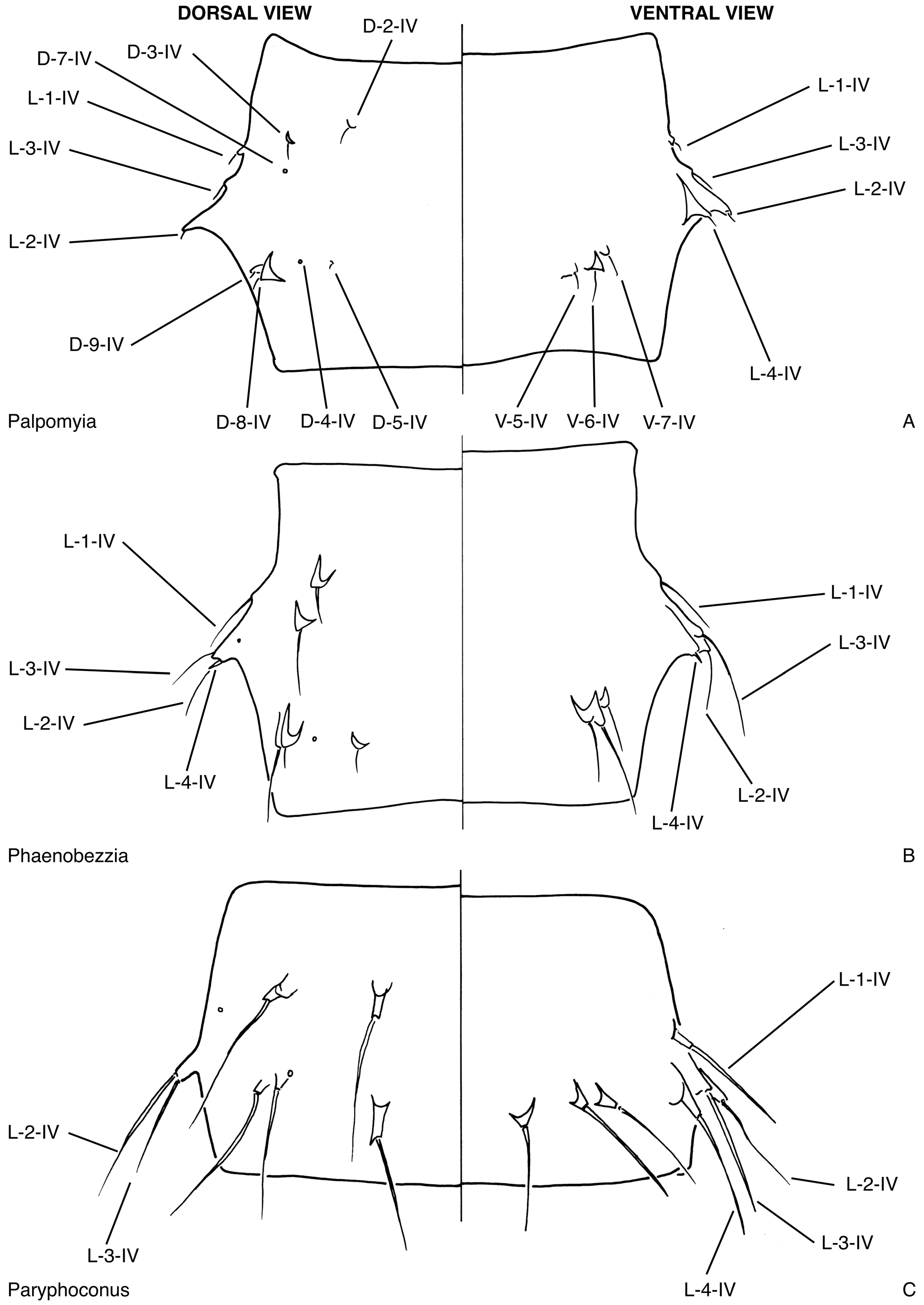
FIGURE 71. Abdominal segments four, in dorsal and ventral view (shagreen not shown). A. Palpomyia occidentalis. B. Phaenobezzia opaca. C. Paryphoconus oliveirai.

FIGURE 78. Abdominal segments 9. A. Palpomyia lineata, male, in dorsal and ventral view (from Grogan & Wirth 1979). B. Palpomyia lineata, female, in dorsal and ventral view (from Grogan & Wirth 1979). C. Palpomyia jonesi, male, in dorsal and ventral view (from Grogan & Wirth 1979. D. Palpomyia jonesi, female, in dorsal and ventral view (from Grogan & Wirth 1979). E. Palpomyia belkini, male, in dorsal and ventral view (from Grogan & Wirth 1979). F. Palpomyia belkini, female, in dorsal and ventral view (from Grogan & Wirth 1979). G. Palpomyia rufa, male, in dorsal and ventral view (from Grogan & Wirth 1979). H. Palpomyia flaviceps, female, in dorsal and ventral view (from Grogan & Wirth 1979). I. Phaenobezzia vacunae, male, in ventral view (from Haeselbarth 1965). J. Phaenobezzia mashonensis, male, in ventral view (from Haeselbarth 1965). K. Phaenobezzia opaca, female, in dorsal and ventral view (from Wirth & Grogan 1982). L. Paryphoconus oliveirai, female, in dorsal view (from Ronderos et al. 2007).

FIGURE 79. Evolution of ecdysial sutures and splits in pupal exuviae of Ceratopogonidae, head and anterior portion of thorax in ventral view. Numbers refer to character states of characters 2 and 8 in text. Red indicates ecdysial splits. A. Leptoconopinae. B. Forcipomyiinae. C. Dasyheleinae. D. Most Ceratopogonini. E. Most Ceratopogonini, Clinohelea. F. Allohelea, Atyphohelea, Parabezzia. G. Heteromyia, Pellucidomyia, Hebetula. H. most Sphaeromiini s. lat., most Palpomyiini, Paryphoconus; some Bezzia are nearly completed fused (as in Fig. 79I), with only the dorsal apotome separate, along with an ecdysial split along the base of the antenna and the dorsal thoracic split. I. Jenkinshelea, Macropeza, Mallochohelea, Probezzia, Neobezzia.
| USNM |
Smithsonian Institution, National Museum of Natural History |
| ZSMC |
Zoologische Staatssammlung |
| ZIN |
Russian Academy of Sciences, Zoological Institute, Zoological Museum |
| CNCI |
Canadian National Collection Insects |
| NYSM |
New York State Museum |
| NMSA |
KwaZulu-Natal Museum |
| IZUG |
Istituto di Zoologia dell'Universita |
| ANIC |
Australian National Insect Collection |
| SAIM |
South African Institute for Medical Research |
| BPBM |
Bishop Museum |
| INPA |
Instituto Nacional de Pesquisas da Amazonia |
No known copyright restrictions apply. See Agosti, D., Egloff, W., 2009. Taxonomic information exchange and copyright: the Plazi approach. BMC Research Notes 2009, 2:53 for further explanation.
1 (by felipe, 2021-06-14 20:35:26)
2 (by ExternalLinkService, 2021-10-20 03:23:19)
3 (by diego, 2021-10-22 14:59:52)
4 (by ExternalLinkService, 2021-10-22 15:18:37)
5 (by ExternalLinkService, 2021-10-22 15:46:02)
6 (by ExternalLinkService, 2022-01-30 07:28:53)
7 (by diego, 2022-05-23 12:43:12)
8 (by admin, 2022-07-14 20:19:55)
9 (by ExternalLinkService, 2022-07-14 20:47:18)
10 (by ExternalLinkService, 2023-11-02 23:08:14)