
Thysanozoon mirtae, Bulnes, Verónica N., Albano, Mariano J., Obenat, Sandra M. & Cazzaniga, Néstor J., 2011
|
publication ID |
https://doi.org/ 10.5281/zenodo.202319 |
|
DOI |
https://doi.org/10.5281/zenodo.6183441 |
|
persistent identifier |
https://treatment.plazi.org/id/8C5F87DF-8C60-FFF0-FF67-FEF9822AF873 |
|
treatment provided by |
Plazi |
|
scientific name |
Thysanozoon mirtae |
| status |
sp. nov. |
Thysanozoon mirtae sp. nov.
Figures 7–9 View FIGURE 7 View FIGURE 8 View FIGURE 9
Diagnosis. Pseudocerotidae with papillated surface, ground colour greenish yellow covered with rounded black spots, more densely distributed on a mid dorsal band. The papillae are elongated, reddish brown; with a marginal band and cerebral area free of papillae. Conspicuous marginal, colourless tentacles. Two cerebral eye clusters present. The tentacular eyes extend from the fore margin, where they are arranged in a single marginal band, backwards up to the base of the tentacles where they are scattered. Male copulatory apparatus double with conspicuous spermiducal vesicles, a single true seminal vesicle, free prostatic vesicle and a slightly asymmetric stylet. The ejaculatory duct joins the prostatic duct proximally at the base of the penis stylet. Male atrium short, narrow and ciliated. Vagina ciliated, without a cement pouch. Female atrium small and ciliated. Separated gonopores. Ventral sucker in the fore body half.
Type material. Holotype: 9.0 μm sagittally sectioned specimen, mounted on 180 slides. Collected by M.J. Albano, 21 February 2009 from the Club Náutico de Mar del Plata. On a mussel and ascidians community, 1–2 m depth. MACN-In 38207.
Etymology. The specific name is dedicated to the memory of Mariano Albano´s mother, Mrs. Mirta R. Elías.
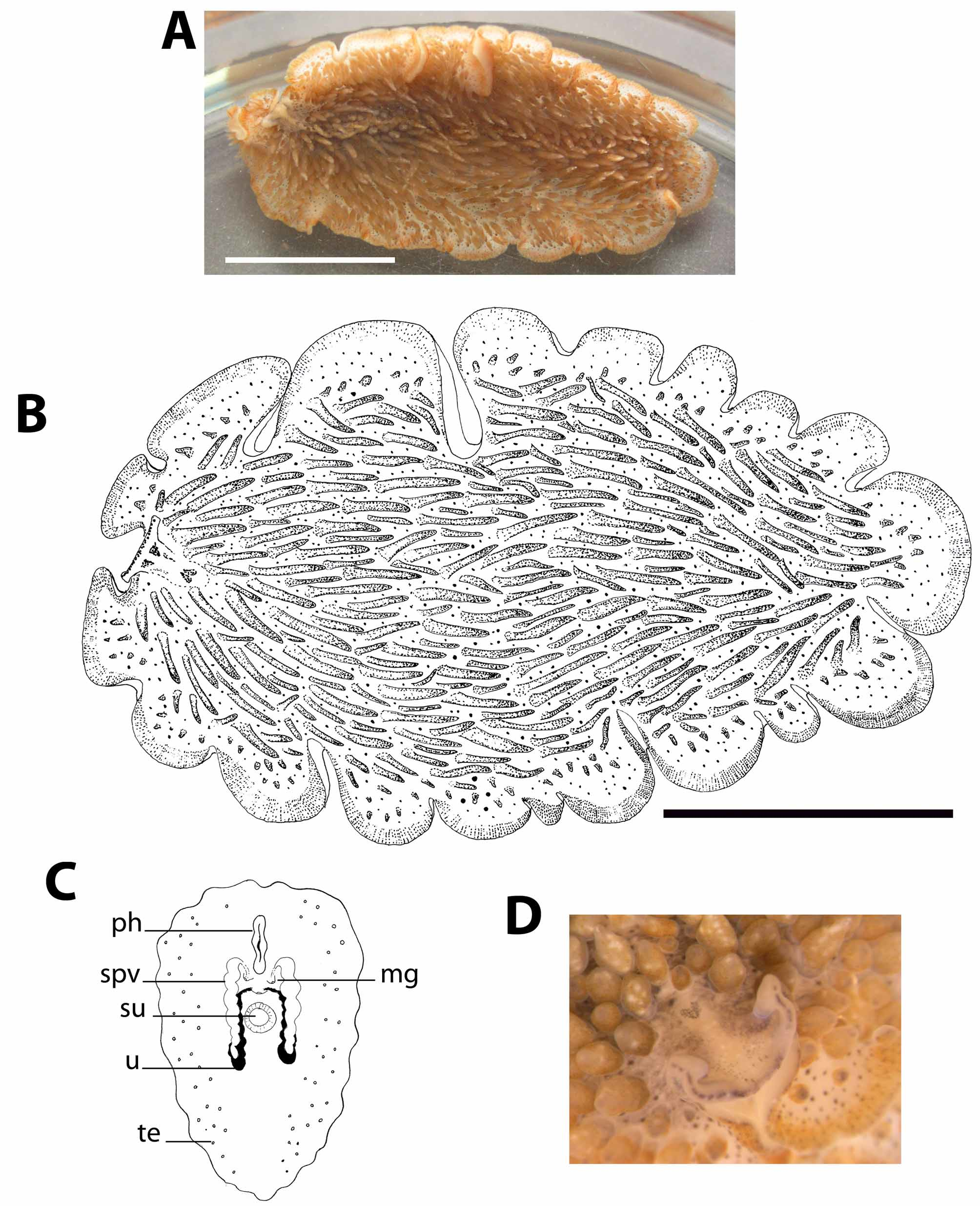
Description. Large, elongated, 36.5 mm long by 20 mm wide alive, 35 mm by 18.4 mm preserved. Marginal tentacles formed by foldings of the fore margin. Dorsal surface covered with elongated reddish brown papillae which are longer and more thickly placed in the centre, declining in length and number to the margins, leaving a marginal band free of papillae.
Ground colour is greenish yellow covered with rounded black spots, more densely distributed on a mid dorsal band. With an interrupted reddish brown marginal band. Marginal tentacles without pigmentation, extending between the tentacle bases and reaching the brain dorsally (figures 7A–7B).
The dorsal tentacular eye spots are numerous, distributed dorsally in a single row and ventrally scattered. The cerebral eyespots form two separated triangular clusters joining anteriorly. Frontal eyes scattered (figure 7D).
Pharynx ruffled, 5.5 mm long (figure 7C). The mouth opens at 14 mm from fore margin, behind the pharynx cavity. The main gut opens in the middle of the pharynx cavity roof and runs distally up to short behind the female system. The intestinal ramifications give out extensions into the dorsal papillae (figure 9C).
Dorsal body wall 103 µm high. The ciliated cellular epidermis bears rhabdites and granular pigmentation. The elongated rhabdites are densely packed on the epidermis of the papillae and are readily seen as small red small rods even in living specimens. The granular pigmentation is distributed mostly in the epidermis free of papillae. Beneath the epidermis a layer of circular muscle fibres is present, followed by a longitudinal muscle layer.
Ventral body wall 110 µm high, without rhabdites but with granular pigmentation. Beneath the basement membrane, there are a layer of circular muscle fibres and an innermost layer of longitudinal muscle fibres.
The ventral epidermis is higher than the ventral one, the ventral circular layer is thinner than the dorsal one, and the ventral longitudinal layer is much higher and dense than the dorsal one.
Body parenchyma immersed in a dense net of parenchymatic muscle fibres, giving the body a strong appearance (figure 9D).
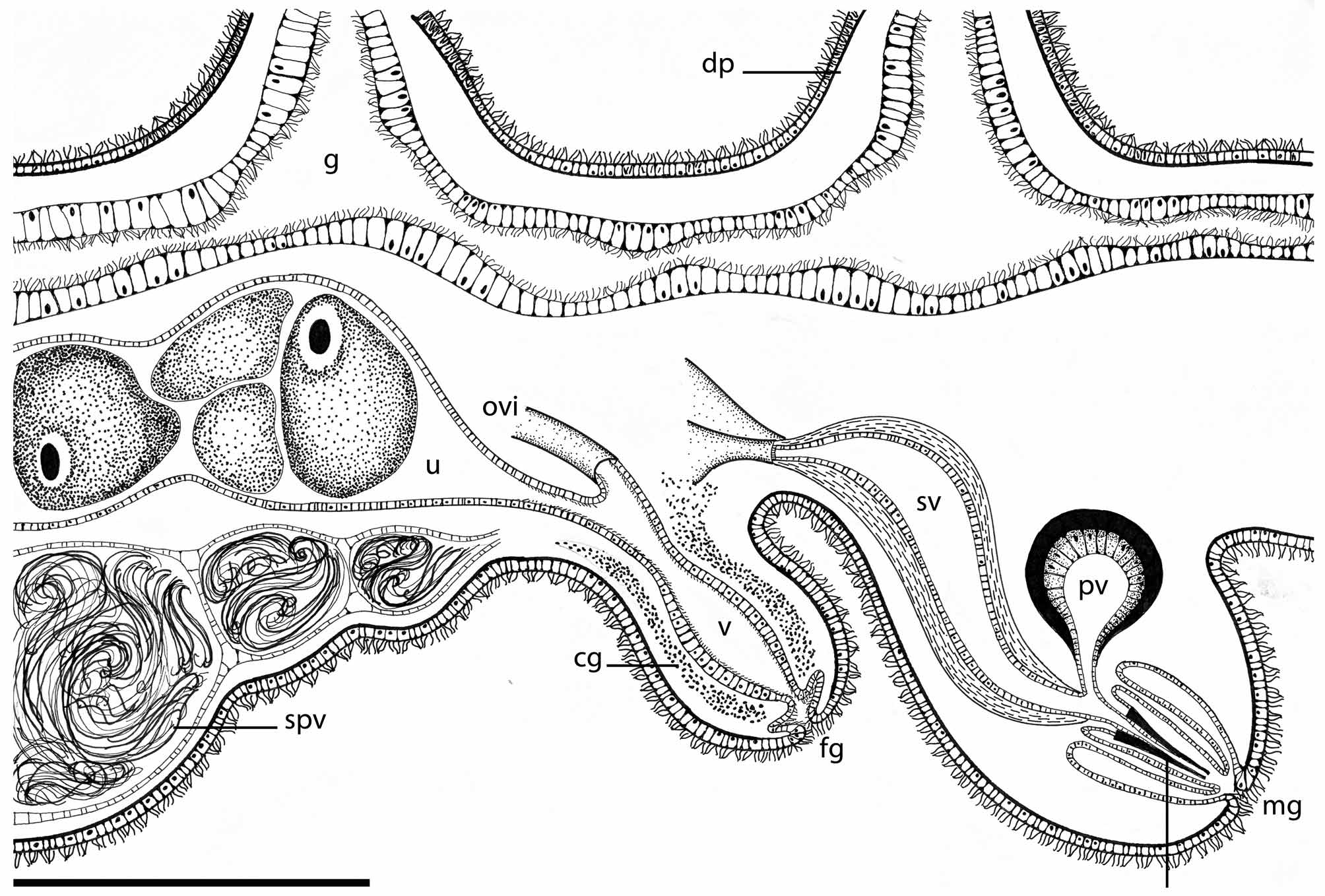
The testes are small, widely spaced and ventrally arranged beyond the uteri. The uteri are dorsal to the testes, but ventral to the gut. They are well developed, filled with numerous eggs and located behind the spermiducal vesicles.
The male copulatory system is double. Each copulatory organ consists of a true seminal vesicle, free prostatic vesicle and penis papillae armed with a stylet (figure 8). The vas deferens runs ventrally from the rear. After it passes the ventral sucker, the vas deferens distends to form a few voluminous spermiducal vesicles (figure 9E) before entering the elongated seminal vesicle. The seminal vesicle has a well developed muscular wall and is arranged dorsally to the male prostatic vesicle and stylet. The rounded prostatic vesicle shows a muscular wall and a very high, smooth glandular inner lining. The ciliated ejaculatory duct follows a short trajectory until reaching the penis papillae, its muscular wall being well developed. The prostatic duct is short and straight, joining the ejaculatory duct at the basis of the penis papilla. The stylet is conical and elongated, 760 µm long, slightly asymmetric and sharp. The male atrium is short and narrow, ciliated and directed backwards, opening to a male gonopore located 12 mm from the fore margin (figure 9B). The second male copulatory organ is similar to the one already described. Both male copulatory organs are symmetrically arranged left and right of the longitudinal body axis.
The female reproductive system is single (figure 8). The oviducts enter separately the vagina from the rear, turning ventrally to open distally into a small female atrium and a median gonopore. The canal is about the same diameter along the whole trajectory. The vagina is surrounded by scarce cement glands. There is no cement pouch. The vagina and atrium are completely ciliated.
Taxonomical discussion. Thysanozoon mirtae sp. nov. is the only known species having slender, elongated dorsal papillae of reddish brown colour on a greenish yellow body covered with rounded black spots with a reddish brown marginal band; the marginal tentacles and cerebral area devoid of pigment complete an exclusive colour pattern. The presence of slender papillae was described for Thysanozoon skottsbergi Bock, 1913 and T. distinctum Stummer-Traunfels, 1895 , while T. minutum Stummer-Traunfels, 1895 has a similar ground body colour. However, the dorsal papillae of T. mirtae sp. nov. differ from the papillae of T. skottsbergi because of the absence of darker pigmentation. The margin of T. distincutum is golden yellow and that of T. minutum is white, sharply different from the reddish brown marginal band of T. mirtae sp. nov. The spermiducal vesicles are thin-walled expansions of the vas deferens serving as storage of sperm ( Faubel 1983, p. 21) different from the spermiducal bulbs, which are more muscularized structures, presumably assisting the propulsion of sperm. The apparent spermiducal vesicles and the strongly developed parenchymatic musculature are two main features characterizing the new species T. mirtae .
It differs readily from the only species of Thyzanozoon with which it was recorded in sympatry, T. brocchii (Risso 1818) , both in colour and anatomy. While T. mirtae is dorsally greenish yellow, covered with translucent, slender reddish brown papillae, with spermiducal vesicles and a well developed mesenchymatic musculature; T. brocchii is dorsally light-brown, covered with, solid, blunt dark brown papillae, without spemiducal vesicles and few mesenchymatic muscle fibres.

No known copyright restrictions apply. See Agosti, D., Egloff, W., 2009. Taxonomic information exchange and copyright: the Plazi approach. BMC Research Notes 2009, 2:53 for further explanation.