
Transversotrema elegans, Hunter & Ingram & Adlard & Bray & Cribb, 2010
|
publication ID |
https://doi.org/ 10.11646/zootaxa.2652.1.2 |
|
publication LSID |
lsid:zoobank.org:pub:CED86721-8335-4A72-A980-485B68ACCF68 |
|
persistent identifier |
https://treatment.plazi.org/id/463B93D6-3ECC-47A1-B13A-2DCB87C375EE |
|
taxon LSID |
lsid:zoobank.org:act:463B93D6-3ECC-47A1-B13A-2DCB87C375EE |
|
treatment provided by |
Felipe |
|
scientific name |
Transversotrema elegans |
| status |
sp. nov. |
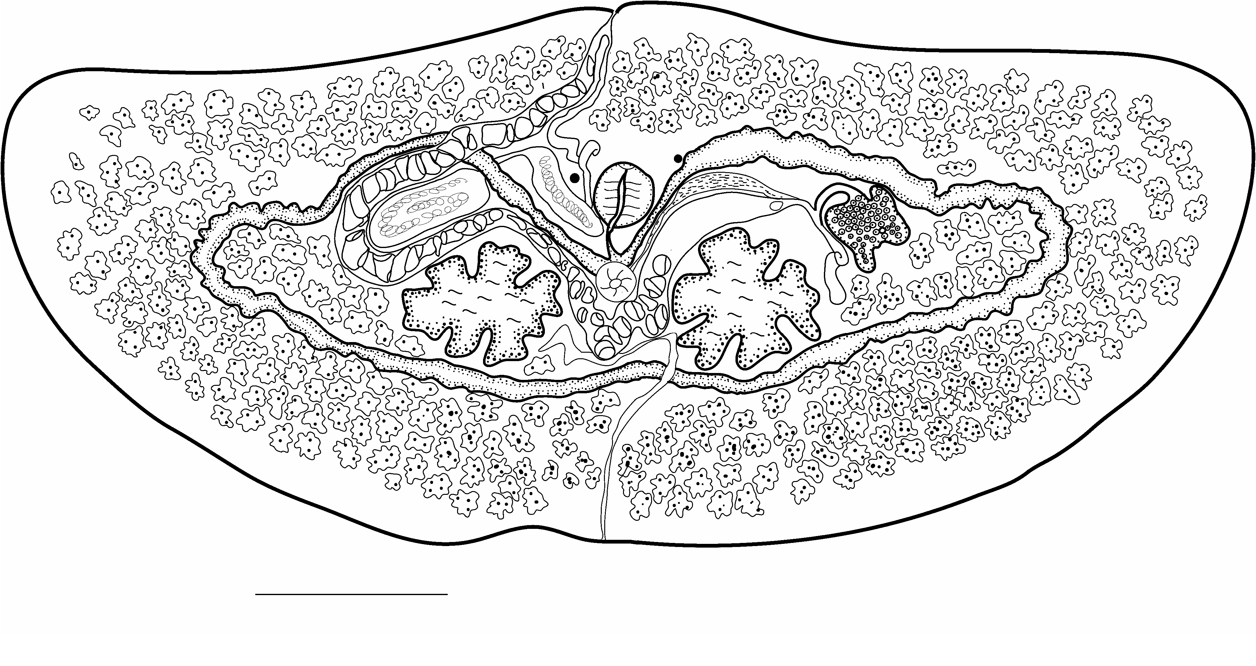
Transversotrema elegans View in CoL n. sp.
( Fig. 3 View FIGURE 3 )
Syn. T. haasi Witenberg, 1944 in part of Cribb et al. (1992)
Type host: Choerodon graphicus (De Vis) Labridae . Graphic tuskfish
Type locality: Heron Island , southern Great Barrier Reef :
Other hosts: Labridae : Gomphosus varius Lacepède Bird wrasse (n =1); Hemigymnus melapterus (Bloch) . Blackeye thicklip (n =5); Choerodon venustus De Vis, Venus tuskfish (n =1)
Site of infection: Beneath the scales
Materials examined: 201 individuals (see Table 2)
GenBank: see Table 1.
Deposited specimens: Holotype and paratypes: Queensland Museum G 231782 – G231787. Specimens previously identified as T. haasi see Cribb (1992) QM GL 12746–55 ; BM (NH) 1991.9.3.1–3 .
Etymology: The Transversotrematidae are visually appealing worms with an elegant swimming action when released from their host, hence the name elegans .
Description: (based on measurements of 21 specimens from labrids from Heron Island). Body transversely elongated, strongly dorsoventrally flattened, 736–2112 (1393) long, 1848–5344 (3358) wide, width/length ratio 2.04–2.75 (2.40). Tegumental spines prominent. Eyespots prominent, 156–393 (273) apart, 6.89–9.66 (8.15%) of body width; no pigment evident other than in eyespots. Ventral sucker well posterior to eyespots, 71–173 (114) long, 77–177 (116) wide. Mouth mid-ventral, inconspicuous. Pharynx between or slightly posterior to eyespots, 90–241 (158) long, 93–226 (155) µm wide. Oesophagus distinct, curved, 64– 778 (148) long. Caecal bifurcation anterior or dorsal to ventral sucker. Caeca form cyclocoel reaching laterally to envelop testes, ovary and some vitelline follicles. Testes opposite, deeply lobed, left, 168–542 (359) long, 168–561 (382) wide; right 161–477 (342) long, 190–600 (391) µm wide. Seminal vesicle formed of lobed, saccular enclosed portion and winding, tubular extracaecal portion. Enclosed portion distinctly lobed or entire, antero-dextral to right testis, constricts distally to form narrow duct that passes ventral to cyclocoel to join tubular portion. Tubular portion of seminal vesicle passes mediad along cyclocoel then turns anteriorly and passes between eyespots dextral to pharynx and passes to common genital pore where it unites with uterus without any specialisation. Common genital pore precisely in midline on anterior margin of worm. Ovary sinistral to left testis, deeply lobed, 84–394 (217) µm long, 97–380 (227) µm wide. Oviduct passes medioposteriorly, unites with Laurer’s canal and duct from oviduct passes vitelline reservoir. Laurer’s canal passes posteriorly to open dorsally close to left testis; median portion dilated, contains sperm or vitelline remnants. Vitelline reservoir immediately anterior to left testis. Vitelline follicles fill almost entire extracaecal space; enclosed follicles in two masses at each lateral extremity, sometimes also scattered in interrupted band along inner margins of posterior cyclocoel, 28–57 (37). Uterus passes medially between anterior half of cyclocoel and testes then between right testis and saccular portion of seminal vesicle. Proximal portions of uterus act as seminal receptacle. Eggs large, 91–130 (107) long, 33–75 (52) wide. Excretory pore opens posteriorly at small notch in middle of posterior margin, bladder extends anteriorly in initially narrow tube which then expands into large sac which passes ventral to cyclocoel anterior to which it becomes laterally directed.
| QM |
Queensland Museum |
| BM |
Bristol Museum |
No known copyright restrictions apply. See Agosti, D., Egloff, W., 2009. Taxonomic information exchange and copyright: the Plazi approach. BMC Research Notes 2009, 2:53 for further explanation.
|
Kingdom |
|
|
Phylum |
|
|
Class |
|
|
Order |
|
|
Family |
|
|
Genus |