
Jeholosaurus shangyuanensis Xu et al. 2000
|
publication ID |
https://doi.org/ 10.5281/zenodo.187103 |
|
DOI |
https://doi.org/10.5281/zenodo.6223696 |
|
persistent identifier |
https://treatment.plazi.org/id/038D87B7-FFB5-543B-88B9-F9EDA303F85A |
|
treatment provided by |
Plazi |
|
scientific name |
Jeholosaurus shangyuanensis Xu et al. 2000 |
| status |
|
Jeholosaurus shangyuanensis Xu et al. 2000
Emended diagnosis (cranial features only: after Xu et al. 2000; Butler et al. 2008). One autapomorphy and a unique combination of character states support the validity of Jeholosaurus . Autapomorphy: presence of a row of small foramina on the lateral surface of the nasal immediately dorsal to the premaxillary articulation. Unique combination of character states: six premaxillary teeth (distinct from all other cerapodans, but present in Lesothosaurus and some ankylosaurs); presence of a foramen enclosed within the quadratojugal (distinct from all ornithischians except Hypsilophodon and Tenontosaurus ); combined presence of nodular ornamentation on the postorbital and jugal (distinct from all ornithopods and non-cerapodan ornithischians, but also present in pachycephalosaurs, Archaeoceratops and Yinlong ); and jugal caudal process bifurcated distally (distinct from all cerapodans except Psittacosaurus , but present in some early ornithischians including Emausaurus , Lesothosaurus and Scelidosaurus ).
Holotype. IVPP V12529 View Materials , a partial skeleton consisting of a skull with adhered mandibles, an articulated series of cervical vertebrae, fragmentary sacrum, articulated sections of caudal vertebrae, and both hindlimbs.
Locality and horizon. Lujiatun, Liaoning Province, People’s Republic of China; Yixian Formation, Early Cretaceous (early Aptian: Swisher et al. 1999; Cretaceous stage boundaries following Gradstein et al. 2004).
Referred specimens. IVPP V12530 View Materials , a skull and associated cervical vertebrae; IVPP V15716 View Materials , a partial skull; IVPP V15717 View Materials , a complete skull; IVPP V15718 View Materials , a complete skull; IVPP V15719 View Materials , a partial skeleton comprising a skull, partial axial column, pelvic girdle and hindlimbs. All referred specimens come from the same locality and horizon as the holotype.
Materials. Jeholosaurus is represented by six skulls that vary in length from 33–99 mm (as measured from the tip of the snout to the caudal margin of the quadrate). The skulls share a consistent set of anatomical features, but do exhibit some variation that is attributable to ontogeny (see Discussion). Combination of information from all six skulls allows the anatomy to be described in reasonable detail. However, several areas of the skull are either unrepresented or remain unprepared and encased in matrix, including the rostral part of the palate and the rostrodorsal part of the braincase.
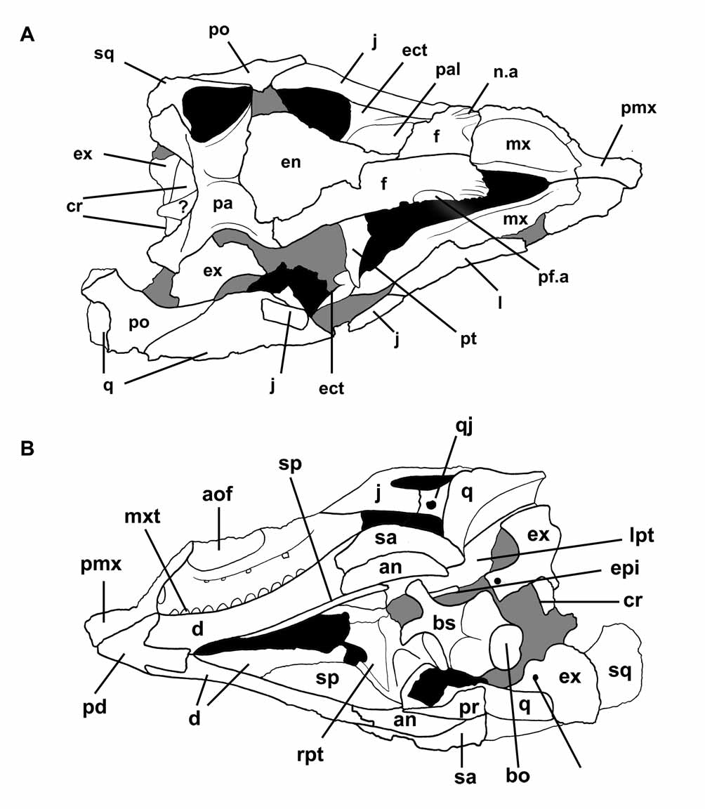
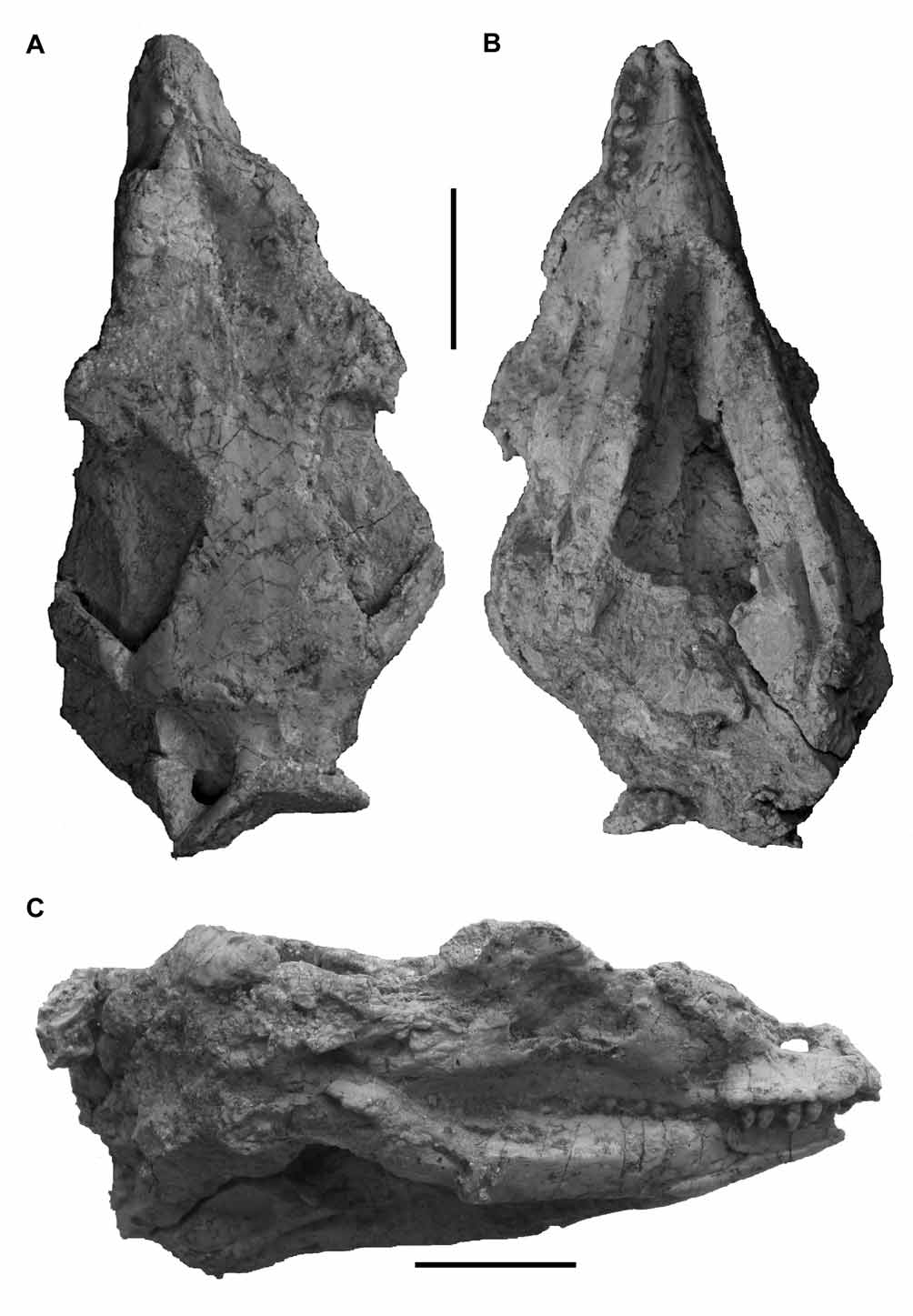
IVPP V12529 View Materials is almost fully prepared, but matrix still obscures the rostral and lateral surfaces of the braincase and the dorsal surface of the palate ( Figs. 1–2 View FIGURE 1 View FIGURE 2 ). The skull has been crushed dorsoventrally, resulting in displacement of the skull roof. Both mandibles are present and adhered to the skull, obscuring details of the palate and tooth rows. The snout is broken dorsally: the nasals are missing, allowing the internal surfaces of the maxillae to be seen ( Figs. 1 View FIGURE 1 A and 2A). Many individual elements are damaged and others are absent, such as the rostral part of the palate and the right prefrontal. Damage to the frontals exposes a well-preserved natural endocast. The braincase is visible in caudal and ventral views: the individual elements are unfused and slightly separated ( Figs. 1 View FIGURE 1 B and 2B).
IVPP V12530 View Materials appears to be complete and articulated, but has not been fully prepared: the occiput, palate and caudal portion of the left hand side of the skull are still covered in matrix ( Figs. 3–4 View FIGURE 3 View FIGURE 4 ). Both mandibles are present and tightly appressed to the skull, though the right mandible is damaged rostrally and the posterior part of the left mandible is encased in sediment. The rostral portion of the snout is slightly crushed and the entire skull exhibits some transverse compression, but the general preservation of the specimen is very good.
IVPP V15716 View Materials is preserved in an unusual fashion. It consists of a thin slab that comprises the right hand side of an articulated skull, together with the adhered right mandible. The medial surface of the slab is encrusted with matrix and reveals no anatomical information: however, the lateral surface is well preserved and reveals some features not present in any other specimens ( Figs. 5–6 View FIGURE 5 View FIGURE 6 ).
IVPP V15717 View Materials is the largest skull specimen known. It is largely complete, but has been crushed dorsoventrally and is broken caudoventrally. Both of the mandibles are present and are in articulation with the skull. This specimen has not been fully prepared: as a consequence, the palate and braincase are obscured ( Fig. 7 View FIGURE 7 ). Unfortunately, the surface preservation is poor and it is difficult to identify the boundaries between many of the individual cranial elements.
IVPP V15718 View Materials is complete and relatively undeformed, and is preserved in articulation with both mandibles, but lacks the tip of the snout, part of the skull roof and the predentary. It is comparable to IVPP V 12530 View Materials in terms of size, but in contrast to the latter specimen the braincase, posterior part of the palate and left hand side of the skull are visible ( Fig. 8 View FIGURE 8 A–D).
IVPP V15719 View Materials represents the smallest skull in the sample. It is undistorted and is associated with both mandibles, but the snout is damaged, the skull table is largely absent and the braincase has not been exposed ( Fig. 8 View FIGURE 8 E–F). It is articulated with a partial, articulated postcranial skeleton.
Description and comparisons. Phylogenetic analysis suggests that Jeholosaurus is a member of the clade Cerapoda ( Butler et al. 2008: see Discussion), which incorporates Ornithopoda, Pachycephalosauria and Ceratopsia ( Sereno 1986). For this reason, comparisons with other taxa are generally limited to members of this clade. However, because Xu et al. (2000) noted that Jeholosaurus was similar to the Chinese neornithischians Agilisaurus ( Peng 1992) and Hexinlusaurus ( He & Cai 1984), these taxa have also been included in the comparisons provided below.
General comments. The skull is low and triangular in lateral view, with the highest point situated dorsal to the orbits. Smaller skulls (e.g. IVPP V12530 View Materials , IVPP V15718 View Materials : Figs. 3 View FIGURE 3 A and 8A) tend to have more strongly vaulted dorsal cranial margins than larger individuals (e.g. IVPP V12529 View Materials , IVPP V15716 View Materials , IVPP V15717 View Materials : Figs. 1 View FIGURE 1 C, 5A and 7C). Rostral to the orbit, the snout slopes ventrally without any significant breaks in slope or changes in angle. The preorbital region of the skull (measured from the tip of the snout to the rostrocaudal border of the orbit) is relatively short in all specimens and accounts for approximately 50% of total skull length. In contrast, there are major changes in the relative orbital diameters of the different specimens, which exhibit a clear ontogenetic signal. In smaller individuals, the orbital diameter is approximately equal to 47–50% of total skull length (e.g. IVPP V12530 View Materials : Fig. 3 View FIGURE 3 A), whereas this is reduced to 33% or less in larger skulls (e.g. IVPP V15716 View Materials : Fig. 5 View FIGURE 5 A). In dorsal view, the skull roof is broadest along its caudal margin and narrows rostrally to terminate in a narrow ‘V’-shaped snout ( Figs. 1 View FIGURE 1 A, 7A and 8B). No soft tissues are preserved with any of the specimens.
The orbit represents the largest of the cranial openings and has a circular to subcircular outline in lateral view ( Figs. 1 View FIGURE 1 C, 3A, 5A, 8A and 8E). The external naris is elliptical or tear-drop shaped in lateral view, with its long axis extending caudodorsally. Its ventral margin lies at the same level as that of the orbit and the maximum length of the external naris is equal to approximately 15% of total skull length. A large antorbital fossa is present: it has a subtrapezoidal outline and the much smaller internal antorbital fenestra is confined to its caudal portion. The infratemporal fenestra is narrow, elongate and subelliptical in outline: its rostroventral margin extends to a point just beneath the orbit. Finally, the supratemporal fenestra is longer than it is wide and has a ‘D’-shaped outline in dorsal view ( Figs. 1 View FIGURE 1 A, 2A and 8B).
In all specimens, the mandible possesses a well-developed coronoid eminence that is almost twice the height of the dentary at its midpoint. A stout retroarticular process is also present and is subtriangular in lateral view (IVPP V12530 View Materials , IVPP V15719 View Materials ). The internal mandibular fenestra can only be seen in IVPP V12529 View Materials , but is largely obscured by matrix and the close proximity of the pterygoids. None of the specimens exhibits an external mandibular fenestra.
The braincase is visible in IVPP V12529 View Materials ( Figs. 1 View FIGURE 1 B and 2B) and IVPP V15718 View Materials ( Fig. 8 View FIGURE 8 C-D) only. In the former, the braincase has moved from its life position and is slightly damaged and disarticulated: the supraoccipital is absent; a caudal rib fragment occludes the foramen magnum; and the opisthotic/exoccipitals have detached from the basisphenoid. In IVPP V15718 View Materials , the braincase has remained in articulation, but the basioccipital condyle has been sheared off. The positions of almost all neurocranial sutures can be determined in both specimens, providing further evidence of their immaturity. IVPP V15718 View Materials has a large foramen magnum with a subcircular outline.
Dermatocranium and splanchnocranium. Premaxilla. The premaxilla consists of a subrectangular main body that supports two caudodorsally projecting processes: a rostrally positioned nasal process and a caudally situated maxillary process ( Figs. 3 View FIGURE 3 A, 4, 5A and 6). The nasal process forms the rostrodorsal border of the external naris: it tapers in thickness as it extends dorsally, both mediolaterally and dorsoventrally, terminating in a fine point. This process contacts its antimere along the midline and has a short overlapping contact with the nasal on its lateral surface. Only a small portion of the narial dorsal margin is formed by the premaxilla: most is composed of the rostral process of the nasal. In contrast, the maxillary process is much better developed and forms the entire caudal margin of the external naris. It is stout and strut-like and tapers to a rounded apex as it curves caudally. The caudal margin of the maxillary process articulates with the rostral margin of the maxilla along its entire length. In larger individuals (IVPP V15716 View Materials : Fig. 6 View FIGURE 6 ) the distal end of the maxillary process articulates with the lachrymal, preventing the maxilla from contacting the nasal, whereas in small specimens (IVPP12530: Fig. 4 View FIGURE 4 ) only a point contact exists between the premaxilla and lachrymal. In IVPP V12530 View Materials ( Fig. 3 View FIGURE 3 A), the rostroventral margin of the process is excavated to form a shallow external narial fossa, but this structure is not present in this position in any other individual (see below).
The body of the premaxilla has a dorsoventrally convex lateral surface that bears a shallow external narial fossa in some individuals (IVPP V15716 View Materials , IVPP V15717 View Materials : Fig. 5 View FIGURE 5 A). The ventral boundary of this fossa extends close to the ventral margin of the premaxilla. In IVPP V15716 View Materials , the rostral margin of the premaxilla is exceptionally rugose and deeply pitted ( Figs. 5 View FIGURE 5 A and 6), as also occurs in Hypsilophodon (e.g. NHM R2477: Galton 1974) and Zephyrosaurus ( Sues 1980) . This area probably served for the attachment of a rhamphotheca. Premaxillary foramina are present at the base of the nasal process (IVPP V15716 View Materials , IVPP V15717 View Materials ), although in IVPP V15716 View Materials they are partly obscured by the abovementioned rugosity. The lateroventral margin of the premaxilla lies at the same level as the maxillary ventral margin, but a small ventral projection is present at the rostralmost tip of the premaxilla (IVPP V15716 View Materials ).
In most respects, the premaxilla of Jeholosaurus is similar to that of non-iguanodontian ornithopods and non-cerapodan neornithischians, including Agilisaurus (ZDM T6011: Peng 1992; Barrett et al. 2005), Bugenasaura ( Galton 1999) , Hypsilophodon (NHM R197: Galton 1974), Orodromeus ( Scheetz 1999) and Zephyrosaurus ( Sues 1980) . It differs substantially from the premaxillae of non-ceratopsid ceratopsians, which possess relatively smaller external narial openings and an articular surface for the rostral bone ( You & Dodson 2004). Moreover, ceratopsian premaxillae have stout maxillary processes that are rostrocaudally and dorsoventrally expanded in comparison with those of Jeholosaurus and non-iguanodontian ornithopods ( You & Dodson 2004).
Six teeth are present in the premaxilla (IVPP V12529 View Materials , IVPP V15716 View Materials , IVPP V15717 View Materials : e.g. Fig. 5 View FIGURE 5 A): breakage prevents determination of the dental formula in other specimens. Six premaxillary teeth are also known in the early ornithischian Lesothosaurus (NHM RUB17: Sereno 1991) and the ankylosaurs Silvisaurus and Cedarpelta ( Eaton 1960; Carpenter et al. 2001). However, all cerapodans have fewer teeth: five are present in Bugenasaura ( Galton 1999) , Changchunsaurus ( Zan et al. 2005) , Hypsilophodon (NHM R2477: Galton 1974), Orodromeus ( Scheetz 1999) and Zephyrosaurus ( Sues 1980) – the same number is also present in Agilisaurus (ZDM T6011: Peng 1992; Barrett et al. 2005); Archaeoceratops (IVPP V11114 View Materials : Dong & Azuma 1997), Liaoceratops (IVPP V12738 View Materials : Xu et al. 2002) and Yinlong (Xu et al. 2006) each possess three teeth; two teeth are present in Chaoyangsaurus (IGCAGS V371: Zhao et al. 1999); and premaxillary teeth are absent in Psittacosaurus (e.g. You & Dodson 2004). A short edentulous region is present rostral to the first premaxillary tooth (IVPP V15716 View Materials : contra Xu et al. 2000): the lack of this feature in IVPP V12529 View Materials probably reflects damage to the tips of the premaxillae. The ventral surfaces of the premaxillae are visible (in part) in both the holotype and IVPP V15717 View Materials (e.g. Fig. 7 View FIGURE 7 B): they meet at the midline to form a small, flat premaxillary palate. Unfortunately, it is not possible to determine the posterior extent of this structure due to the presence of the mandibles. Damage to the dorsal portion of the snout in IVPP V12529 View Materials allows the premaxillae to be seen in dorsomedial view. The medial margin of the element possesses a short, caudally directed process that overlaps the premaxillary process of the maxilla.
Maxilla. In lateral view, the maxilla is an elongate bone consisting of a rod-like tooth-bearing ramus and a caudodorsally extending ascending process that arises from the rostrodorsal margin of the element (e.g. Figs. 1 View FIGURE 1 C, 3A, 5A and 8A). The tooth row is medially inset to form a deep buccal emargination that extends for the entire length of the maxilla, as in almost all ornithischians ( Galton 1973a), though this is not developed to the same extent as in Bugenasaura ( Galton 1999) . Three to four large elliptical nutrient foramina lie within the buccal emargination, forming a row that extends parallel to the maxillary ventral margin. The tooth-bearing ramus maintains an approximately even height along most of its length, but the caudal third tapers gradually towards its termination, with the sloping dorsal surface of this section forming the articular region for the jugal. A subnarial fossa and foramen is present on the rostral margin of the maxilla, adjacent to the junction with the premaxilla (IVPP V12529 View Materials , IVPP V15716 View Materials , IVPP V15717 View Materials ). The tooth row continues to the rostral margin of the maxilla and there is no significant diastema (contra Xu et al. 2000: there is an empty alveolus positioned rostral to the first erupted tooth maxillary tooth in IVPP V12529 View Materials and IVPP V15716 View Materials ). The ascending process has a broad base and a subtriangular outline in lateral view. It tapers dorsally to its contact with the lachrymal and does not contact the nasal (see above). Unfortunately, it is not possible to determine if the lachrymal fits into a slot on the maxilla (contra Xu et al. 2000; see also Butler 2005:182). The ascending process has a smooth and flat lateral surface.
The maxilla is excavated by an extensive antorbital fossa, which is bounded rostrally by the ascending process, ventrally by the main maxillary body and caudally by the lachrymal ( Figs. 4 View FIGURE 4 and 6 View FIGURE 6 ). The lateral margin of the fossa is partially bordered by a low thin sheet of bone, the lateral lamina, which arises from the dorsolateral margin of the tooth-bearing ramus. Some of the nutrient foramina that open into the buccal emargination open medially into the base of the antorbital fossa, piercing the lateral lamina (IVPP V12529 View Materials , IVPP V12530 View Materials , IVPP V15718 View Materials ). The caudal margin of the ascending process is excavated to form a thin sheet of bone that — together with a similar thin sheet arising from the lachrymal — forms the internal wall of the antorbital fossa (the medial lamina). The maxilla forms the greatest part of the medial lamina and the boundary between the maxilla and lachrymal forms a low ridge in this area (IVPP V12530 View Materials ). A small internal antorbital fenestra is positioned in the caudal part of the antorbital fossa and lies on the boundary between the maxilla and lachrymal (e.g. Fig. 5 View FIGURE 5 A).
A small portion of the medial maxillary surface is visible in IVPP V12529 View Materials ( Figs. 1 View FIGURE 1 B and 2B). It bears a prominent longitudinal shelf that extends for the entire length of the element. Rostrally, immediately posterior to their contact with the premaxillae, the opposing maxillary shelves contact each other along the midline for a short distance. This contact would have excluded the vomers from contact with the premaxillae. The area ventral to the shelf and dorsal to the tooth row is strongly concave dorsoventrally, the shelf itself has a planar dorsal surface and the area dorsal to the shelf is weakly concave dorsoventrally. The presence or absence of alveolar foramina cannot be determined. Each maxilla contains between 12-15 teeth, depending on the size of the individual (tooth number increases with increasing skull length).
In overall morphology, the maxilla resembles those of Hexinlusaurus (ZDM T6001: He & Cai 1984; Barrett et al. 2005), non-iguanodontian ornithopods (e.g. Bugenasaura [ Galton 1999], Changchunsaurus [ Zan et al. 2005], Hypsilophodon [NHM R197, R2477: Galton 1974], Leaellynasaura [NMV P185991: Rich & Vickers-Rich 1989], Orodromeus [ Scheetz 1999], Parksosaurus [ Galton 1973b], Yandusaurus [GCC V20501 View Materials : He & Cai 1984] and Zephyrosaurus [ Sues 1980]), some non-ceratopsid ceratopsians ( Liaoceratops [ Xu et al. 2002] and Yinlong [Xu et al. 2006]) in retaining an open antorbital fenestra and a distinct ascending process.
Lachrymal. The lachrymal is similar to that of Agilisaurus (ZDM T6011: Peng 1992; Barrett et al. 2005), Changchunsaurus ( Zan et al. 2005) and Hypsilophodon (NHM R197, NHM R2477: Galton 1974) and forms an inverted ‘L’ shape in lateral view ( Figs. 3 View FIGURE 3 A, 5A, 8A and 8F). It consists of a ventral and dorsal process: together these processes support a thin, rostrally directed medial sheet of bone. This sheet forms the caudal part of the inner wall of the antorbital fossa (the medial lamina). The lateral margins of each process also extend rostroventrally for a short distance to partially overhang the caudal portion of the antorbital fossa. An angle of approximately 120 degrees separates the two processes ventrally. The base of the ventral process is rostrocaudally expanded and articulates with the maxilla rostrally and the jugal caudally. This process first narrows in rostrocaudal width as it extends rostrodorsally and then expands again as it reaches the junction with the dorsal process. In caudal view, a large lachrymal duct enters the dorsal part of the ventral process. The dorsal process extends rostrally and slightly ventrally from its junction with the ventral process and tapers to a blunt rounded apex. The dorsal margin of this process contacts the prefrontal and nasal: rostrally it contacts the ascending process of the maxilla (and also the premaxilla in some individuals: IVPP V15716 View Materials ).
Palatine. The left palatine is present in IVPP V12529 View Materials , but is only partially exposed in dorsal view ( Figs. 1 View FIGURE 1 A and 2A). It is a sheet like element with a gently concave dorsal surface. A small hook-like process from its lateral margin articulates with the base of the lachrymal and the maxilla. The palatine is not visible in any other specimen.
Ectopterygoid. Both ectopterygoids are preserved in IVPP V12529 View Materials , but each is only visible in dorsal view and the medially positioned portions of the bones are still encased in matrix ( Figs. 1 View FIGURE 1 A and 2A). They extend toward the midline from their articulation with the medial surface of the maxillary process of the jugal. The exposed portions are rod-like elements that lack any significant curvature: however, the central section is constricted with respect to the articular region for the jugal.
Epipterygoid. Small, slender cylindrical elements present in IVPP V12529 View Materials and IVPP V15716 View Materials may represent epipterygoids ( Figs. 2 View FIGURE 2 B, 5A and 6). Neither is well exposed and no other details are discernable.
Pterygoid. The pterygoid (only exposed in IVPP V12529 View Materials and IVPP V15718 View Materials ) is a complex element consisting of a planar central region that supports three principal rami: a palatine ramus, the transverse flange and the quadrate ramus ( Figs. 1 View FIGURE 1 B, 2B and 8C). Unfortunately, most of the palatine ramus and the rostral and rostrodorsal surfaces of the other rami are obscured (by the presence of matrix and other articulated cranial elements). Consequently, the pterygoids are only visible in caudal and ventral views. The visible portion of the central region tapers rostrally to form the palatine ramus. A second process, the pterygoid contribution to the transverse flange, arises from the caudolateral portion of the main body. It extends laterally and increases in dorsoventral thickness toward its distal end. The final process, the quadrate ramus, arises from the caudodorsomedial corner of the main body and is oriented perpendicular to the ventral surface of the palate. This process forms a vertically inclined sheet that is directed caudolaterally and which extensively overlaps the quadrate. The ventral margin of the quadrate ramus is slightly thickened into a low ridge. A shallow excavation is present on the caudal surface of the pterygoid at the point where the main body, quadrate ramus and transverse flange converge. This concavity represents the articular surface for the basipterygoid process.
Jugal. The jugal consists of three processes: a rostrally directed maxillary process; a caudodorsally inclined postorbital process; and a caudally oriented quadratojugal process ( Figs. 1 View FIGURE 1 C, 3A, 5A, 8A and 8F). These processes converge to form a strap-like main body that forms most of the caudoventral margin of the skull. The lateral surface of the jugal is mildly convex dorsoventrally and strongly convex rostrocaudally: the latter causes the jugals to bow laterally in dorsal view ( Figs. 1 View FIGURE 1 A and 8B). In lateral view, the surface of the main body is divided into two subequal surfaces that are separated from each other by a distinct change in slope. This break in slope does not create a distinct ridge (as occurs in ceratopsians: e.g. Psittacosaurus [ You & Dodson 2004] and Yinlong [Xu et al. 2006]), but does form a low rounded eminence that is continuous with the base of the postorbital process. As a result, the rostral part (comprised of the maxillary process) faces rostrolaterally, while the caudal part (comprised of the quadratojugal process) faces caudolaterally. A similar condition occurs in pachycephalosaurs ( Maryanska & Osmólska 1974; Butler et al. 2008). In IVPP V15716 View Materials , the main body of the jugal is ornamented and bears a cluster of small nodes and surface striations ( Fig. 5 View FIGURE 5 B): remnants of this ornamentation can also be seen on the poorly preserved left jugal of IVPP V15717 View Materials and in an incipient form in IVPP V12529 View Materials . However, no ornamentation is present in this region in smaller specimens (IVPP V12530 View Materials , IVPP V15718 View Materials , IVPP V15719 View Materials ) suggesting that this feature is under ontogenetic control. Nodular ornamentation is also present in some ceratopsians (e.g. Archaeoceratops [IVPP V11114 View Materials : You & Dodson 2003], Chaoyangsaurus [IGCAGS V371: Zhao et al. 1999], Xuanhuaceratops [ Zhao et al. 2006] and Yinlong [Xu et al. 2006]), pachycephalosaurs ( Maryanska & Osmólska 1974; Sues & Galton 1987; Butler & Zhao 2009) and Changchunsaurus ( Zan et al. 2005) , but is absent in Hypsilophodon (NHM R197: Galton 1974) and Yandusaurus (GCC V20501 View Materials : He & Cai 1984). Jeholosaurus lacks the prominent jugal boss that is present in Orodromeus ( Scheetz 1999) , Zephyrosaurus ( Sues 1980) and many ceratopsians (e.g. Psittacosaurus , Liaoceratops , Archaeoceratops: Xu et al. 2002 ; You & Dodson 2003, 2004).
The ventral border of the jugal is almost straight in lateral view and is not stepped. The maxillary process has a subtriangular outline and tapers to a sharp point rostrally. It is excluded from the margin of the antorbital fossa by contact between the maxilla and lachrymal. The dorsal margin of the maxillary process forms the ventral orbital margin and makes a small contact with the lachrymal rostrally: its ventral margin has a long oblique contact with the maxilla. Medially, the surface of this process is gently concave both dorsoventrally and rostrocaudally.
The postorbital process is the longest: it is rostrocaudally narrow and tapers in width as it extends dorsally. It forms the caudoventral orbital margin and the rostroventral margin of the infratemporal fenestra. This process bears a well-developed groove along its rostrolateral surface that forms the articular surface for the ventral process of the postorbital. The postorbital process reaches dorsally to a point almost level with the ventral margin of the frontal process of the postorbital. The caudomedial surface of the postorbital process bears an elongate longitudinal groove, which gives the process a crescentic transverse cross-section, with the open face of the crescent facing posteromedially.
Finally, the quadratojugal process is rostrocaudally short and overlaps the lateral surface of the quadratojugal. The quadratojugal process increases in height as it extends caudally, expanding to a greater degree than the main jugal body. In both IVPP V12529 View Materials and IVPP V12530 View Materials the process bifurcates distally into short ventral and dorsal branches ( Figs. 1 View FIGURE 1 C, 3A and 4). These branches follow the contours of the dorsal and ventral margins of the quadratojugal, respectively. The bone surfaces of the jugal margins around the bifurcation are smooth and composed of finished bone, suggesting that this is a genuine feature and not the result of breakage. Unfortunately, the jugals in all other specimens are broken caudally. Distal bifurcation of the jugal is absent in Agilisaurus (ZDM T6011: Peng 1992; Barrett et al. 2005), Changchunsaurus ( Zan et al. 2005) , Hypsilophodon (NHM R197: Galton 1974), Leaellynasaura (NMV P185991: Rich & Vickers-Rich 1999) and the non-ceratopsid ceratopsians Archaeoceratops (IVPP V11114 View Materials : You & Dodson 2003), Chaoyangsaurus (IGCAGS V371: Zhao et al. 1999), Liaoceratops (IVPP V12738 View Materials : Xu et al. 2002) and Yinlong (Xu et al. 2006) , but is present in several species of Psittacosaurus (e.g. P. mongoliensis and P. meileyingensis: Sereno et al. 1988 ) and some early ornithischians, including the basal thyreophorans Emausaurus ( Haubold 1990) and Scelidosaurus (NHM R1111), and Lesothosaurus (NHM RU B23). Unfortunately, the caudal end of the jugal is unknown in many non-iguanodontian ornithopods (e.g. Bugenasaura [ Galton 1999], Orodromeus [ Scheetz 1999], Parksosaurus [ Galton 1973b], Thescelosaurus [ Galton 1997] and Zephyrosaurus [ Sues 1980]) so the distribution of this feature among cerapodans is currently unclear.
Quadratojugal. In lateral view, the quadratojugal is a subtriangular, plate-like element with a gently convex surface ( Figs. 1 View FIGURE 1 C, 3A, 5A and 8A). Due to the presence of the overlapping jugal, the rostral part of the bone is only exposed in two specimens (IVPP V15716 View Materials , IVPP V15718 View Materials ): however, this demonstrates that the amount of overlap between these two elements was considerable and that the rostral part of the quadratojugal tapers towards its apex. The small subcrescentic area of the quadratojugal that is exposed in the remaining specimens probably represents only half of the total surface area of the element.
The quadratojugal contributes to the caudoventral margin of the infratemporal fenestra. Its caudal margin abuts the rostroventral surface of the quadrate but does not extend to contact the squamosal. The caudoventral extremity of the quadratojugal develops into a tab-like process that overlaps the ventrolateral surface of the quadrate: this process almost reaches the caudal border of the skull and approaches the craniomandibular joint (IVPP V12529 View Materials , IVPP V12530 View Materials , IVPP V15716 View Materials : see Fig. 4 View FIGURE 4 ). This is similar to the condition in Agilisaurus (ZDM T6011: Peng 1992; Barrett et al. 2005), Changchunsaurus ( Zan et al. 2005) , Orodromeus ( Scheetz 1999) , Parksosaurus ( Galton 1973b) and some ceratopsians (e.g. Psittacosaurus [ You & Dodson 2004] and Yinlong [Xu et al. 2006]), but differs from that present in Hypsilophodon (NHM R197: Galton 1974) in which the quadratojugal does not extensively overlap the quadrate shaft. A large, circular foramen pierces the central part of the quadratojugal ( Figs. 1 View FIGURE 1 C, 3A, 5A and 8A), as also occurs in Hypsilophodon (NHM R197: Galton 1974) and some individuals of Tenontosaurus ( Winkler et al. 1997; R.J. Butler, pers. comm., 2009). This foramen is absent in non-ceratopsid ceratopsians (e.g. Archaeoceratops [IVPP V11114 View Materials : You & Dodson 2003], Liaoceratops [IVPP V12738 View Materials : Xu et al. 2002] and Yinlong [Xu et al. 2006]), Agilisaurus (ZDM T6001: Peng 1992; Barrett et al. 2005), Changchunsaurus ( Zan et al. 2005) , Orodromeus ( Scheetz 1999) and Parksosaurus ( Galton 1973b) .
Quadrate. In caudal view, the quadrate consists of an elongate and transversely compressed columnar shaft. The shaft is narrowest dorsally, in the region of the quadrate head, but expands mediolaterally in its ventral portion to form the condyles of the craniomandibular joint. In lateral view, the shaft is bowed slightly rostrally so that its caudal margin is concave ( Fig. 5 View FIGURE 5 A). The head of the quadrate is bluntly rounded and has a subtriangular transverse cross-section. Few details of the ventral end of the quadrate are visible. However, the distal end of the quadrate is divided into two articular condyles, which appear to be subequal in mediolateral width. However, the medial condyle extends slightly more ventrally than the lateral condyle. The articular surface of the latter faces ventrolaterally, while that of the medial condyle is more ventrally oriented. In IVPP V12529 View Materials , a shallow depression covers the posterior surface of the ventral quadrate shaft at a point situated just dorsal to the condyles: however, this feature cannot be observed in any of the other specimens. The level of the jaw joint is only slightly lower than the maxillary tooth row in lateral view.
The rostral surface of the shaft supports two wing-like processes that each extend along most of the length of the quadrate: a laterally positioned and rostrally directed quadratojugal wing and a medially positioned and rostromedially extending pterygoid wing. These processes enclose a long shallow concavity. The lateral surface of the quadratojugal wing and the medial surface of the pterygoid wing are both concave rostrocaudally and dorsoventrally; the medial surfaces exhibit the converse morphology. The medial surface of the pterygoid wing is extensively overlapped by the pterygoid, obscuring most if its surface in caudal view.
Squamosal. All of the available squamosals are broken or deformed: however, combining information from IVPP V12529 View Materials , IVPP V15716 View Materials and IVPP V15718 View Materials allows this element to be described in full ( Figs. 1 View FIGURE 1 A, 1C, 5A and 8A–B). The squamosal forms the caudolateral corner of the skull and consists of a central main body that supports four divergent processes. The external surface of the main body faces caudodorsally and is gently concave mediolaterally. It is smooth and bears no ornamentation. Jeholosaurus lacks any indication of a parietosquamosal shelf, thereby differing from all ceratopsians (including the earliest ceratopsian Yinlong: Xu et al. 2006 ).
The postorbital process is subtriangular in lateral view and extends rostrally from the lateral portion of the main body. The lateral surface of the process is excavated, forming a triangular groove that receives, and is overlapped by, the caudal process of the postorbital. In dorsal view, a second process extends medially from the caudomedial corner of the main body, forming an angle of approximately 90 degrees with the postorbital process. It is relatively short, extends toward the midline and is overlapped by the caudolateral process of the parietal. In dorsal view, the squamosal forms the caudolateral border of the supratemporal fenestra.
Two further processes are visible in lateral view, which both originate from the caudolateral corner of the main body: a rostroventral process and a caudoventral process. The rostroventral process represents the longest process of the squamosal. It articulates with the rostral margin of the quadrate and extends ventrally and slightly rostrally from the main body of the squamosal. The rostroventral process is elongate, slender and tapers distally. However, it does not contact the quadratojugal ventrally, allowing the quadrate to enter the caudal margin of the infratemporal fenestra. The caudoventral process is shorter than the rostroventral process, but is also slender and tapers to its distal termination. It extends ventrally to contact the caudodorsal margin of the quadrate shaft. Together, the two ventral processes bound the articular region for the head of the quadrate. Dorsal to this area, the lateral surface of the squamosal (at the junction of the postorbital, rostroventral and caudoventral processes) is excavated to form a deep, smoothly concave sulcus that would have housed the origin of the M. adductor mandibulae superficialis: this region also forms the caudodorsal corner of the infratemporal fenestra.
Parietal. Parietals are present in IVPP V12529 View Materials ( Figs. 1 View FIGURE 1 A and 2A), IVPP V12530 View Materials ( Figs. 3 View FIGURE 3 A and 4), IVPP V15717 View Materials ( Fig. 7 View FIGURE 7 A) and IVPP V15718 View Materials ( Fig. 8 View FIGURE 8 B). They are similar to those of many other basal cerapodans, including Hypsilophodon (NHM R197, NHM R2477: Galton 1974) and Zephyrosaurus ( Sues 1980) , but are not extended in to the parietosquamosal shelf present in ceratopsians (e.g. You & Dodson 2004). In all specimens, the parietals appear to be fused and bear a low, but well-defined, sagittal crest that extends along the midline of the two elements. There is no indication of a pineal opening. Rostrally, the parietals are notched at the midline to receive a short triangular process from the caudal margin of the frontals (only visible on IVPP V15717 View Materials ). Lateral to this point, the margin of the parietal is rostrally convex, to produce a sinuous articulation with the frontals in dorsal view. The paired parietals are strongly constricted in dorsal view. With the exception of the sagittal ridge, the remainder of the parietal dorsal surface is flat, but becomes strongly convex laterally as the parietal turns ventrally through an angle of approximately 90 degrees to form the dorsal sidewall of the braincase. The lateral surface of the parietal is saddle-shaped.
The rostrolateral corner of each parietal flares laterally, to form a large, rostrocaudally expanded process that extends to meet the postorbital laterally and the frontal rostrally. A second process emerges from the caudolateral margin of the parietal and extends outward to contact the squamosal. The caudolateral process is approximately the same length as the rostrolateral process, but is significantly narrower rostrocaudally. It is visible in caudal view and forms a flat plate whose ventromedial surface articulates with the supraoccipital. In dorsal view, the parietal forms the medial margin of the supratemporal fenestra and the majority of the rostral and caudal margins of this opening.
Postorbital. Postorbitals are preserved in all available specimens, but are generally incomplete or poorly preserved. However, combination of information from several specimens allows this element to be described in full. The postorbital is a triradiate bone consisting of a central body that supports three divergent processes: a rostral process that contacts the frontal and parietal; a caudal process that contacts the squamosal; and a ventral process that articulates with the jugal. The dorsomedial margin of the element forms the rostrolateral corner of the supratemporal fenestra, the rostral margin of the ventral process and ventral margin of the rostral process form the caudolateral corner of the orbit, and the ventral margin of the caudal process and caudal margin of the ventral process form the rostrodorsal corner of the infratemporal opening.
In lateral view, the surface of the main body is gently concave and the three processes diverge from each other at angles of approximately 120 degrees. In IVPP V15716 View Materials ( Figs. 5 View FIGURE 5 A and 6), the lateral surface of the main body is strongly rugose and bears low nodular ornament similar to that present on the jugal (see above). Such ornamentation is absent in Agilisaurus (ZDM T6011: Peng 1992; Barrett et al. 2005) and Hexinlusaurus (ZDM T6001: He & Cai 1984; Barrett et al. 2005), all ornithopods (e.g. Changchunsaurus [ Zan et al. 2005], Hypsilophodon [NHM R192, NHM R197, NHM R2477: Galton 1974], Orodromeus [ Scheetz 1999], Parksosaurus [ Galton 1973b], Thescelosaurus [ Galton 1997] and Zephyrosaurus [ Sues 1980]) and some ceratopsians (e.g. Liaoceratops [IVPP V12738 View Materials : Xu et al. 2002] and Psittacosaurus [ Sereno et al. 1988; You & Dodson 2004]), but is present in Archaeoceratops (IVPP V11114 View Materials ), Yinlong (Xu et al. 2006) and pachycephalosaurs ( Maryanska & Osmólska 1974; Sues & Galton 1987; Butler & Zhao 2009). In IVPP V12530 View Materials the rostral process is the longest and also the deepest dorsoventrally ( Figs. 3 View FIGURE 3 A and 4). It maintains its height along most of its length and ends in a bluntly rounded apex. In dorsal view, this process is thickened mediolaterally and bears two articular regions: a medially directed eminence that contacts the parietal and the rostrally directed apex of the process, which articulates with the frontal. In larger individuals (e.g. IVPP V15716 View Materials and IVPP V15717 View Materials ), this process is reduced in relative length due to the reduction in relative orbit size (e.g. Fig. 6 View FIGURE 6 ). Contact between the postorbital and parietal excludes the frontal from the margin of the supratemporal fenestra. The ventral and caudal processes are more slender than the rostral process and both taper distally to a sharp narrow tip in lateral view. The ventral process is inclined slightly rostrally at its distal end and extends for approximately 50% of the total length of the postorbital bar and is extensively underlapped by the postorbital process of the jugal. The posterior process is relatively short, subtriangular in outline and is flattened mediolaterally.
Frontal. IVPP V15717 View Materials is the only specimen that possesses complete frontals ( Fig. 7 View FIGURE 7 A), but many of the articulations with other cranial elements are indistinct and difficult to determine due to poor preservation. However, the partial frontals preserved in IVPP V12529 View Materials ( Figs. 1 View FIGURE 1 A and 2A), IVPP V12530 View Materials ( Figs. 3 View FIGURE 3 A and 4) and IVPP V15718 View Materials ( Fig. 8 View FIGURE 8 B) resolve many of these problems. In dorsal view, the frontal is an elongate, subrectangular element that comprises the majority of the skull roof. It narrows rostrally and expands caudally to reach its maximum width adjacent to its contact with the parietal. The frontal has a maximum length to width ratio of approximately 3.0, as also occurs in Agilisaurus (3.0, ZDM T6011: Peng 1992), Hypsilophodon (3.2, NHM R197, NHM R2477: Galton 1974) and Zephyrosaurus (3.0: Sues 1980). This contrasts with noniguanodontian ornithopods and ceratopsians that have relatively stout frontals with length to width ratios of approximately 2.0 (e.g. Liaoceratops , 2.2 [IVPP V12738 View Materials : Xu et al. 2002]; Orodromeus , 2.2 [ Scheetz 1999]; Psittacosaurus , 1.8 [ Sereno et al. 1988]; Thescelosaurus , 1.9 [ Galton 1997]; Yinlong , 1.8 [Xu et al. 2006]): this ratio is 2.2 in Hexinlusaurus (ZDM T6001: He & Cai 1984). Proceeding rostrally, the lateral margin contracts towards the midline and then expands laterally in its anterior part, to form the concave outline of the orbital margin. The midline contact with the other frontal is straight: it is unfused in IVPP V12529 View Materials and IVPP V15718 View Materials , but may be fused in IVPP V15717 View Materials (though in the latter case the absence of a clear articulation may be due to poor surface preservation). The dorsal surface of the frontal is arched rostrocaudally, especially in smaller specimens (IVPP V15718 View Materials ), and is gently convex mediolaterally (the surface is almost planar in IVPP V15717 View Materials , but this is probably the result of dorsoventral compression). Caudally, the frontals contact the parietals: laterally, the caudal margin of the frontals is rostrally convex. However, a small triangular process arising from the caudomedial corner of the frontals intervenes between the parietals.
A prominent sulcus, situated in the rostrolateral corner of the element and visible in both dorsal and lateral views, represents the articular region for the prefrontal. Immediately anterior to this sulcus, the rostrolateral tip of the dorsal surface bears several short but prominent ridges, which form the articular area for the caudal process of the nasal. The rostral margins of the frontals are not well preserved in any of the available specimens and the exact nature of the contact with the nasals cannot be determined. However, it does appear that the medial sections of the frontals did intervene between the nasals along the midline for a short distance.
In lateral view, the frontal is dorsoventrally thickest in its caudal part and reduces in thickness rostrally. The caudolateral surface bears an elongate subtriangular sulcus for the reception of the rostral process of the postorbital: this structure is also visible in dorsal view. The frontal comprises approximately 50% of the dorsal orbital margin. A small section of the ventral surface is exposed in the orbit of several specimens: it is strongly concave rostrocaudally and the orbit is backed by a low, but sharp, ridge medially.
Nasal. The element identified as the nasal by Xu et al. (2000:fig. 1) is part of the left frontal: consequently, the nasals are not preserved in the holotype, but they are present (at least in part) in all other individuals. In dorsal view, the nasal is a subtriangular element consisting of three processes: rostral, lateral and caudal ( Fig. 8 View FIGURE 8 B). The medial border of the nasal forms a straight contact for its antimere and the junction between the two elements is marked by a well-developed longitudinal depression (e.g. Figs. 3 View FIGURE 3 A, 4, 7A and 8B). This depression has been enhanced by crushing in many specimens, but clearly represents a genuine feature of the skull. A thin, tapering rostral process forms the caudal part of the dorsal margin of the external naris. It curves ventrally in lateral view and extends to meet the nasal process of the premaxilla. The laterodorsal surface of the nasal (i.e. that portion lateral to the midline depression) is gently convex rostrocaudally.
The base of the lateral process is rostrocaudally broad, but the process tapers as it extends ventrally: it contacts the maxillary process of the premaxilla along its rostral margin and the lachrymal and prefrontal caudally. Its surface is mediolaterally convex, giving the entire nasal a gently arched morphology. Rostrally, the lateral margin of this process bears a row of three small foramina that are situated just dorsal to the junction with the maxillary process of the premaxilla ( Fig. 3 View FIGURE 3 B). The rostralmost foramen is the largest: the other foramina are reduced in size. The rest of this surface is smooth and unornamented. This row of foramina appears to be present in all specimens and does not represent either a preservational or pathological artefact. These foramina appear to be absent in all other cerapodans for which appropriate material is known (e.g. Archaeoceratops [IVPP V11114 View Materials : You & Dodson 2003], Hypsilophodon [NHM R197, NHM R2477: Galton 1974: though it should be noted that some small foramina are irregularly distributed over the surface of the nasal in NHM R2477, but they do not form the regular row seen in Jeholosaurus , nor do they occur in other specimens], Liaoceratops [IVPP V12738 View Materials : Xu et al. 2002] and Yinlong [Xu et al. 2006]) as well as the noncerapodan ornithischians Agilisaurus [ZDM 6011: Peng 1992; Barrett et al. 2005] and Hexinlusaurus [ZDM T6001: He & Cai 1984; Barrett et al. 2005]. Possession of this row of foramina probably represents an autapomorphy for Jeholosaurus ( Xu et al. 2000) . The caudal process tapers towards its apex and overlaps the rostrolateral corner of the frontals. Its lateral surface forms an extensive contact with the prefrontal. Unfortunately, the boundary between the nasals and frontals is damaged in most of the available specimens and it not possible to determine the nature of the contact between them, though it does appear that the frontals intervened between the nasals for a short distance (IVPP V12530 View Materials , IVPP V15718 View Materials ).
Prefrontal. In lateral and dorsal views, the prefrontal is a strap-like element that forms the rostrodorsal margin of the orbit ( Fig. 3 View FIGURE 3 A). It is narrow and elongate, is broadest rostrally and reduces in thickness caudally. It articulates with the lachrymal ventrally, the nasal medially and the frontal caudomedially. The lateral surface of the bone is gently convex rostrocaudally.
Neurocranium. Basisphenoid. In both IVPP V12529 View Materials ( Figs. 1 View FIGURE 1 B and 2B) and IVPP V15718 View Materials ( Fig. 8 View FIGURE 8 C) the basisphenoid is visible in ventral view. It is widest caudally and the caudal margin of the element forms the rostral portions of the basal tubera, with the basisphenoid extending slightly lateral to the basioccipital component of the tubera. The ventral surface of the basisphenoid bears a strong concavity that covers most of this surface. The bone tapers in width rostrally and supports the basipterygoid processes on its rostrolateral margins. These processes are short, blunt-ended, and have distal ends are that slightly expanded relative to their shafts. They extend ventrolaterally and slightly rostrally. In IVPP V12529 View Materials , the processes diverge from each other at an angle of approximately 80 degrees; in IVPP V15718 View Materials this angle is around 40 degrees. This difference probably results from distortion of the holotype specimen.
Basioccipital. The basioccipital comprises the majority of the basioccipital condyle, which projects caudally and slightly ventrally. In caudoventral view, the condyle has an elliptical outline, a smoothly convex surface (IVPP V12529 View Materials : Figs. 1 View FIGURE 1 B and 2B) and is supported by a short but distinct neck that is constricted at midlength. A prominent crest or flange of bone extends along the ventral midline of the condylar neck and terminates at the level of the basal tubera. A similar feature is also present in Hypsilophodon (NHM R192, NHM R2477), Orodromeus ( Scheetz 1999) , Yinlong (Xu et al. 2006) and Zephyrosaurus ( Sues 1980) . Rostral to the condylar neck, the basioccipital expands transversely and dorsoventrally to form the caudal portions of the basal tubera. The tubera form a narrow transversely oriented ridge that is divided along the ventral midline by a shallow notch. The dorsal surface of the basioccipital floors the caudal part of the endocranial cavity and forms the ventral margin of the foramen magnum.
Exoccipital/opisthotic. This is a compound ossification and the boundary between the two elements cannot be determined: superficially, they appear to be separate in IVPP V12529 View Materials ( Figs. 1 View FIGURE 1 B and 2B), but this is due to breakage rather than lack of fusion. The exoccipital/opisthotic forms the lateral boundary of the foramen magnum ( Fig. 8 View FIGURE 8 D). The exoccipital portion consists of a stout pedicle that would have articulated with the basioccipital and thereby formed the dorsolateral portions of the occipital condyle. This pedicle is pierced by a single large foramen that probably represents the combined exit for the branches of cranial nerve XII ( Fig. 2 View FIGURE 2 B). Dorsal to the pedicle the opisthotic flares laterally to form the paraoccipital process. This extends ventrolaterally to contact the squamosal and quadrate at its distal end. The central section of the process is dorsoventrally constricted, but it expands again near to its distal end, forming a broader fan-shaped structure: however, the processes are not pendant. Only a small potion of the lateral surface of the exoccipital/ opisthotic is visible (in IVPP V12529 View Materials ), which bears one large opening that probably represents the metotic foramen. Another small foramen representing the post-temporal fossa is enclosed with the paraoccipital process close to its dorsal margin.
Supraoccipital. Although the supraoccipital is present in IVPP V15718 View Materials ( Fig. 8 View FIGURE 8 D), it is poorly preserved and its contacts with the surrounding elements are unclear. As far as can be determined, it has a rhomboidal outline in caudal view and contacts the parietals dorsolaterally and the exoccipital/opisthotics ventrolaterally. The supraoccipital is approximately as wide as it is tall. A strong ridge extends along the midline of the element and the areas lateral to the ridge bear deep, dorsoventrally elongate depressions. The ventral border is smoothly concave and forms the dorsal margin of the foramen magnum.
Mandible. Predentary. A predentary is preserved in IVPP V12529 View Materials ( Figs. 1 View FIGURE 1 C and 2B), IVPP V15716 View Materials ( Fig. 5 View FIGURE 5 A) and IVPP V15717 View Materials ( Fig. 7 View FIGURE 7 B), but is only visible in ventral and lateral views in each specimen. It is an arrow-shaped element in ventral view with a triangular main body that tapers to a sharp point rostrally. The caudodorsal corners of the predentary each support a well-developed lateral process that tapers caudally and articulates with the rostrodorsal surface of the dentary. A longitudinal groove extends along the outer surface of each lateral process from a point near the rostral tip of the predentary to the sulcus that separates the lateral process and ventral process. A small foramen is situated in each groove, close to the posterior margin of the main body. The ventral process is long, straight, unilobate and expands slightly mediolaterally towards its caudal termination. A unilobate ventral process is also present in Hypsilophodon (NMH R2470: Galton 1974), whereas that of Changchunsaurus exhibits a small bifurcation near its distal end ( Zan et al. 2005; R.J. Butler, pers. comm., 2008). The dorsal margin of the predentary is slightly shorter than the ventral margin of the premaxilla: the total length of the predentary is approximately 1.2 times the length of the premaxilla (slightly shorter than estimated by Xu et al. 2000). This differs from the condition in Hypsilophodon in which the predentary is likely shorter than the premaxilla ( Galton 1974). However, Archaeoceratops (IVPP V11114 View Materials : You & Dodson 2003) and Changchunsaurus ( Zan et al. 2005) each possess a relatively long predentary with an elongate ventral process: consequently, elongation of the predentary cannot be used as a diagnostic feature of Jeholosaurus (contra Xu et al. 2000).
Dentary. Dentaries are present and complete in almost all of the specimens ( Figs. 1 View FIGURE 1 , 3 View FIGURE 3 A, 5A, 7B–C, 8A and 8F), but the dentary tooth rows are frequently obscured by their apposition to the skull (e.g. IVPP V12529 View Materials , IVPP V15716 View Materials , IVPP V15717 View Materials ) so an accurate tooth count is not always possible. Minimum numbers of 12 teeth are present in the left dentaries of IVPP V12530 View Materials and IVPP V15718 View Materials and the right dentary of IVPP V15719 View Materials , and at least 13 teeth are present in the right dentary of IVPP V15718 View Materials . The dentary is a slender, elongate element that comprises approximately 68% of the total length of the mandible (IVPP V12529 View Materials : Fig. 1 View FIGURE 1 C). It is similar to those of numerous non-cerapodan ornithischians and non-iguanodontian ornithopods, including Agilisaurus (ZDM T6011: Peng 1992), Bugenasaura ( Galton 1999) , Changchunsaurus ( Zan et al. 2005) , Hypsilophodon (NHM R197, NHM R2477: Galton 1974) and Thescelosaurus ( Galton 1997) . In ventral view, the rostral end of the dentary is rotated medially to meet its partner at the midline, forming a spout-shaped symphysis. In lateral view, the dentary tapers rostrally to form a subtriangular process that articulates with the predentary. IVPP V15716 View Materials exhibits a deep groove on its rostroventral surface for the reception of the ventral predentary process (which is occluded in the other specimens). The dorsal and ventral margins of the bone extend subparallel to each other along most of their length, but at a point around twothirds of the distance from its rostral end the dentary deepens. It reaches its maximum depth at the caudal end of the tooth row at the point where it contributes to the rostral border of the coronoid eminence. The dentary terminates in a caudodorsally projecting process that forms the leading edge of the coronoid eminence.
In lateral view, the first dentary tooth is slightly inset from the rostral tip of the dentary and the tooth row is strongly recessed to form a deep buccal emargination, though this is not developed to the same extent as in Bugenasaura ( Galton 1999) . The buccal emargination is bounded laterally by a bony shelf that bears a row of large subcircular nutrient foramina. Several small foramina are situated close to the rostral end of the dentary near to its junction with the predentary. The lateral surface of the dentary is smooth and unornamented, in contrast to the condition in some ceratopsians ( Archaeoceratops [IVPP V11114 View Materials : You & Dodson 2003], Xuanhuaceratops [ Zhao et al. 2006]) and pachycephalosaurs ( Stegoceras: Sues & Galton 1987 ), which bear rugose and nodular ornament. Few details of the medial surface are available, but where exposed (e.g. IVPP V15718 View Materials ) it is dorsoventrally convex and an open Meckelian canal extends along the ventral surface, which broadens caudally. Damage to IVPP V12529 View Materials allows parts of the dentaries to be seen in dorsomedial view, revealing the presence of circular alveolar foramina.
Splenial. Splenials are exposed in IVPP V12529 View Materials ( Figs. 1 View FIGURE 1 B and 2B), IVPP V15718 View Materials ( Fig. 8 View FIGURE 8 C) and IVPP V15719 View Materials ( Fig. 8 View FIGURE 8 E), though in the latter only its ventral border is visible. The splenial is a flat, plate-like element with an arrowhead shaped outline that overlaps the caudal part of the medial surface of the dentary. It tapers rostrally and its caudal boundary extends close to the rostral border of the internal mandibular fenestra. The caudal part of the splenial bifurcates into dorsal and ventral processes; the former projects caudodorsally and the latter extends parallel to the ventral margin of the mandible.
Surangular. In all specimens the dorsal part of the surangular is obscured in lateral view by its proximity to the skull: however, the dorsal margin is visible in medial view in IVPP V12529 View Materials . In lateral view, the surangular is a crescentic element that articulates with the dentary rostrally and the angular ventrally ( Figs. 1 View FIGURE 1 , 3 View FIGURE 3 A, 5A and 8A). It comprises the dorsal two-thirds of the coronoid eminence. Its dorsal margin is thickened relative to the rest of the element and forms the dorsal border of the internal mandibular fenestra in medial view. A large circular foramen pierces its lateral surface near to the craniomandibular joint. Caudal and dorsal to the foramen a small subtriangular process arises from the dorsal margin of the surangular to form part of the lateral margin of the jaw articulation. The lateral surface of this process bears another small foramen. Caudal to this point, the surangular tapers in dorsoventral height to form the retroarticular process, which is upturned dorsally at its caudal termination. The surangular lacks the tubercular or nodular ornamentation present in Archaeoceratops (IVPP V11114 View Materials : You & Dodson 2003) and Stegoceras ( Sues & Galton 1987) .
Angular. In lateral view, the angular is a strap-like bone that forms the caudoventral portion of the mandible ( Figs. 1 View FIGURE 1 , 3 View FIGURE 3 A, 5A and 8A). It is gently arched along its length and tapers both rostrally and caudally. It comprises approximately one third of the total height of the coronoid eminence. In IVPP V12529 View Materials , a small portion of the angular is visible in medial view situated immediately ventral to the prearticular, but no other details are available. The angular lacks the nodular surface texture present in Archaeoceratops (IVPP V11114 View Materials ) and Yinlong (Xu et al. 2006) and the more prominent tubercles possessed by Liaoceratops (IVPP V12738 View Materials : Xu et al. 2002) and Stegoceras ( Sues & Galton 1987) .
Prearticular. The prearticular is partially visible in IVPP V12529 View Materials ( Figs. 1 View FIGURE 1 B and 2B). The exposed part consists of a short cylindrical rostral section that forms the lower border of the internal mandibular fenestra and which terminates in a dorsally and transversely expanded caudal section. No other details can be determined.
Accessory elements. The right palpebral is preserved in IVPP V15716 View Materials ( Fig. 5 View FIGURE 5 A) and IVPP V15719 View Materials ( Fig. 8 View FIGURE 8 F). In both dorsal and lateral views, it is a slender rod-like element that tapers caudally, as in the noniguanodontian ornithopods Bugenasaura ( Galton 1999) , Hypsilophodon (NHM R197: Galton 1974), Orodromeus ( Scheetz 1999) and Thescelosaurus ( Galton 1997) , the non-neoceratopsian ceratopsian Yinlong (Xu et al. 2006) , and the non-cerapodan neornithischian Hexinlusaurus (ZDM T6001: He & Cai 1984; Barrett et al. 2005). It is elongate, but in contrast to the condition in Agilisaurus, in which the caudal end of the palpebral articulates with the postorbital (ZDM T6011: Peng 1992; Barrett et al. 2005), it does not traverse the entire diameter of the orbit. In lateral view, the palpebral is almost straight, but in dorsal view the shaft is bowed laterally. The lateral surface of the palpebral is dorsoventrally convex, while the medial surface is correspondingly concave. The base of the palpebral is transversely expanded into a footplate that consists of rostrally and medially projecting processes that together form an articular area for contact with the prefrontal. In contrast, the palpebrals of Zephyrosaurus ( Sues 1980) and Psittacosaurus (e.g. Sereno et al. 1988) are stouter and have a subtriangular outline in lateral and/or dorsal view.
A small thin cylindrical element preserved near to the caudoventral border of the quadrate in IVPP V15716 View Materials may represent the stapes ( Figs. 5 View FIGURE 5 A and 6). No footplate is preserved and no other details are discernable.
Two thin rod-like structures are associated with the caudal end of the left mandible in IVPP V12530 View Materials ( Figs. 3 View FIGURE 3 A and 4) and lie between the articulated mandibles in IVPP V15719 View Materials ( Fig. 8 View FIGURE 8 E). They are gently bowed along their lengths and probably represent the ceratobranchials. Another possible ceratobranchial is also present in IVPP V15717 View Materials .
Dentition. Premaxillary teeth. The premaxillary dentition is not arranged en echelon ( Figs. 5 View FIGURE 5 A and 7B–C): each tooth is widely separated from its neighbour, as also occurs in other cerapodans (e.g. Changchunsaurus [ Zan et al. 2005], Hypsilophodon [NHM R2477: Galton 1974] and Yinlong [Xu et al. 2006]). The tooth crowns are not procumbent. Due to incomplete preparation, or apposition of the lower jaws, the premaxillary teeth are only visible in labial view. Crowns are mesiodistally expanded with respect to the roots and have a subconical shape, with bulbous crown bases. They taper apically and are slightly recurved, though both of the crown margins are gently convex in labial view. None of the teeth bear denticles. Many of the crown apices in IVPP V15716 View Materials and IVPP V15717 View Materials exhibit small wear facets that are either present on the labial surface of the crown apex or truncate the apex entirely. These were probably formed by contact with the predentary rhampthotheca or food items during food gathering and mastication. Neither the crowns nor the roots are ornamented: the enamel is smooth, unwrinkled and lacks ridges or striations: however, some teeth bear short grooves that are adjacent to either the mesial or distal crown margins. The roots have an elliptical cross-section with the long axis of this ellipse directed mesiodistally.
Maxillary teeth. Unfortunately, the maxillary teeth are visible in labial view only ( Fig. 1 View FIGURE 1 D): the lingual surfaces are usually obscured by the dentary teeth or by matrix. The teeth are smallest mesially, reach their maximum apicobasal and mesiodistal lengths in the centre of the tooth row, and decline in size distally. Maxillary tooth crowns are mesiodistally expanded relative the roots and have a low triangular outline. They are arranged en echelon and replacement teeth can be seen emerging from the alveolar margin in some cases: there appears to be only one column of functional teeth per tooth position. The base of the crown is swollen to produce a distinct cingulum. There is no distinct primary ridge, but the centre of each crown bears a low apicobasally extending eminence, as also occurs in the early ornithischian Lesothosaurus (e.g. NHM RUB17, NHM R8501, NHM R11956: Sereno 1991), the non-cerapodan neornithischian Othnielosaurus ( Galton 2007), the ornithopod Yandusaurus (GCC V20501 View Materials : He & Cai 1984) and various other small ornithischians of u nc erta in sy ste m atic p os itio n in clu din g ‘ G on gb us au rus wu ca iw ane ns is ’ (IV P P V 8 30 2), Micropachycephalosaurus (IVPP V5542: Butler & Zhao 2009) and Drinker ( Galton 2007) . Both the mesial and distal crown margins are supported by well-developed ridges that converge with the cingulum and extend to a point approximately 50% of the distance to the crown apex. The remainder of the crown surface bears numerous apicobasally extending ridges that each support marginal denticles. The number of these ridges present varies and is dependent on tooth size: 6–7 ridges (excluding the mesial and distal bounding ridges) are present on the larger, centrally positioned teeth. Denticles are relatively large, triangular in outline and are oriented at approximately 45 degrees to the crown margins. Some individuals show evidence of tooth wear along the mesial and distal crown margins: in some cases the denticles are absent and the margins of the crown are bevelled. However, the actual wear facets are not exposed in any specimens.
Dentary teeth. As far as can be determined, the dentary teeth are essentially identical to those in the maxilla. They are exposed in labial view in IVPP V12530 View Materials , IVPP V15718 View Materials and IVPP V15719 View Materials , but are often poorly preserved: the labial surfaces are all obscured by the maxillary teeth. The only clear difference between the maxillary and dentary dentitions relates to the ridges on the ornamented crown surface, which are less distinct on dentary teeth than on the corresponding maxillary tooth crowns.
Endocast. IVPP V12529 View Materials possesses a natural endocast, which has been exposed by breakage of the frontals ( Fig. 1 View FIGURE 1 A). The endocast reveals two elliptical parasagittal eminences (the cerebral lobes), which are divided by a shallow groove, and an elongate olfactory tract that tapers rostrally.


| IVPP |
Institute of Vertebrate Paleontology and Paleoanthropology |
No known copyright restrictions apply. See Agosti, D., Egloff, W., 2009. Taxonomic information exchange and copyright: the Plazi approach. BMC Research Notes 2009, 2:53 for further explanation.