
Proteocephalus membranacei Troncy, 1978
|
publication ID |
https://doi.org/ 10.5281/zenodo.166251 |
|
DOI |
https://doi.org/10.5281/zenodo.5680903 |
|
persistent identifier |
https://treatment.plazi.org/id/03AA87FE-FFA2-BA65-4DE7-E37BFB344D0E |
|
treatment provided by |
Plazi |
|
scientific name |
Proteocephalus membranacei Troncy, 1978 |
| status |
|
Proteocephalus membranacei Troncy, 1978
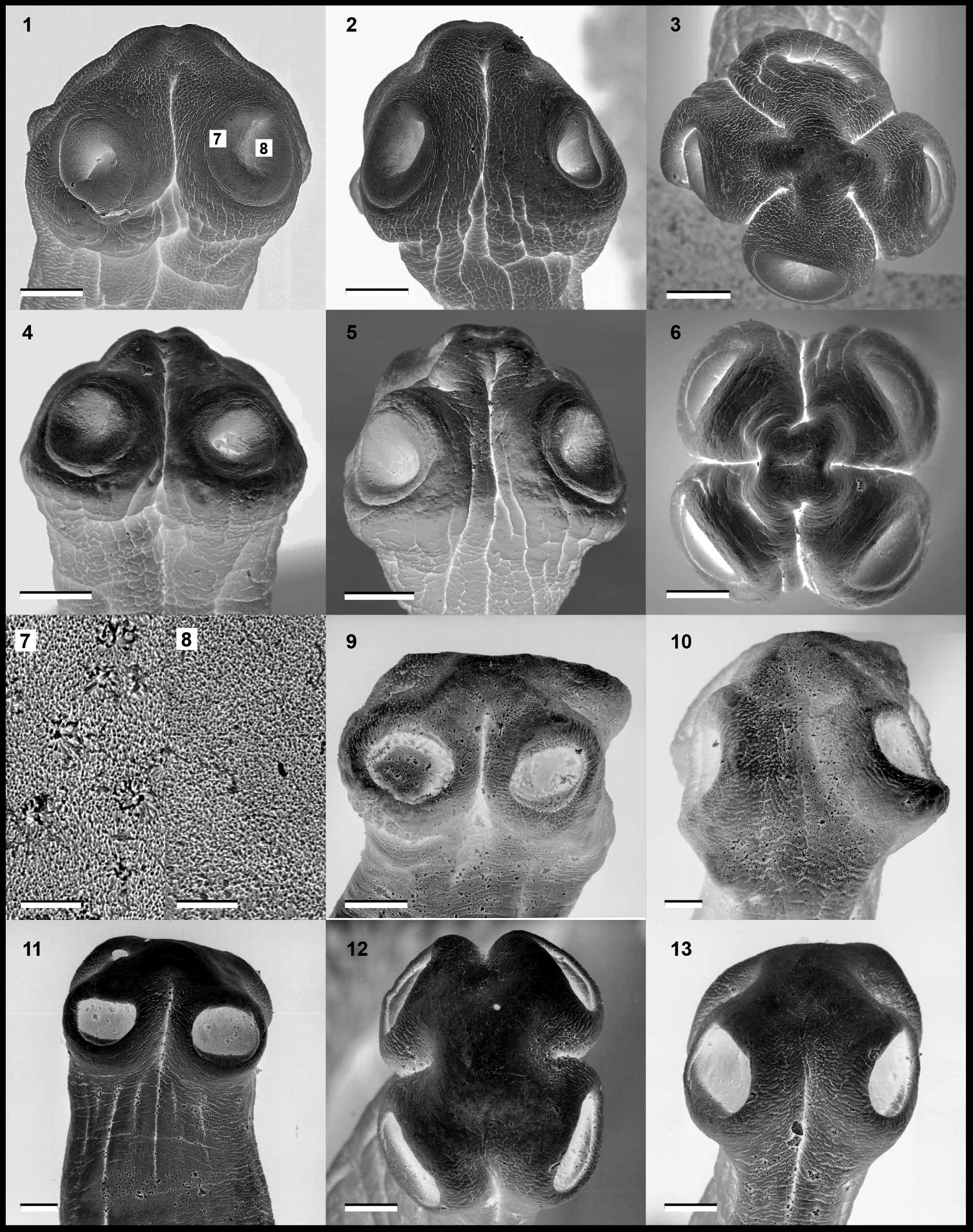
( Figures 9–13 View FIGURES 1 – 13 , 34, 35 View FIGURES 25 – 35 , 39, 40 View FIGURES 36 – 40 ; see also Troncy 1978 —figs. 6 [ P. membranacei ] & 7 [ P. largoproglottis ])
Syn.: Proteocephalus largoproglottis Troncy, 1978 (new synonym)
Type-and only host. Synodontis membranacea (Geoffrey Saint Hillaire, 1809) ( Siluriformes : Mochokidae ).
Type locality. Lake Chad in Ndjamena, Chad.
Type material. MNHNP (1116H).
Geographical distribution. Lake Chad.
Redescription. Based on type specimens; measurements of P. largoproglottis in parentheses. Proteocephalidae, Proteocephalinae. Strobila with acraspedote proglottides, 154–162 (70–86) mm long and up to 2.8 (1.6) mm wide, consisting of 110–121 (138) proglottides: 57–66 (71) immature, only 1–3 (2) mature, 25–27 (22) pregravid and 14–38 (43) gravid. Proglottides variable in shape, from almost rectangular to much longer than wide (much wider than long to slightly longer than wide in gravid proglottides).
Longitudinal internal musculature well developed, forming wide band of isolated muscle fibres. Osmoregulatory canals, including ventral ones, difficult to observe, visible only in some cross sections of mature proglottides ( Fig. 34 View FIGURES 25 – 35 ).
Scolex unarmed, slightly wider than neck, 500–525 (600) wide, rounded ( Figs. 9–11, 13 View FIGURES 1 – 13 ), with four uniloculate suckers, opening sublaterally on dorsal and ventral sides of scolex ( Figs. 10–13 View FIGURES 1 – 13 ). Apical organ widely oval, thinwalled, 35–50 (35–40) long and 30–35 (30–35) wide, difficult to observe due to high concentration of glands cells in apex of scolex. Proliferative zone 0.4–1.6 mm long and 270–375 wide. Scolex and proliferative zone uniformly covered with short, dense papilliform filitriches.
Testes medullary, spherical to oval, 65–85 long and 50–65 wide, 200–250 (150–200) in number according to original description (precise number could not be counted reliably in type specimens), usually in one layer, with a few testes in second incomplete layer in cross section ( Fig. 35 View FIGURES 25 – 35 ). Testes forming two fields confluent anteriorly, present also in gravid proglottides. External vas deferens coiled, occupying narrow field, reaching, but never overlapping, midline of proglottis aporally. Cirrus-sac elongated, thin-walled, 130–160 (210–270) long and 60–100 wide (length/width ratio = 2.5–4.1). RLCS = 12–16% (14–17%). Internal vas deferens thin-walled; ejaculatory duct thick-walled, long, forming several loops; cirrus unarmed, long, may occupy more than half-length of cirrussac. Genital pore irregularly alternating, almost equatorial, situated at 42–47% (46–52%) of proglottis length. Genital atrium shallow.
Ovary medullary, bilobed. OV = 55–59% (56–60%). Mehlis’ glands about 100–105 in diameter. Vagina anterior 60% (40%) or posterior 40% (60%; n = 40) to cirrus-sac, with concentration of chromophilic cells in its distal part, surrounded by weakly developed muscles fibres near genital pore. Vitelline follicles medullary in two longitudinal bands on both sides of proglottis, occupying almost its total length.
Uterus medullary, with type 1 development according to de Chambrier et al. (2004), with lumen of uterus appearing in last immature proglottides, gradually extending to form tubular structure. Eggs appear simultaneously with formation of lateral, thick-walled diverticula lined with chromophilic cells. In gravid proglottides, lateral diverticula remain thin-walled, 15–23 (9–16) in number on each side, occupying up to 71% of proglottis width; they open by two or three uterine longitudinal orifices.
ratio of the length of the cirrus–sac to the width of the proglottis (in %); 2 ratio of the total width of the ovary to the width of the proglottis (in %); 3 ratio of the distance of the genital from the anterior margin of the proglottis to the total length of the proglottis (in %); 4 anterior/posterior position of the distal part of the vagina to the cirrus–sac (expressed in % as minimum–maximum for individual specimens); 5 number of diverticula on each side. Number of testes of P. membranacei and P. largoproglottis after Troncy (1978).
Eggs with membraneous outer envelope with paired lateral auricular projections ( Figs. 39, 40 View FIGURES 36 – 40 ), 47–58 long and 25–27 wide; embryophore thick, spherical to slightly subspherical, long 23–25 long and 21–24 wide, containing granular, usually incomplete internal envelope ( Figs. 39, 40 View FIGURES 36 – 40 ); oncospheres oval, 12–15 long and 10–12 wide, with six embryonic hooks about 6 long ( Figs. 39, 40 View FIGURES 36 – 40 ).
Remarks. Quality of type material of both species described by Troncy (1978), i.e. Proteocephalus largoproglottis and P. membranacei (MNHNP 1115H and 1116H), is poor and it was not possible to use it for making new illustrations ( Troncy 1978 provided only a few line drawings of both species—see his figs. 6 & 7). However, it was possible to observe internal morphology and to reveal that both taxa are indistinguishable in all but one morphological characteristics. Among others, they share an identically shaped scolex, with a very small, thin-walled apical organ and a similar structure of the inner longitudinal musculature; their osmoregulatory canals, including ventral ones, are indistinguishable in mature, pregravid and gravid proglottides, and their eggs with lateral processes are also identical.
Both species only differ by the shape of their proglottides, those of P. membranacei being elongate, much longer than wide, whereas those of P. largoproglottis being short and wide (see figs. 6A, B and 7A, B, D in Troncy 1978). However, this difference seems to reflect a different state of contraction of the worms when they were fixed. The tapeworms described as P. membranacei were probably already dead when fixed, because they were too relaxed and thus became more elongate; their internal organs are difficult to observe because of decomposition of the tissues. Troncy (1978) admitted poor quality of these specimens (“État de conservation médiocre”). In contrast, worms described as P. largoproglottis are strongly contracted and thus their proglottides are much wider than long. Contraction of the material was also mentioned by Troncy (1978—“Les spécimens sont en général rétractés”).
Previous studies on Proteocephalus tapeworms (e.g. those parasitic in European fish– Scholz and Hanzelová, 1998), revealed a considerable intraspecific variability of several taxa in proglottis shape. Similarly, proglottides of congeneric cestodes from African catfish, namely P. glanduligerus from Clarias spp. (Scholz et al. 2009) and P. synodontis (this study) were highly variable in their shape (even in the same worm). Therefore, the shape of proglottides is not considered to represent a character that would justify the validity of the tapeworms described by Troncy (1978) as two separate species. As a result, P. largoproglottis , the description of which appeared later (pp. 542–546), is synonymized with P. membranacei , which was described first (pp. 538–542).


No known copyright restrictions apply. See Agosti, D., Egloff, W., 2009. Taxonomic information exchange and copyright: the Plazi approach. BMC Research Notes 2009, 2:53 for further explanation.