
Psednos microstomus, Stein, David L., 2012
|
publication ID |
https://doi.org/ 10.5281/zenodo.283120 |
|
DOI |
https://doi.org/10.5281/zenodo.6173286 |
|
persistent identifier |
https://treatment.plazi.org/id/03B487D1-FFBD-AF75-FF6F-10C2FE79168F |
|
treatment provided by |
Plazi |
|
scientific name |
Psednos microstomus |
| status |
sp. nov. |
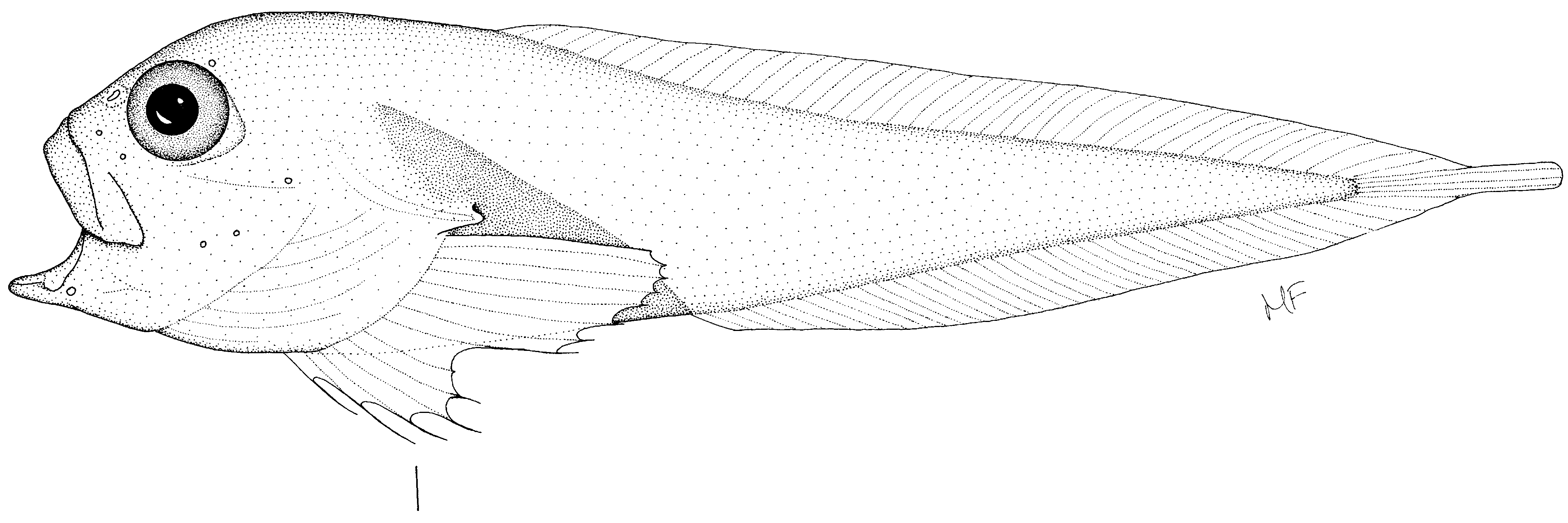
Psednos microstomus View in CoL n. sp.
Figs. 31 View FIGURE 31 , 32 View FIGURE 32
Holotype. NMNZ P.039410, ripe female, 34 mm SL, 39 mm TL, 33°30.09' S, 170°01.45' E, R/V Tangaroa, Stn. TAN 0308/129, Reinga Ridge ( NZ EEZ), 31 May 2003, 1158– 1230 m.
Diagnosis. Vertebrae 46, dorsal fin rays 43. Mouth small, upper jaw ~26% HL, its angle 70–80 degrees. Mandibular teeth small, simple sharp canines; premaxillary teeth tiny, irregularly arranged, near symphysis only. Opercular flap small, gill opening short. Body not humpbacked, spine straight.
Description. Counts: V 46, D 43, A ~37, C 5, P 14, radials unknown, gr unknown, pc unknown. Ratios: HL 32.5% SL, sn 7.5, E 8.9, orbit ~9.5, uj 8.4, lj 7.5, po 17.6, go 2.7, bd 26.9, bdA 14.9, preD 31.6, preA 50.7, sna 31.3, aAf 30.4. In % HL: sn 22.9, E 27.5, orbit ~29.4, uj 25.7, lj 22.9, go 8.2, po 54.1, bd 82.6, bdA 45.9, preD 97.2, preA 156.0, sna 96.3, aAf 93.6.
Head about 1/3 SL, dorsal profile rising gradually to occiput; snout short, not protruding anteriorly. Nostrils single, nasal rosette large, anterior to dorsal half of eye, almost touching orbit. Mouth terminal, small; when open, mouth opening almost round, retroarticular angle not obvious. Premaxillary teeth tiny, sharp canines arranged anteriorly in irregular rows forming a narrow band less than three teeth wide, posteriorly irregularly bi- or uniserial; difficult to find and see. Premaxillary symphyseal gap present, wide, a shallow symphyseal notch barely present. Mandibular teeth distinctly larger, smallest conical, gradually becoming larger medially. Largest teeth stout and sharp but not conical. Teeth arranged in about 20 oblique rows of up to 7 teeth each, forming a narrow band. Symphyseal gap absent. Orbits large, eyes prominent, not entering dorsal profile of head but forming part of interorbital region of head. Interorbital space damaged, apparently less than eye diameter. Gill flap triangular, tiny, gill opening short, almost pore like, flap oriented horizontally above dorsalmost pectoral fin ray and posterior to its base. Opercle long, slender, curved gradually over 70–80°, its posterior end sharp, visible through skin of opercular flap. Branchial cavity supported by six long, clearly visible branchiostegal rays curving posterodorsally. Symphyseal pores small, fragile, distinctly separated by a distance of about two pore diameters. Other cephalic pores damaged, not found.
Pectoral fin upper ray about even with horizontal through posterior of oral cleft. Pectoral fin base low on body, about even with posterior end of upper jaw and not far anterior to a vertical through dorsal fin origin. Pectoral fin rays 8+1+5, upper lobe rays closely spaced, notch deep with a single ray distinctly more widely spaced dorsally and ventrally from upper and lower lobe rays. Rudimentary rays absent. Longest rays in each lobe slender, fragile, their ends apparently free. Pectoral girdle not examined.
Trunk of body not dorsally curved or humpbacked, abdominal vertebrae forming a shallow, rather than a strong dorsal curve. Body deepest just behind orbit, stout, tapering evenly and rapidly to caudal fin. Vertebrae 10+36; on radiograph, first neural spine appears double; remaining neural spines single. Dorsal fin origin far posterior, between vertebrae 6–7, well behind head and base of pectoral fin, almost at posterior end of upper pectoral lobe, slightly anterior to posterior end of abdominal cavity. Origin of anal fin between vertebrae 10–11, distinctly posterior to end of peritoneum and body cavity rather than at the end as usual in other species; preanal fin length almost exactly half SL. Anus position about 30% SL anterior to anal fin origin, about below middle of pectoral fin base and anterior part of gill opening. Abdominal cavity long, triangular in side view, extending about 1.2 times head length behind pectoral symphysis. Internal organs incompletely examined owing to specimen fragility and size. Hypural elements fused, caudal fin of five (3/2) rays, about 50% overlapped by terminal dorsal and anal fin rays.
Fresh color of head including mouth, branchial cavity, and abdomen, black except for occipital region, remainder of remaining skin transparent, musculature visible through skin. Body (musculature) color in alcohol tan, mouth and tongue black, branchiostegal membranes dark brown, peritoneum black, visible through whitish body wall; rectal portion of intestine brown. Stomach pale, pyloric caeca unknown.
The specimen is apparently ripe or nearly ripe, with a few (ca 10?) large eggs about 1.7 mm diameter.
Distribution. Known only from the holotype, taken to the northwest of New Zealand at about 1158–1230 m.
Etymology. The specific epithet microstomus from the Greek mikros, small, and stoma, mouth, denoting the small mouth of the species.
Comparisons. Psednos microstomus differs distinctly from all other Psednos described herein. Not only its small mouth (upper jaw 26 vs longer than 36% HL) but also its short spine (vertebrae 46, 10 abdominal, vs 53 or more, 10 or more abdominal) and consequent few dorsal and anal fin rays (43, ~37 vs 45, 40 or more), its tiny teeth visible only at high magnifications (vs small teeth more easily seen), straight spine and consequently absent hump (vs anteriorly curved spine), black orobranchial cavity (vs dusky or brown dotted) shallow body depth (83 vs 120% HL), and many other proportions set it apart. In appearance it is similar to P. carolinae Stein 2005 , but differs in number of vertebrae (49 vs 38), number of dorsal and anal fin rays (43, ~37 vs 33, 26), tooth arrangement (banded vs biserial), and many more characters. In number of vertebrae and caudal fin rays, it is similar to P. cf. dentatus from Chile ( Stein 2005), but it differs in almost all other available characters: mouth angle (70–80° vs 50°), head length (33 vs 23% SL), upper jaw length (26 vs 45% HL), predorsal length (97 vs 120% HL), and stomach color (pale vs brown).
Comments. The unusual morphology of P. microstomus emphasizes the need for a thorough review of Psednos . The number of known species has grown rapidly in the past decade (from five to 37 including those in this paper). The characters originally defining the genus were presence of an oblique mouth, humped spinal column, disconnected infraorbital and temporal sensory canals, and coronal and additional temporal pores. Now, there appear to be only two defining characters: the oblique mouth and lack of the sensory canal connection. Only the second of these appears to be unique to the genus; several species of Paraliparis also have oblique mouths. Psednos species are also apparently holopelagic and of small size, but these last two characteristics also occur in some Paraliparis species and in several other genera ( Nectoliparis Gilbert and Burke 1912 , Lipariscus Gilbert 1915 ). Psednos appears to include at least three species groups, and some species lack both the coronal pore and the occipital hump. Analysis of all the species could clarify the relationships of the group and lead to an improved definition of the genus.

| NMNZ |
Museum of New Zealand Te Papa Tongarewa |
No known copyright restrictions apply. See Agosti, D., Egloff, W., 2009. Taxonomic information exchange and copyright: the Plazi approach. BMC Research Notes 2009, 2:53 for further explanation.