
Protoneura sanguinipes Westfall, 1987
|
publication ID |
https://doi.org/ 10.11646/zootaxa.4361.1.1 |
|
publication LSID |
lsid:zoobank.org:pub:53489D29-C68F-44FD-9EA2-CFCA7B949630 |
|
DOI |
https://doi.org/10.5281/zenodo.6030159 |
|
persistent identifier |
https://treatment.plazi.org/id/D2332A59-FF8C-4E74-FF5D-FEE4FAAE697A |
|
treatment provided by |
Plazi |
|
scientific name |
Protoneura sanguinipes Westfall, 1987 |
| status |
|
Protoneura sanguinipes Westfall, 1987 View in CoL
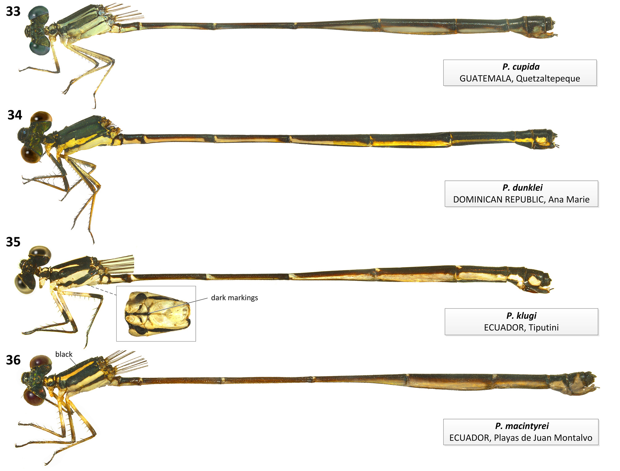
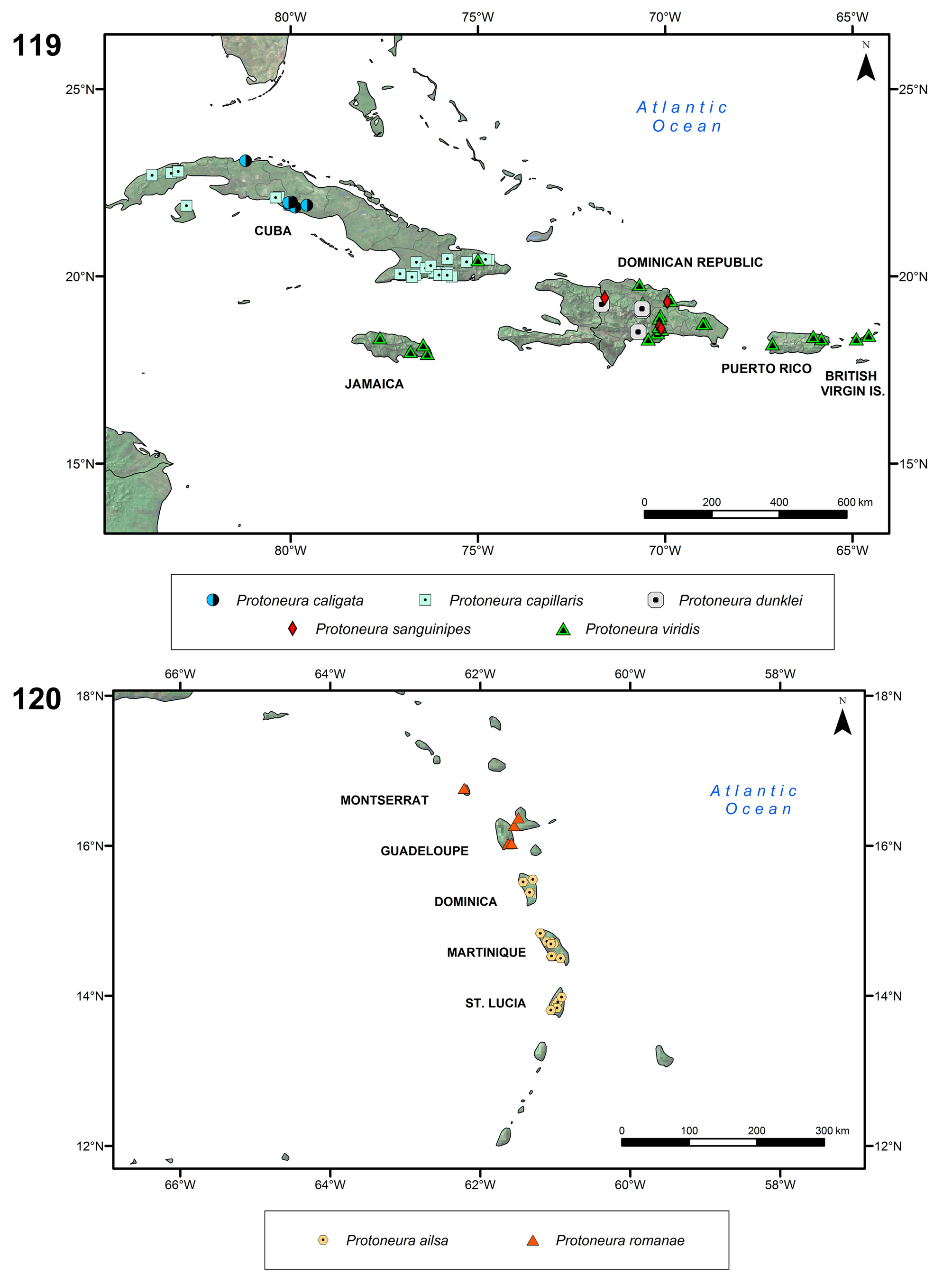
Figs. 17 View FIGURES17–20 (♂ habitus), 41 (♀ habitus), 63 (♀ mes. plate), 87 (gen. lig.), 113 (♂ app.), 119 (map)
Protoneura sanguinipes Westfall, 1987: 93 View in CoL –97, Figs. 1–4 View FIGURES 1–4 (description of ♂ and ♀, illustrations of ♂ appendages and ♀ mesostigmal plates);— Paulson (2004: 176; listed as range restricted);— Flint et al. (2006: 73; Dominican Republic);— Westfall & May (2006: 431, 433–435, 439, 440, Figs. 225B, 226F; in key to northern representatives of genus, characterization of adults, illustrations of ♂ S10, ♀ pronotum and mesostigmal plates);— Paulson (2009f; IUCN assessment);— Garrison et al. (2010: 379, 381, Figs. 2515, 2517; illustrations ♂ S10, ♀ S8–10);— Meurgey (2013: 300, 306; distribution).
Primary types. Holotype ♂. Arroyo Bermejo, 4 km NNE of Hatillo and Autopista Duarte, Distrito Nacional, Distrito Nacional, 10 viii 1983, R.W. Garrison leg. [ FSCA # 727 View Materials ].
Specimens examined. 19 ♂, 23 ♀: DOMINICAN REPUBLIC : 1 ♂ 1 ♀, A. Busck leg. [ USNM]; Prov. Dajabón : 1 ♂ 2 ♀ (paratypes), Masacre River, Balneario El Salto, Loma de Cabrera {19°25' N, 71°36'51'' W, 252 m}, 20–23 v 1973, D. & M. Davis leg. [ FSCA]; Distrito Nacional GoogleMaps : 1 ♂ 1 ♀, Isabela River, about 2 km E of Berrio (18°39'49'' N, 70°9'28'' W), 10 viii 2001, D.E. Perez-Gelabert leg. [ USNM] GoogleMaps ; 8 ♂ 11 ♀ paratypes (8 pairs in tandem), Arroyo Bermejo, 4 km NNE of Hatillo , Autopista Duarte (18°36'2'' N, 70°6'16'' W, 90 m), 10 viii 1983, R.W. Garrison leg. [RWG] GoogleMaps ; 1 ♂ 1 f # paratypes (in tandem), same data but [ CSCA] GoogleMaps ; 3 ♂ 3 ♀, paratypes (in tandem), same data but [ FSCA] GoogleMaps ; 1 ♂ 1 ♀ paratypes (in tandem), same data but [ UMMZ] GoogleMaps ; 1 ♂ 1 ♀, paratypes (in tandem), same data but [ USNM] GoogleMaps ; 1 ♂ 1 ♀, same data but 6 vi 1989, J.J. Daigle leg. [RWG] GoogleMaps ; 1 ♂ 1 ♀, same data but [ USNM]. GoogleMaps
Characterization. Male: Epicranium and dorsum of thorax dark metallic copper with green reflections, dorsum of S1, distal ring on S2–5, and S6–10 black, remainder orange-brown, with a dorsal orange spot on S9 distal half; pale colors reddish orange and yellow ( Fig. 17 View FIGURES17–20 ). Pronotum black with lateral corner of middle lobe orange red. Mesepisternum and mesepimeron dark metallic copper; metepisternum dark with large yellow spot on ventral half; metepimeron pale yellow with anterodorsal corner black. Coxa and trochanter dark with margins orange; remainder of leg entirely reddish orange; legs and venter of thorax pruinose; tibial spurs shorter than twice intervening spaces. Genital ligula lacking lateral lobes, with a triangularly convex distal margin and laterodistal corners of distal segment not projected ventrally ( Fig. 87 View FIGURES 85–91 ). Cercus about as long as S10 length and paraproct, longer than wide in lateral view, with a longitudinal split along dorsal portion of external surface, delimiting a medial sclerotized branch which ends on a broad triangular tip directed medioventrally located at about midlength of cercus ( Fig. 113 View FIGURES 112–114 ); remainder of cercus foliaceous, medially concave, with a small triangular tooth at ventrobasal edge, and a long, curved thick pointed tooth at mediobasal edge directed medioposteriorly. Paraproct about as long as S10 length, narrowing to half its basal width at distal 1/3, and concave medially ( Fig. 113 View FIGURES 112–114 ). TL 37–38.5; Hw 18– 18.5.
Female: As male but dark color black with metallic green to copper reflections, pale areas orange or yellow; pronotum black with margins yellow; legs with coxae, trochanters, and base of outer surface of femora yellow, extensor surface of femora dark brown, remainder of legs pale orange; pale lateral area of S9 extended dorsally medially to about 1/2 of segment height ( Fig. 41 View FIGURES 41–44 ). Middle lobe of pronotum lacking pronounced lateral depressions; posterior lobe directed dorsally, trilobed with medial lobe entire and smoothly convex ( Fig. 63a View FIGURES 60–63 ). Mesostigmal plate oval with medial margin elevated and with a shallow central concavity; mesanepisterum with oblique carina arising from posteromedial corner of mesostigmal plate and ending midway to middorsal carina ( Fig. 63b View FIGURES 60–63 ). TL 32.5–36; Hw 17.5–21.
Diagnosis. Male cercus morphology, with a medial sclerotized branch which ends on a tooth directed medioventrally and remainder of cercus foliaceous, medially concave, with a small triangular tooth on outer ventrobasal edge, and a long, curved thick pointed tooth at medial ventrobasal edge directed medially ( Figs. 113 View FIGURES 112–114 ), is shared with P. caligata , P. capillaris , P. dunklei and P. viridis ( Figs. 100 View FIGURES 100–102 ; 102; 106; 117). Within this group, P.
sanguinipes resembles P. dunklei by the medial sclerotized branch of male cercus ending on a broad triangular tip located at about midlength of cercus ( Figs. 106 View FIGURES 106–108 ; 113) and by distal margin of genital ligula triangularly convex with laterodistal corners not projected ventrally ( Figs. 80 View FIGURES 79–84 ; 87); in P. caligata , P. capillaris , and P. viridis the medial branch ends on a tooth directed medioventrally located at cercus tip ( Figs. 100 View FIGURES 100–102 ; 102; 106; 117), and distal margin of genital ligula is straight with laterodistal corners projected ventrally ( Figs. 74 View FIGURES 71–78 ; 76; 93). Protoneura sanguinipes differs from males of all four species by the entirely reddish orange femora, tibiae and tarsi ( Fig. 17 View FIGURES17–20 ), which have various extents of dark colors in the other species ( Figs. 4 View FIGURES 1–4 ; 6; 21). Female can be distinguished from all congeners except P. dunklei by its trilobed posterior lobe of pronotum directed dorsally and with medial lobe entire and smoothly convex ( Fig. 63a View FIGURES 60–63 ) and by the oval mesostigmal plate ( Fig. 63b View FIGURES 60–63 ). It differs from P. dunklei by flexor surface of tibiae pale ( Fig. 41 View FIGURES 41–44 ; dark in P. dunklei , Fig. 34 View FIGURES 33–36 ).
Habitat and biology. Females oviposit on sedges near banks while still in tandem with males ( Westfall 1987). Distribution. Dominican Republic ( Fig. 119 View FIGURES 119–120 ). Assessed as Endangered by IUCN ( Paulson 2009f).


No known copyright restrictions apply. See Agosti, D., Egloff, W., 2009. Taxonomic information exchange and copyright: the Plazi approach. BMC Research Notes 2009, 2:53 for further explanation.
|
Kingdom |
|
|
Phylum |
|
|
Class |
|
|
Order |
|
|
Family |
|
|
Genus |
Protoneura sanguinipes Westfall, 1987
| Ellenrieder, Natalia Von & Garrison, Rosser W. 2017 |
Protoneura sanguinipes
| Garrison 2010: 379 |
| Flint 2006: 73 |
| Westfall 1987: 93 |