
Protoneura dunklei Daigle, 1990
|
publication ID |
https://doi.org/ 10.11646/zootaxa.4361.1.1 |
|
publication LSID |
lsid:zoobank.org:pub:53489D29-C68F-44FD-9EA2-CFCA7B949630 |
|
DOI |
https://doi.org/10.5281/zenodo.6030145 |
|
persistent identifier |
https://treatment.plazi.org/id/D2332A59-FFB6-4E4E-FF5D-F92FFA976CFF |
|
treatment provided by |
Plazi |
|
scientific name |
Protoneura dunklei Daigle, 1990 |
| status |
|
Protoneura dunklei Daigle, 1990 View in CoL
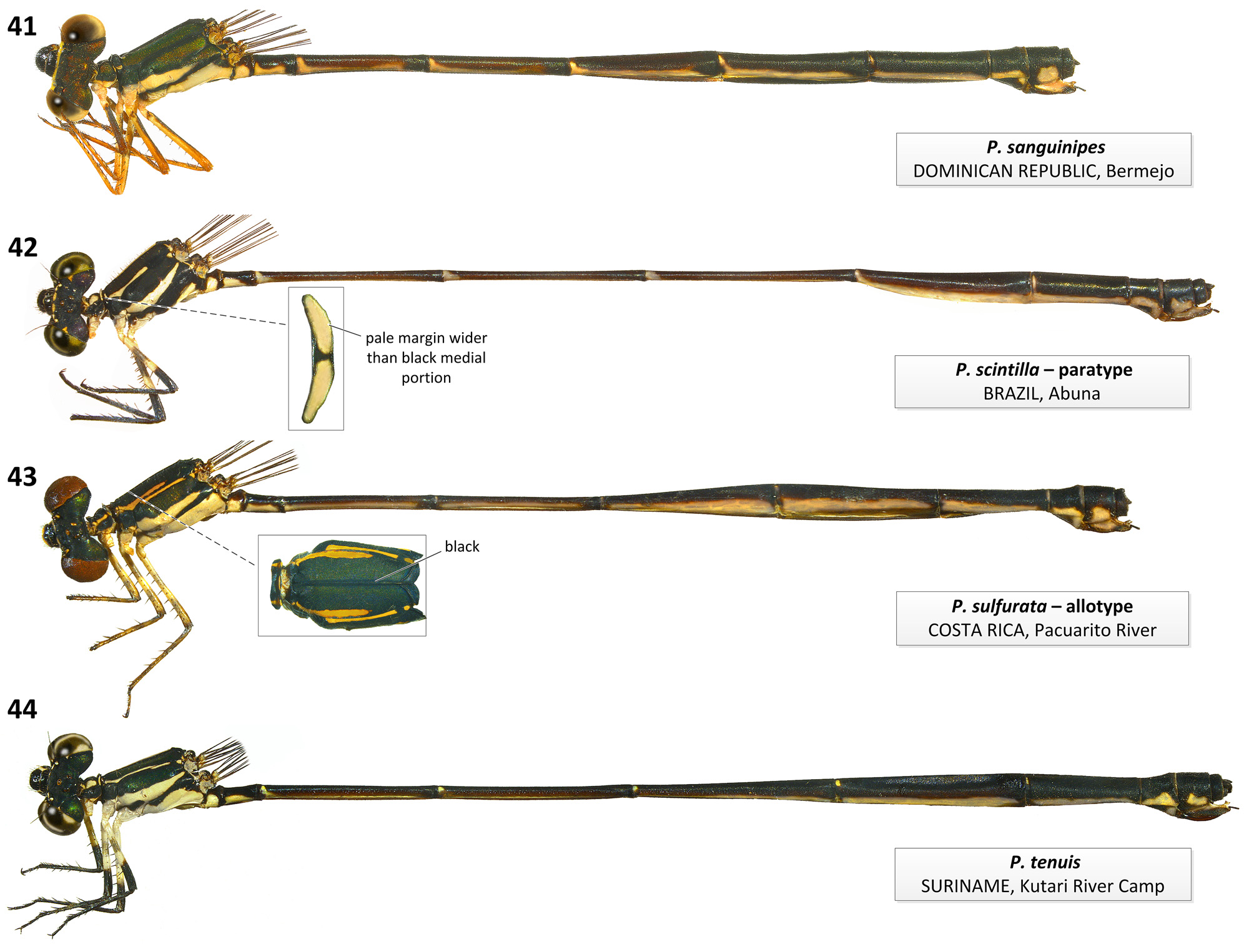
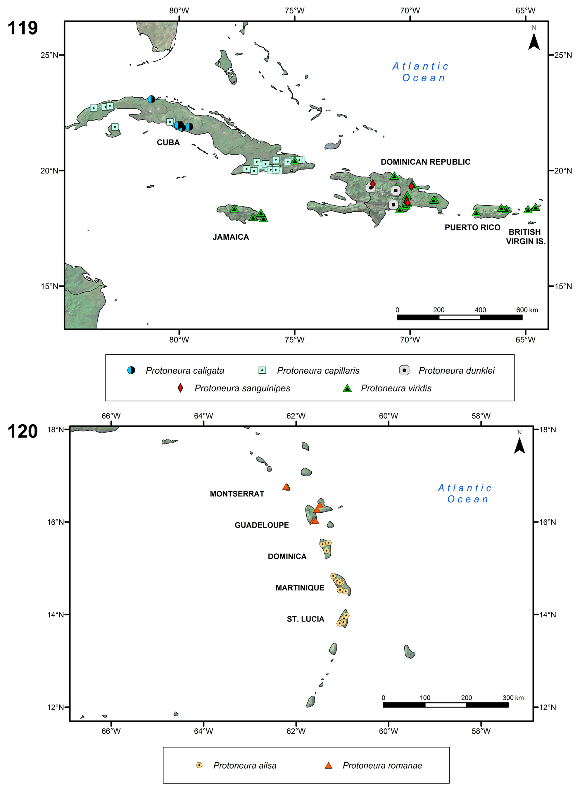
Figs. 10 View FIGURES 9–12 (♂ habitus), 34 (♀ habitus), 56 (♀ mes. plate), 80 (gen. lig.), 106 (♂ app.), 119 (map)
Protoneura dunklei Daigle, 1990: 81 View in CoL –83, Figs. 1–4 View FIGURES 1–4 (description of ♂ and ♀, illustrations of ♂ S10 and ♀ mesostigmal plates, diagnosis from P. sanguinipes View in CoL );— Paulson (2004: 176; listed as range restricted);— Flint et al. (2006: 73; Dominican Republic);— Westfall & May (2006: 431, 433–435, 439, 440, Figs. 225A, 226E; in key to northern representatives of genus, characterization of adults, illustrations of ♂ S10, ♀ pronotum and mesostigmal plates);— Pessacq (2008: 527; in phylogenetic analysis);— Paulson (2009e; IUCN assessment);— Garrison et al. (2010: 379);— Meurgey (2013: 300, 306; distribution).
Protoneura sanguinipes nec Westfall, 1987 — Westfall (1987: 94; paratype 798 from Villa Anacaona). Misidentification.
Primary types. Holotype ♂. Dominican Republic, La Vega Province, Arroyo Ana Marie , 8 vi 1989, S.W. Dunkle leg. [ FSCA].
Specimens examined. 15 ♂ 11 ♀: DOMINICAN REPUBLIC, Dajabón Prov. : 1 ♂ paratype, Villa Anacaona, 3 vi 1986, R. Miller & L. Stange leg. {19°15' N, 71°42' W} [ FSCA]; La Vega Prov. GoogleMaps : 3 ♂ 2 ♀, paratypes, cloud forest 10.9 km SE of highway Duarte on road to Jarabacoa, on grounds of trickles of Vacational Turístico Racquet Club about 0.7 km from entrance (19°8'3'' N, 70°37'11'' W, 552 m), 8–9 vi 1989, S.W. Dunkle leg. [FSCA]; 4 ♂ 2 ♀, same data but 24–28 v 1991 [ FSCA] GoogleMaps ; 1 ♂ 1 ♀ paratypes, same data but Ana Marie stream, 8 vi 1989, S.W. Dunkle leg. {19°6' N, 70°36' W} [RWG] GoogleMaps ; 3 ♂ 3 ♀, paratypes, same data but [RWG] GoogleMaps ; 1 ♂ 1 ♀, paratypes [USNM]; 1 ♂ 1 ♀, paratypes, same data but 9 vi 1989, J.J. Daigle leg. [RWG] GoogleMaps ; 1 ♂, Salto Baiguate, 5 km S of Jarabacoa (19°5'33'' N, 70°36'55'' W, 580 m), 9 v 1995, O.S. Flint Jr. leg. [ USNM] GoogleMaps ; 1 ♀, small stream 5 km SE of Jarabacoa on road to El Río, 29 v 1989, S.W. Dunkle leg. [ FSCA].
Characterization. Male: Epicranium and dorsum of thorax black with metallic purple reflections, abdomen black except for very narrow basal pale orange ring and middorsal hair thin longitudinal stripe on S3–7 and pair of small dorsolateral orange spots on S9; pale colors reddish orange, orange and yellow ( Fig. 10 View FIGURES 9–12 ). Pronotum black with lateral margin of middle lobe orange. Mesepisternum and mesepimeron black; metepisternum black with large orange to yellow spot on ventral half; metepimeron pale yellow with anterior and dorsal margins black. Coxa and trochanter black with margins orange, basal 5/6 of outer surface of femur black, distal 1/6 of femur and outer surface of tibia orange-red; tibial spurs shorter than twice intervening spaces. Genital ligula lacking lateral lobes, with a triangularly convex distal margin and laterodistal corners of distal segment not projected ventrally ( Fig. 80 View FIGURES 79–84 ). Cercus about as long as S10 length and paraproct, longer than wide in lateral view, with a longitudinal split along dorsal portion of external surface, delimiting a medial sclerotized branch which ends on a broad triangular tip directed medioventrally located at about midlength of cercus ( Fig. 106 View FIGURES 106–108 ); remainder of cercus foliaceous, medially concave, with a small triangular tooth at ventrobasal edge, and a long, curved thick pointed tooth at mediobasal edge directed medioposteriorly. Paraproct about as long as S10 length, narrowing to half its basal width at distal 1/ 3, and concave medially ( Fig. 106 View FIGURES 106–108 ). TL 41–43; Hw 20–22.
Female: As male but black color with metallic green to copper reflections, pale areas yellow; pronotum black with margins yellow; with a yellow spot on anterodorsal edge of mesepimeron and metepimeron; legs with coxae, trochanters, and base of outer surface of femora yellow, most of femora dark brown, tibiae pale brown; pale lateral area of S9 extended dorsally medially to about 1/3 of segment height ( Fig. 34 View FIGURES 33–36 ). Middle lobe of pronotum lacking pronounced lateral depressions; posterior lobe directed dorsally, trilobed with medial lobe entire and smoothly convex. Mesostigmal plate oval with medial margin elevated and with a shallow central concavity; mesanepisterum with oblique carina arising from posteromedial corner of mesostigmal plate and ending midway to middorsal carina ( Fig. 56 View FIGURES 55–59 ). TL 38–38.5; Hw 21.5–22.5.
Diagnosis. Male cercus, with a medial sclerotized branch which ends on a tooth directed medioventrally and remainder of cercus foliaceous, medially concave, with a small triangular tooth on outer ventrobasal edge, and a long, curved thick pointed tooth at medial ventrobasal edge directed medially ( Fig. 106 View FIGURES 106–108 ), is shared with P. caligata , P. capillaris , P. sanguinipes , and P. viridis ( Figs. 100 View FIGURES 100–102 ; 102; 113; 117). Within this group, P. dunklei resembles P. sanguinipes by the medial sclerotized branch of male cercus ending on a broad triangular tip located at about midlength of cercus ( Figs. 106 View FIGURES 106–108 ; 113) and by distal margin of genital ligula triangularly convex with laterodistal corners not projected ventrally ( Figs. 80 View FIGURES 79–84 ; 87); in P. caligata , P. capillaris , and P. viridis the medial branch ends on a tooth directed medioventrally located at cercus tip ( Figs. 100 View FIGURES 100–102 ; 102; 117), and distal margin of genital ligula is straight with laterodistal corners projected ventrally ( Figs. 74 View FIGURES 71–78 ; 76; 93). Protoneura dunklei differs from males of all four species by the black basal half of femora outer surface ( Fig. 10 View FIGURES 9–12 ), which is pale in the other species ( Figs. 4 View FIGURES 1–4 ; 6; 17; 21). Female can be distinguished from all congeners except P. sanguinipes by its trilobed posterior lobe of pronotum directed dorsally and with medial lobe entire and smoothly convex (as in Fig. 63a View FIGURES 60–63 ) and by oval mesostigmal plate ( Figs. 56 View FIGURES 55–59 ; 63b View FIGURES 60–63 ). It differs by flexor surface of tibiae dark ( Fig. 34 View FIGURES 33–36 ; pale in P. sanguinipes , Fig. 41 View FIGURES 41–44 ).
Habitat and biology. Seepage headwaters of small, forested montane streams, where males perch about 3 m from the ground on tips of leaves and branches of shrubs located in sunny openings along densely shaded streamsides, and tandem pairs hover over muddy masses of fibrous roots near water’s edge ( Daigle 1990).
Distribution. Dominican Republic ( Fig. 119 View FIGURES 119–120 ). Assessed as Least concern by IUCN ( Paulson 2009e).


No known copyright restrictions apply. See Agosti, D., Egloff, W., 2009. Taxonomic information exchange and copyright: the Plazi approach. BMC Research Notes 2009, 2:53 for further explanation.
|
Kingdom |
|
|
Phylum |
|
|
Class |
|
|
Order |
|
|
Family |
|
|
Genus |
Protoneura dunklei Daigle, 1990
| Ellenrieder, Natalia Von & Garrison, Rosser W. 2017 |
Protoneura dunklei
| Garrison 2010: 379 |
| Flint 2006: 73 |
| Daigle 1990: 81 |