
Platystigma martinezi (Machado, 1985) Machado, 1985
|
publication ID |
https://doi.org/ 10.11646/zootaxa.4242.3.4 |
|
publication LSID |
lsid:zoobank.org:pub:4FCEE7BF-C562-4550-9D42-F7B86678DBBB |
|
DOI |
https://doi.org/10.5281/zenodo.6042205 |
|
persistent identifier |
https://treatment.plazi.org/id/891587DD-FFC0-9B29-D2A2-FC62FD3C634A |
|
treatment provided by |
Plazi |
|
scientific name |
Platystigma martinezi (Machado, 1985) |
| status |
comb. nov. |
Platystigma martinezi (Machado, 1985) n. comb.
Figures 9 View FIGURES 8 – 13 , 15 View FIGURES 14 – 19 , 21 View FIGURES 20 – 24 , 38 View FIGURES 37 – 41 , 44, 45 View FIGURES 42 – 51 , 53–55 View FIGURES 52 – 53 View FIGURES 54 – 61
Mecistogaster martinezi Machado View in CoL (1985: 35, 854); Davies & Tobin (1984: 59); Garrison & Ellenrieder (2016: 20), Bridges (1994: VII–147); Tsuda (2000: 58); Corbet (2004: 44, 46, 146, 593, 596, 619); Heckman (2008: 207); Garrison, Ellenrieder & Louton (2010: 391); Schorr & Paulson (2016); Pinto (2016: 21).
Material examined. Holotype ♀ and two paratypes ♀: Bolivia . Department of Santa Cruz, Buena Vista, Ichilo Province (17° 27' 32" S, 63° 44' 07" W), Tacu Pallido, II.1951, A. Martinez leg. Additional ♂: Department of Santa Cruz, 450 m, February – April, no year. Types deposited in the ABMM Collection. Male deposited in FSCA. GoogleMaps
Redescription of the female holotype. Head: Labrum laterally yellow with large black spot occupying its middle 1/3. Anteclypeus brown, whitish yellow medially. Postclypeus black; base of mandible and genae yellow. Antefrons yellow with median black spot. Upper part of head black, antennal scape black surrounded with yellow at base. Area between vertex and eye with pair of yellow streaks. Rear of head yellow bordered with black posteriorly.
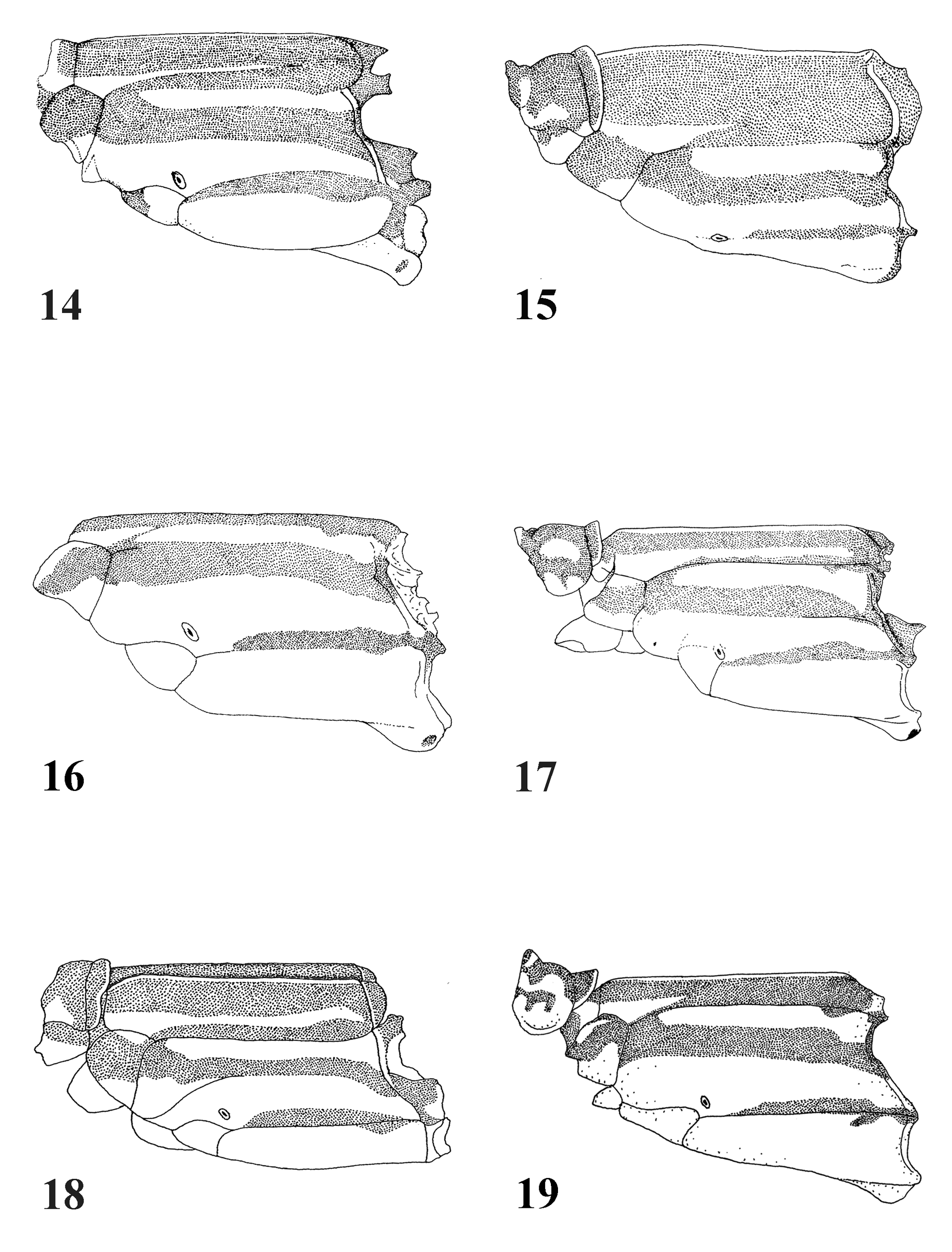
Thorax. Prothorax: Pronotum ( Fig. 9 View FIGURES 8 – 13 ) black surrounded by reddish-yellow stripe interrupted at anterior lobe. Posterior prothoracic lobe bordered with yellow. Propleuron reddish yellow ventrally, black dorsally. Pterothorax: Middorsal carina yellowish brown. Antehumeral pale stripe subtriangular, reaching up to 1/3 of mesepisternum ( Fig. 15 View FIGURES 14 – 19 ). Venter yellowish with anterior and posterior black spots ( Fig. 21 View FIGURES 20 – 24 ).
Wings. Both wings with apical milky-white area reaching level of IR 1 in Fw and IR 2 in Hw. At high saturation, white areas become pale blue. Pseudostigma poorly indicated in both wings with eight pale yellow cells in Fw and 13 in Hw, more intensely yellow at high saturation. Venation: Px in Fw 31, in Hw 29, RP2 arising at Px 12 in Fw, at Px 10 in Hw. IR1 arising at Px 18 in Fw, at Px 15 in Hw. RP2 branching from RP at less than half distance from subnodus to tip of wing in both wings. Petiolation in Fw originating distally to CuP by a distance about the length of CuP; in Hw about 1½ time this length.
Legs. Fore legs missing. Middle and hind legs with extensor surface of femora light brown, black distally. Tibiae with extensor surface yellowish brown, flexural surface black. Tarsi black.
Abdomen. S1–9 dorsally black, laterally brownish yellow, S10 and cerci black. Ovipositor mainly brownish yellow, lateral valve black.
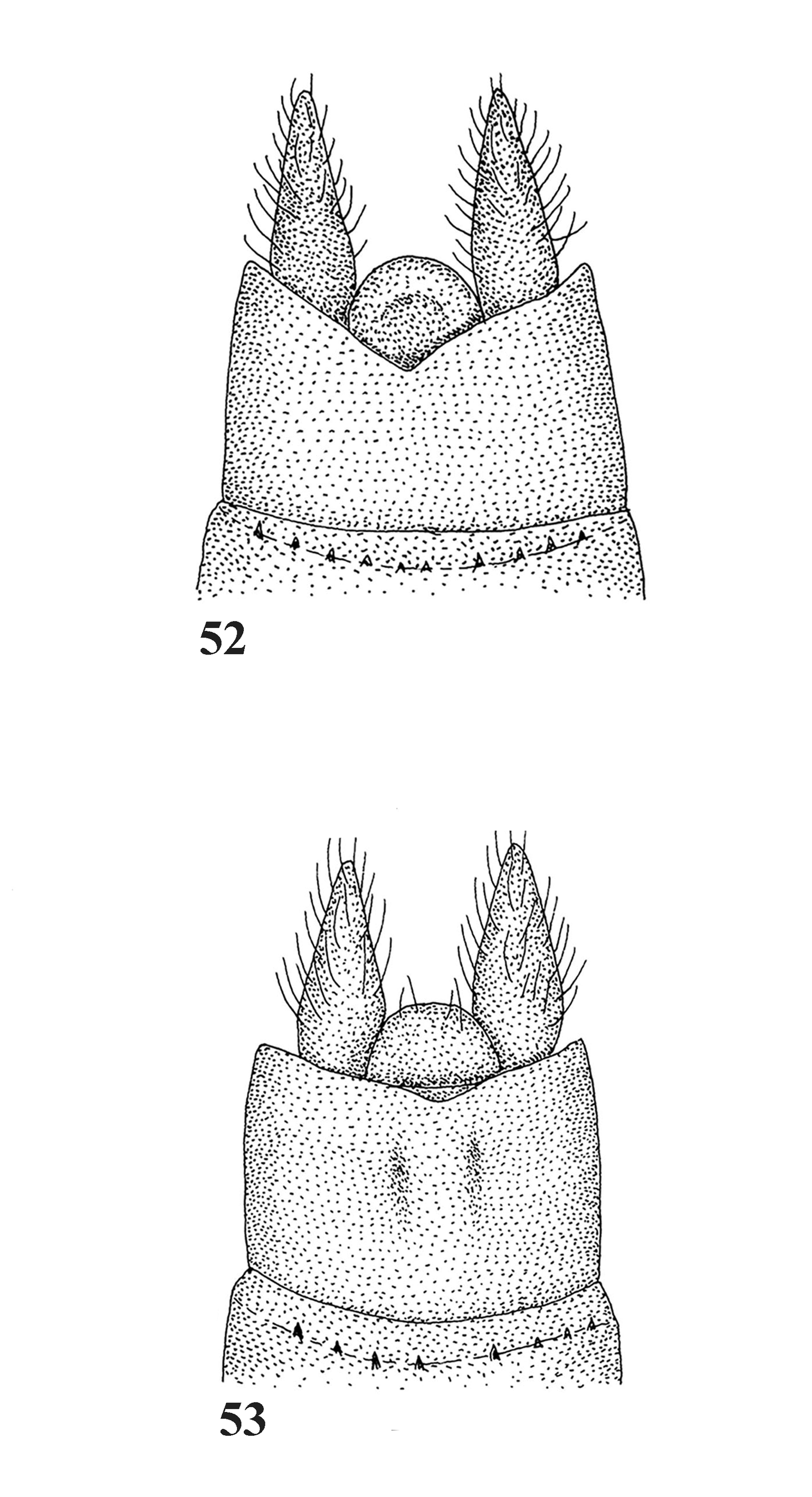
Structural characters. Median and lateral portions of posterior prothoracic lobe not separated and its posterior border almost straight ( Fig. 9 View FIGURES 8 – 13 ). Supplementary tooth of tarsal claws absent. Cerci shorter than S 10 in dorsal view ( Fig. 53 View FIGURES 52 – 53 ), parallel, conical, with many white hairs. Ovipositor short, extending posteriorlly to level of hind border of S10. Ventral border of lateral valve with sclerotized triangular plate provided with row of very small denticles of equal sizes.
Measurements (mm): Hw 46, abdomen 54.
Description of the male. Head. Lacking.
Thorax. Prothorax: as in female. Pterothorax with pale and black markings as described for female ( Fig. 15 View FIGURES 14 – 19 ). Antehumeral pale stripe subtriangular, limited to lower ¼ of sclerite. Venter yellowish white with anterior and posterior small black spots.
Wings. Border of wings with subapical elevation. Fw and Hw hyaline with pale apical area milky-white, including venation. Pseudostigma grayish white with crossveins brown and six cells. At high saturation, apical white area becomes light-blue and pseudostigma more evident. Venation: Px in Fw 30, in Hw 28, RP 2 in Fw arising near Px 11, in Hw at Px 9. IR2 arising at level of Px 16 in Fw Px, 14 in Hw. IR1 arising near Px 17 in Fw, at Px 14 in Hw. RP branching from RP1 at less than half distance from subnodus to tip of wing. Petiolation distal to CuP by a distance about the length of CuP in Fw and Hw.
Legs. Femora with flexural surface light-brown, extensor surface black.
Abdomen. S1–9 dorsally black, laterally yellow. S10 and cerci black.
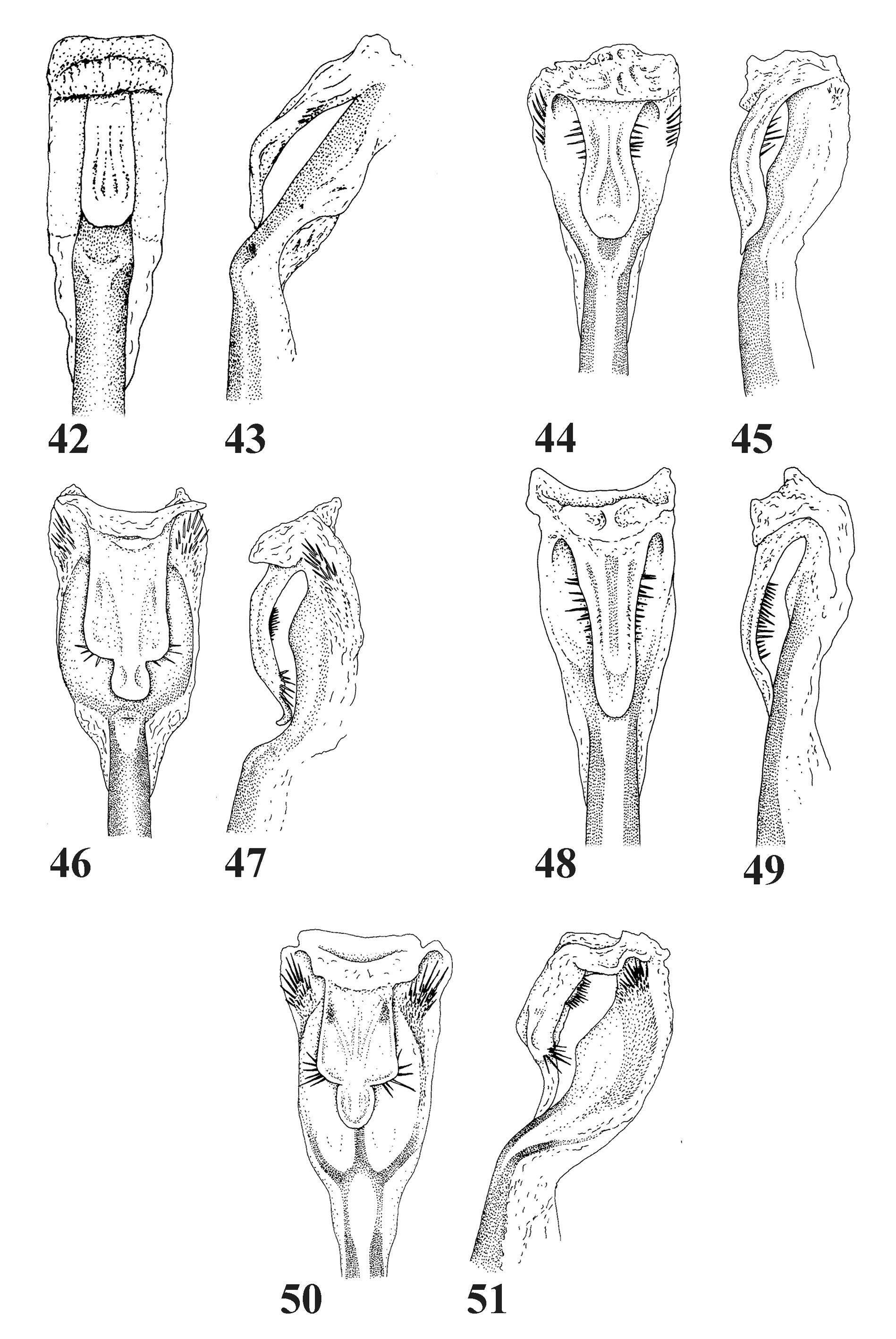
Structural characters. Posterior prothoracic lobe with no separation between median and lateral portions, posterior border slightly convex. Cleft of anterior lamina shaped as ogival Gothic arch ( Fig. 38 View FIGURES 37 – 41 ). Anterior hamuli with ear-like basal plate, and posteromedial angle slightly sclerotized. Distal portion of batilliform lamina curved ventrally with sclerotized apex. Posterior hamuli curved anteromedially ( Fig. 38 View FIGURES 37 – 41 ). Penis segment 3 in ectal view subrectangular, slightly narrowed medially ( Fig. 44 View FIGURES 42 – 51 ); in lateral view with median row of spines ( Fig. 45 View FIGURES 42 – 51 ). Penis segment 2 with row of spines near commissure ( Fig. 44 View FIGURES 42 – 51 ). Hind border of S10 with small concavity ( Fig. 54 View FIGURES 54 – 61 ). Cerci shorter than S10, subcylindrical and tapering distally, in lateral view ( Fig. 55 View FIGURES 54 – 61 ), with convergent apex ( Fig. 54 View FIGURES 54 – 61 ), in dorsal view ( Fig. 54 View FIGURES 54 – 61 ).
Measurements (mm): Hw 44, abdomen 52.
Remarks. The male described here is a specimen borrowed from the Florida State Collection of Arthropods, where it had been identified by M. J. Westfall, Jr., as Mecistogaster martinezi . Inside the glassine envelope there is a label written: “it corresponds to the description of the female. Det S. W. Dunkle 1989.” This identification, which we believe to be correct is corroborated by the fact that this specimen was collected in the same Department (Santa Cruz) of Bolivia as the holotype. Platystigma martinezi belongs to the group of species in which the border of Hw of the male has a subapical elevation, and which also includes P. astictum and P. buckleyi . It differs from these species mainly by the presence of a pale apical area in both wings of male and female. Although close to P. astictum , P. martinezi differs from it by the presence of a row of spines in the penis segment 3 that lack in P. astictum . The female cerci of P. martinezi had not been described previously. It is remarkably similar to that of P. astictum , especially in the presence of much hair. The oviposition of P. martinezi (misidentified as jocaste ) has been observed by Machado & Martinez (1982) and will be compared with that of P. astictum at the end of this paper.
Platystigma minimum sp. nov.
Figures 11 View FIGURES 8 – 13 , 17 View FIGURES 14 – 19 , 23 View FIGURES 20 – 24 , 29, 30 View FIGURES 29 – 32 , 40 View FIGURES 37 – 41 , 48, 49 View FIGURES 42 – 51 , 58, 59 View FIGURES 54 – 61
Material examined. Holotype ♂: Brazil, Acre State, Rio Branco (09° 58' 13"S, 67° 48' 00"W), 4.VII.1989, collected in Taboca (area rich in bamboo). L. Bedê leg. Type deposited in ABMM Collection. GoogleMaps
Etymology. From Latin, minimus – a – um, the smallest. A reference to its very small size.
Description of the male holotype. Head. Labium yellow, labrum black with pair of large yellow rounded spots. Base of mandible and genae yellow, anteclypeus yellow, postclypeus black, antefrons yellow. Upper part of head with yellow marking posteromedially to antennae bases and smaller yellow marking between it and each lateral ocellus. Rear of head yellow.
Thorax. Prothorax ( Fig. 11 View FIGURES 8 – 13 ): anterior lobe yellow bordered with dark brown, with pair of oblong yellow spots disposed transversally. Middle lobe reddish brown with elongated yellow stripe dilated posteriorly. Posterior lobe brown with border yellow. Propleuron dorsally reddish brown and ventrally yellow. Pterothorax: mesepisternum ( Fig. 17 View FIGURES 14 – 19 ) black with antehumeral pale stripe occupying whole extension of sclerite. Posthumeral pale stripe not reaching lower part of sclerite. Venter pale with middle narrow black stripe dilated anteriorly, and posteriorly forming triangular spot ( Fig. 23 View FIGURES 20 – 24 ).
Legs. Femora and tibiae brown with flexural surface yellow. Tarsi black.
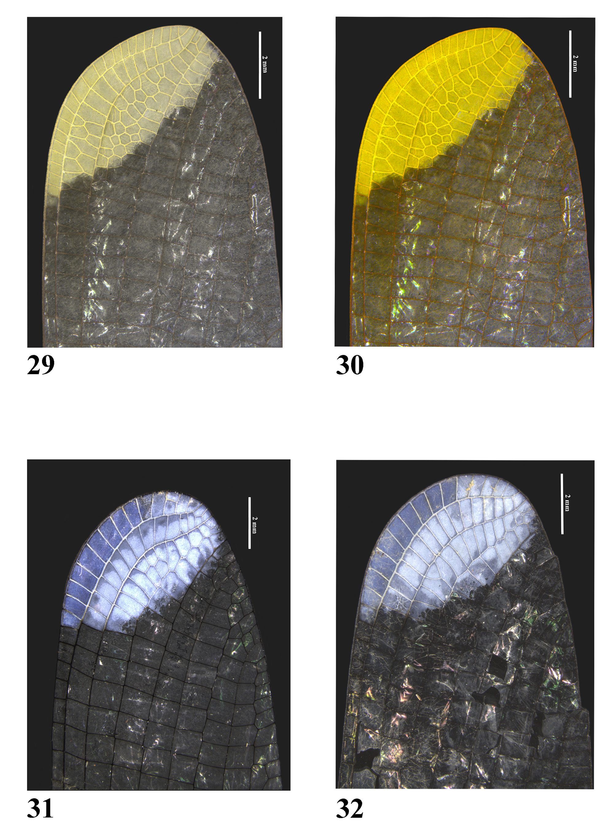
Wings. Fw and Hw slightly infuscated ante-apically, with large light-yellow apical area and medial border oblique, reaching the level of RP2 ( Fig. 29 View FIGURES 29 – 32 ). Pseudostigma hardly distinguishable from yellow area posterior to RA. At high saturation the yellow color becomes more intense ( Fig. 30 View FIGURES 29 – 32 ). Venation: Px 30 in Fw; in Hw 28, RP2 arising near Px 8 in Fw, at Px 9 in Hw. IR1 arising at Px 16 in Fw, at Px 17 in Hw, RP 2 in both wings branching from RP by less than half distance from subnodus to tip of wing. Petiolation originated distal to CuP by a distance about 1.5X the length of CuP in both wings.
Abdomen. S1–2 dorsally dark brown, laterally yellow. S3–8 dorsally black, ventrolaterally yellow. S9 black with pair of ventrolateral yellow streaks adjacent to intersegmental sutures. S10 and cerci black.
Structural characters. Median and lateral portions of posterior prothoracic lobe not separated and its posterior border slightly convex ( Fig. 11 View FIGURES 8 – 13 ). Border of Hw without subapical elevation. Anterior hamuli with ear-like basal plate, alula very slightly sclerotized, except at ventral border ( Fig. 40 View FIGURES 37 – 41 ). Anterior lamina with a medial elongated elevation, with apex sclerotized ( Fig. 40 View FIGURES 37 – 41 ) and provided with a comb-like column of spines visible only in medial view ( Fig. 40 View FIGURES 37 – 41 ). Cleft of anterior lamina shaped as ogival Gothic arch. Distal portion of batilliform lamina well visible, curved ventrally, with sclerotized apex. Posterior hamuli not sclerotized, curved anteromedially. Penis segment 3 in ectal view ( Fig. 48 View FIGURES 42 – 51 ) subrectangular with a comb-like row of lateral spines ( Figs. 48–49 View FIGURES 42 – 51 ). Cerci in lateral view ( Fig. 59 View FIGURES 54 – 61 ) about same length as S10, subcylindrical and tapering distally. In dorsal view with apex convergent ( Fig. 58 View FIGURES 54 – 61 ).
Measurements (mm). Hw 32, abdomen 48.
Female: unknown.
Remarks. Platystigma minimum is the smallest species of the family Pseudostigmatidae , a position previously occupied by Anomisma abnorme McLachlan, 1877 . It can be readily identified by its small size, large apical light yellow area in both wings and anterior lamina with a medial elongated elevation, with apex sclerotized and provided with a comb-like column of spines.


| FSCA |
Florida State Collection of Arthropods, The Museum of Entomology |
No known copyright restrictions apply. See Agosti, D., Egloff, W., 2009. Taxonomic information exchange and copyright: the Plazi approach. BMC Research Notes 2009, 2:53 for further explanation.
|
Kingdom |
|
|
Phylum |
|
|
Class |
|
|
Order |
|
|
Family |
|
|
Genus |
Platystigma martinezi (Machado, 1985)
| Machado, Angelo B. M. & Soldati Lacerda, Déborah S. 2017 |
Mecistogaster martinezi
| Garrison 2016: 20 |
| Pinto 2016: 21 |
| Garrison 2010: 391 |
| Heckman 2008: 207 |
| Tsuda 2000: 58 |
| Davies 1984: 59 |