
Neanthes helenae ( Kinberg, 1865 )
|
publication ID |
https://doi.org/ 10.5852/ejt.2021.760.1443 |
|
publication LSID |
lsid:zoobank.org:pub:917481FF-7C89-4B0F-8C91-77E616271ECC |
|
DOI |
https://doi.org/10.5281/zenodo.5122988 |
|
persistent identifier |
https://treatment.plazi.org/id/03D687C6-FFB0-F53E-FDF5-FA2AFEC9F89A |
|
treatment provided by |
Felipe |
|
scientific name |
Neanthes helenae ( Kinberg, 1865 ) |
| status |
|
Neanthes helenae ( Kinberg, 1865) View in CoL
Neanthes helenae Kinberg, 1865: 172 View in CoL [type locality: Jamestown, Saint Helena Island].
Nereis (Neanthes) nanciae Day, 1949: 445 View in CoL , fig. 4a–h.
Neanthes helenae View in CoL – Hartman 1959: 250 (species list). — Wilson 1984: 225 (species list). — Villalobos- Guerrero & Idris 2021: 559 View Cited Treatment (table 1), 561 (table 2).
Neanthes nanciae View in CoL – Hartman 1959: 250. — Fauchald 1972: 410 (group IIB 2a). — Wilson 1984: 226 (group IIB 2a) (all in species list).
Type material
Syntypes
ST HELENA • 2 atokous (one ♀); Jamestown; 16°00′ S, 5°40′ W; 27.5 m depth; Eugenie Expedition 1851–53 leg.; sta. 173; poor condition; SMNH Type-457 GoogleMaps • 1 spec., permanent slide of atokous ♀; same data as for syntypes; parapodium 24 th; SMNH Type-457 , housed at the NHMUK [apparently loaned to Arthur Willey in this museum]. GoogleMaps
Type material of Nereis (Neanthes) nanciae Day, 1949
Holotype
ST HELENA • 1 atokous; Dockyard , Crown Point and Jamestown ; 73 m depth; some date between 7 May to 3 Aug. 1945; J. Colman leg.; upper intertidal; NHMUK 1950.1.5.72-95 .
Paratypes
ST HELENA • several atokous; same collection data as for holotype; NHMUK 1950.1.5.72-95b .
Comparative material
Syntypes of Neanthes indica brunnea ( Day, 1957)
MOZAMBIQUE • 1 atokous; Inhambane, Morrumbene Estuary ; J.H. Day leg.; NHMUK 1961.16.23 • 1 atokous; same collection data as for preceding; SAM-A-20983 .
Description
COLOUR AND MEASUREMENTS. Syntype atokous, complete but fragmented into two parts, in poor condition, 26.5 mm TL, 3.3 mm L15, 1 mm W15, with 89 chaetigers. Body pale yellow, lacking pigmentation patterns, stained with Shirlastain A to observe edges, scars and paragnaths ( Fig. 3A–D View Fig ).
PROSTOMIUM. Campanulate, as long as wide ( Fig. 3B View Fig ); anterior end broad, distally complete; anterolateral gap beside palpophore narrow, as wide as antennal diameter; dorsal groove distinct, shallow, running mid-subdistally. Nuchal organs exposed, broad, twice as wide as diameter of posterior pair of eyes.
PALPOPHORES. Sub-conical, 1.2 times wider than long ( Fig. 3B View Fig ), as long as four-fifths of entire prostomium; with distinct sub-distal transverse groove. Palpostyles ovoid, thick, with diameter as wide as two-fifths of palpophore.
ANTENNAE. Tapered, thick, long, extending forwards beyond tip of palpophore and posteriorly to halflength of prostomium; antennae separated, with gap as wide as basal diameter of antennae ( Fig. 3B View Fig ).
EYES. Paired eyes purplish but gradually fading, arranged in a trapezoid form; gap between both pairs two-thirds as wide as diameter of posterior pair of eyes ( Fig. 3B View Fig ); anterior pair of eyes oval, 1.6 times as wide as basal diameter of antennae, gap between both eyes 3.5 times as wide as diameter of eyes, with lens barely distinct, translucent, covering 20% of eye; posterior pair of eyes rounded, 1.7 times as wide as basal diameter of antennae, with lens distinct, translucent, placed in middle of eye and covering 20% of it.
APODOUS ANTERIOR SEGMENT. Segment 4 times wider than long, 1.5 times as long as chaetiger 1, with rounded anterior margin and ventrolateral projections; dorsum without marked transverse wrinkle.
TENTACULAR CIRRI. Cirri of right flank dehiscent, remaining slightly thickened, smooth ( Fig. 3B View Fig ); posterodorsal cirri broken, extending posteriorly to chaetiger 6, but according to original description, extending to chaetiger 10; antero-dorsal cirri broken, extending posteriorly to chaetiger 3; postero-ventral cirri extended over opposite side of prostomium; antero-ventral cirri slightly longer than postero-ventral cirri and extending beyond twice length of palpophore; dorsal cirrophores cylindrical, ventral cirrophores ring-shaped, postero-dorsal cirrophores 1.5 times as long as antero-dorsal cirrophores, antero-ventral cirrophores 1.2 times as wide as postero-ventral cirrophores.
PHARYNX. Everted, in poor condition, damaged, jaws and several areas missing, only areas V and VI remain. Pharynx of second syntype described here, everted, in poor condition, damaged, jaws missing; paragnaths on maxillary and oral rings amber, barely distinct but stained here ( Fig. 3C–D View Fig ), consisting of conical, p-bars, and merged paragnaths; plate-like basements absent. Area I: 1, small cone; areas IIa: 31 and IIb: 38, three slightly regular rows of uneven cones in eyebrow-shaped patch, subdistal cones larger, fang-shaped in outer-most row ( Fig. 3C View Fig ); area III: 18, four irregular rows of uneven cones in sub-oval patch, with three distinct laterally-isolated cones, distal cones smaller; areas IVa: missing and IVb: 26, L-shaped patch with proximal half consisting of two regular rows and distal half with four slightly regular rows of uneven cones and four long and slender merged paragnaths (3–4 times longer than wide) located near jaw; area V: 1 (1), small conical paragnath placed on same level between most distal and proximal paragnaths on area VI ( Fig. 3C View Fig ); areas VIa: 7 (5) and VIb: 6 (5), oblique transverse row of uneven p-bars becoming shorter outwards (six in VIa, four in VIb; Fig. 3C View Fig ) and one single outermost cone ( Fig. 3C View Fig ); areas VII–VIII: 31, two bands of cones, with anterior band consisting of two transversely aligned rows (furrow row with one small cone and ridge row with one fang-shaped cone on each region) ( Fig. 3D View Fig ), and posterior band with two transverse rows ( Fig. 3D View Fig ). Areas VI–V–VI ridge pattern, π- shaped. Gap between area VI and areas VII–VIII narrow, as wide as palpostyle.
PAIRED OESOPHAGEAL CAECA. Unknown.
PARAPODIA. Without glandular, dorsal patches. Notopodia consisting of dorsal cirrus, dorsal ligule (distal and proximal), notopodial prechaetal lobe, and median ligule in biramous parapodia. Neuropodia consisting of neuroacicular ligule with inferior and postchaetal lobes, ventral ligule, and ventral cirrus; superior lobe absent throughout.
DORSAL CIRRI. Cirriform, long, extending markedly beyond distal region of dorsal ligule throughout ( Fig. 3E–I View Fig ); dorsal cirri 3–3.5 times as long as proximal region of dorsal ligule in anteriormost and anterior parapodia ( Fig. 3E–F View Fig ), 2–2.5 times as long as that in following parapodia ( Fig. 3G–I View Fig ); attached basally to dorsal ligule in anteriormost parapodia ( Fig. 3E View Fig ), medially in anterior, middle and posterior parapodia ( Fig. 3F–I View Fig ), two-thirds in posteriormost parapodia.
DORSAL LIGULE. Proximal region even throughout, except slightly humped from middle parapodia towards posterior end; shorter than distal region of dorsal ligule in anteriormost parapodia ( Fig. 3E View Fig ), as long as that in anterior, middle and posterior parapodia ( Fig. 3F–I View Fig ), 1.5 times as long as that in posteriormost parapodia; two irregular glandular patches covering partially proximal region of dorsal ligule in anteriormost and anterior parapodia ( Fig. 3E–F View Fig ) and entirely covering that in following parapodia ( Fig. 3G–I View Fig ). Distal region well developed, becoming slightly longer towards posterior end; digitiform in anteriormost parapodia ( Fig. 3E View Fig ), bluntly rounded in anterior parapodia ( Fig. 3F View Fig ), conical in middle parapodia ( Fig. 3G–H View Fig ), bluntly conical in remaining parapodia ( Fig. 3I View Fig ); as long as or slightly shorter than median ligule in anterior parapodia ( Fig. 3F View Fig ), longer than that in following parapodia; projecting beyond notoacicula throughout; one irregular glandular patch covering entirely distal region of dorsal ligule ( Fig. 3F–I View Fig ).
NOTOPODIAL PRECHAETAL LOBE. Present from parapodia 5 to parapodia 14 ( Fig. 3F View Fig ); bluntly rounded, as long as median ligule in anterior parapodia, then reducing and tapering gradually to notoacicular process that disappears in about parapodia 32.
MEDIAN LIGULE. Digitiform in anteriormost parapodia, bluntly rounded in anterior parapodia ( Fig. 3F View Fig ), conical in middle parapodia ( Fig. 3G–H View Fig ), bluntly conical in following parapodia ( Fig. 3I View Fig ), becoming narrower from middle parapodia towards posterior end.
NEUROACICULAR LIGULE. Smaller than ventral ligule in anteriormost parapodia ( Fig. 3E View Fig ), as long as in following parapodia ( Fig. 3F–I View Fig ), 1.5–2 times as wide as ventral ligule throughout.
NEUROPODIAL INFERIOR LOBE. Slightly developed and longer than neuroacicular ligule in first four parapodia, absent in following chaetigers.
NEUROPODIAL POSTCHAETAL LOBE. Digitiform, longer than neuroacicular ligule in first 5 parapodia ( Fig. 3E View Fig ), then reducing gradually to disappear in parapodium 12.
VENTRAL LIGULE. Well developed throughout; digitiform, thick in anteriormost parapodia ( Fig. 3E View Fig ), bluntly rounded, thick in anterior parapodia ( Fig. 3F View Fig ), digitiform in following chaetigers; smaller than median ligule throughout, more distinct in middle and posterior parapodia ( Fig. 3H–I View Fig ); shorter than distal region of dorsal ligule in parapodia 1 and 2 ( Fig. 3E View Fig ).
VENTRAL CIRRI. Cirriform, slender; as long as ventral ligule in anteriormost parapodia ( Fig. 3E View Fig ), twothirds as long as that in following parapodia ( Fig. 3F, H View Fig ).
ACICULAE. Colour mostly faded by long-term preservation. Notoaciculae absent in first two chaetigers ( Fig. 3E View Fig ). Neuroaciculae extending similarly to distal end of notoaciculae throughout, with proximal half 1.3–1.5 times as wide as notoaciculae.
NOTOCHAETAE. All homogomph spinigers ( Fig. 3J View Fig ); 10–13 spinigers present in anterior parapodia, 6–9 spinigers in middle parapodia, 4–5 spinigers in posterior parapodia and 3 spinigers in posteriormost parapodia.
SUPRACICULAR NEUROCHAETAE. Consisting of homogomph spinigers and heterogomph falcigers ( Fig. 3K– L View Fig ), both present throughout; 4–5 spinigers present in anteriormost parapodia, 6–7 spinigers in anterior parapodia, 4–5 spinigers in middle parapodia, 1–2 spinigers in posterior and posteriormost parapodia; 2 falcigers present in anteriormost parapodia, 2–3 falcigers in anterior parapodia, 1–2 falcigers in middle parapodia, 1 falciger in posterior and posteriormost parapodia.
SUBACICULAR NEUROCHAETAE. Consisting of heterogomph spinigers ( Fig. 3M View Fig ) and heterogomph falcigers ( Fig. 3N–P View Fig ), both present throughout; 3 spinigers present throughout, sometimes 4 spinigers in middle parapodia; 8 falcigers present in anteriormost parapodia, 8–12 falcigers in anterior parapodia, 3–6 falcigers in middle parapodia, 1–3 falcigers in posterior and posteriormost parapodia.
BLADES. Both homogomph ( Fig. 3J View Fig ) and heterogomph ( Fig. 3M View Fig ) spinigers with blades of medium and long size (B/A ratio 6.6–11.2), finely serrated towards toothed edge, evenly spaced, except homogomph spinigers from middle parapodia towards posterior end with coarse proximal teeth.Blades of heterogomph falcigers in anteriormost parapodia slender, convex, long (B/A ratio 2.2–2.7), terminal tooth blunt with inconspicuous tendon ( Fig. 3N View Fig ), serrations present in about two-fifths (0.60–0.65) of total blade length; blades in following parapodia thicker, convex, short (B/A ratio 0.9–1.6), terminal tooth incurved with distinct tendon in supracicular fascicle ( Fig. 3K–L View Fig ), blunt with inconspicuous tendon in subacicular fascicle ( Fig. 3O–P View Fig ), serrations present in about one-fifth to one-quarter (0.22–0.28) of total blade length. Shaft of supracicular falcigers thicker than subacicular ones in middle and posterior parapodia; camerated, with cavity divided sub-distally into two longitudinal partitions, supracicular rarely divided into three. Fused heterogomph falcigers present in supracicular fascicle, fusing from parapodia 23, simple in parapodia 35 to parapodia 65, separating gradually to non-fused in posteriormost parapodia.
PYGIDIUM. Distinctly projected, bluntly rounded, wider than long ( Fig. 3Q View Fig ), with anal cirri as long as last 11 chaetigers; cirrophores of anal cirri barely developed.
Description of holotype and two paratypes of Nereis (Neanthes) nanciae
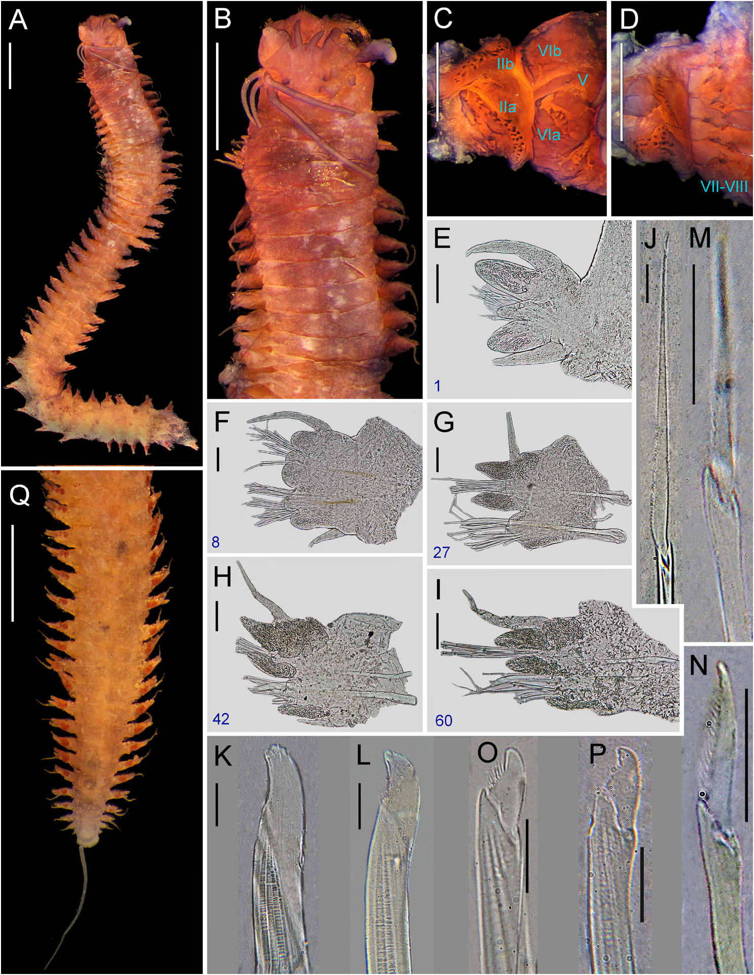
COLOUR AND MEASUREMENTS. All complete atokous (one paratype female), in good condition, 20–22.5 mm TL, 6.3–7.6 mm L15, 1– 1.6 mm W15, with 60–64 chaetigers. Body brownish, distal half of prostomium, apodous anterior segment and few anterior chaetigers with reddish-brown pigmentation ( Fig. 4A–B View Fig ), smallest specimen forming faint X-shaped pattern on dorsum of anterior segments.
PROSTOMIUM. Campanulate, as long as wide or slightly longer than wide ( Fig. 4B View Fig ); anterior end broad, distally complete; anterolateral gap beside palpophore narrow, as wide as antennal diameter; dorsal groove distinct, shallow, running mid-subdistally. Nuchal organs deeply embedded, broad, 1.7–2 times as wide as diameter of posterior pair of eyes.
PALPOPHORES. Sub-conical, as wide as long to slightly wider as long ( Fig. 4B View Fig ), subequal to entire length of prostomium; with distinct sub-distal transverse groove. Palpostyles ovoid, thick, with diameter as wide as two-fifths to half of palpophore.
ANTENNAE. Tapered, thick, long, extending forwards beyond tip of palpophore and posteriorly to half to two-fifths length of prostomium; antennae separated, with gap subequal to three-quarters as wide as basal diameter of antennae ( Fig. 4B View Fig ).
EYES. Paired eyes black, arranged in a trapezoid form; gap between both pairs one-quarter to threequarters as wide as diameter of posterior pair of eyes ( Fig. 4B View Fig ); anterior pair of eyes oval to rounded, 1.5–1.7 times as wide as basal diameter of antennae, gap between both eyes 3–3.5 times as wide as diameter of eyes, with lens barely distinct, translucent, covering 10–20% of eye; posterior pair of eyes rounded, 1.2–1.5 times as wide as basal diameter of antennae, with lens distinct, translucent, placed in middle of eye and covering 15–25% of it.
APODOUS ANTERIOR SEGMENT. Segment 3.5–4 times wider than long, 1.5 times as long as chaetiger 1, with rounded anterior margin and distinct ventrolateral projections ( Fig. 4C–D View Fig ); dorsum without marked transverse wrinkle.
TENTACULAR CIRRI. Thick, smooth ( Fig. 4B View Fig ); postero-dorsal cirri extending posteriorly to chaetiger 7–8; antero-dorsal cirri extending posteriorly to chaetiger 3; postero-ventral cirri extended over opposite side of prostomium; antero-ventral cirri subequal or slightly longer than postero-ventral cirri and extending beyond 1.5–2 times length of palpophore; dorsal cirrophores cylindrical, ventral cirrophores ring-shaped, postero-dorsal cirrophores 1.5 times as long as antero-dorsal cirrophores, antero-ventral cirrophores subequal or slightly wider than postero-ventral cirrophores.
PHARYNX. Non-everted. Jaws with distal quarter brownish, remaining yellow amber, with 9–10 welldeveloped and sharp denticles ( Fig. 4E View Fig ); pulp cavity with two canals ( Fig. 4F View Fig ). Reddish-brown paragnaths on maxillary and oral rings ( Fig. 4G–H View Fig ), consisting of cones, p-bars, and merged paragnaths; plate-like basements absent. Area I: 1–2, cones; areas IIa: 20–27 and IIb: 19–27, two slightly regular rows of uneven cones in eyebrow-shaped patch, subdistal cones larger, fang-shaped in outer-most row ( Fig. 4G View Fig ); area III: 18–28, two to four irregular rows of uneven cones in sub-oval or sub-rectangular patch, with two to four distinct laterally-isolated cones, distal cones smaller; areas IVa: 24–45 and IVb: 26–48, L-shaped patch ( Fig. 4G View Fig ) with proximal half consisting of two regular rows and distal half with three or four slightly regular rows of uneven cones and four to six long and slender (3–4 times longer than wide) merged paragnaths located near jaw; area V: 1 (1), coarse conical paragnath placed slightly behind level of paragnaths on area VI ( Fig. 4G View Fig ); areas VIa: 5–8 and VIb: 5–7, oblique transverse row of uneven p-bars becoming shorter outwards (four to seven in VIa, four to six in VIb; Fig. 4H View Fig ) and one single outermost cone ( Fig. 4H View Fig ); areas VII–VIII: 28–33, two bands of cones ventrally well separated, with anterior band consisting of two transversely aligned rows (furrow and ridge rows with one coarse cone on each region), and posterior band with two transverse slightly displaced rows ( Fig. 4G View Fig ). Areas VI–V– VI ridge pattern, π-shaped. Gap between area VI and areas VII–VIII narrow, as wide as palpostyle.
PAIRED OESOPHAGEAL CAECA. Present.
PARAPODIA. Without glandular, dorsal patches. Notopodia consisting of dorsal cirrus, dorsal ligule (distal and proximal), notopodial prechaetal lobe, and median ligule in biramous parapodia. Neuropodia consisting of neuroacicular ligule with inferior and postchaetal lobes, ventral ligule, and ventral cirrus; superior lobe absent throughout.
DORSAL CIRRI. Cirriform, long, extending markedly beyond distal region of dorsal ligule throughout ( Fig. 4I–M View Fig ); dorsal cirri 3–3.5 times as long as proximal region of dorsal ligule in anteriormost and anterior parapodia ( Fig. 4I–J View Fig ), 2–2.5 times as long as that in following parapodia ( Fig. 4K–M View Fig ); attached basally to dorsal ligule in anteriormost parapodia ( Fig. 4I View Fig ), medially in anterior, middle and posterior parapodia ( Fig. 4J–L View Fig ), two-thirds in posteriormost parapodia ( Fig. 4M View Fig ).
DORSAL LIGULE. Proximal region even towards posterior end, except slightly humped from middle parapodia towards posterior end; shorter than distal region of dorsal ligule in anteriormost parapodia ( Fig. 4I View Fig ), as long as that in anterior, middle and posterior parapodia ( Fig. 4J–L View Fig ), 1.5 times as long as that in posteriormost parapodia ( Fig. 4M View Fig ); two irregular glandular patches covering partially proximal region of dorsal ligule in anteriormost parapodia ( Fig. 4I View Fig ) and entirely covering that in following parapodia ( Fig. 4J–M View Fig ). Distal region well developed, becoming slightly longer towards posterior end; digitiform in anteriormost parapodia ( Fig. 4I View Fig ), bluntly rounded in anterior parapodia ( Fig. 4J View Fig ), conical to bluntly conical in following parapodia ( Fig. 4K–M View Fig ); as long as or slightly shorter than median ligule in anterior and middle parapodia ( Fig. 4J–L View Fig ), longer than that in following parapodia; projecting beyond notoacicula throughout; one irregular glandular patch covering entirely distal region of dorsal ligule.
NOTOPODIAL PRECHAETAL LOBE. Present from parapodia 5 to parapodia 14–16 ( Fig. 4J View Fig ); bluntly rounded, as long as median ligule in anterior parapodia, then reducing and tapering gradually to notoacicular process that disappears in about parapodia 34–37.
MEDIAN LIGULE. Digitiform in anteriormost parapodia, bluntly rounded in anterior parapodia ( Fig. 4J View Fig ), digitiform to bluntly conical in following parapodia ( Fig. 4K–M View Fig ), becoming narrower from middle parapodia towards posterior end.
NEUROACICULAR LIGULE. Smaller than ventral ligule in anteriormost and posteriormost parapodia ( Fig. 4I, M View Fig ), as long as in remaining parapodia ( Fig. 4J–L View Fig ), parapodia 1.5–2 times as wide as ventral ligule throughout.
NEUROPODIAL INFERIOR LOBE. Slightly developed and longer than neuroacicular ligule in first four or five parapodia, absent in following chaetigers.
NEUROPODIAL POSTCHAETAL LOBE. Digitiform, longer than neuroacicular ligule in first 5 parapodia ( Fig. 4I View Fig ), then reducing gradually to disappear in parapodia 10–12.
VENTRAL LIGULE. Well developed throughout; digitiform, thick in all parapodia, except bluntly rounded, thick in anterior parapodia ( Fig. 4J View Fig ); smaller than median ligule throughout, more distinct from middle parapodia towards posterior end ( Fig. 4L–M View Fig ); shorter than distal region of dorsal ligule in parapodia 1 and 2 ( Fig. 4I View Fig ).
VENTRAL CIRRI. Cirriform, slender; as long as ventral ligule in anteriormost parapodia ( Fig. 4I View Fig ), twothirds to three-quarters as long as that in following parapodia ( Fig. 4J–M View Fig ).
ACICULAE. Mostly black with basal end uncoloured in anterior chaetigers, one-half paler in posterior chaetigers ( Fig. 4L View Fig ). Notoaciculae absent in first two chaetigers ( Fig. 4I View Fig ). Neuroaciculae extending similarly to distal end of notoaciculae throughout, with proximal half 1.3–1.5 times as wide as notoaciculae.
NOTOCHAETAE. All homogomph spinigers; 11–14 spinigers present in anterior parapodia, 7–9 spinigers in middle parapodia, 4–5 spinigers in posterior parapodia and 2–3 spinigers in posteriormost parapodia.
SUPRACICULAR NEUROCHAETAE. Consisting of homogomph spinigers and heterogomph falcigers ( Fig. 4N View Fig ), both present throughout; 3–5 spinigers present in anteriormost parapodia, 5–7 spinigers in anterior parapodia, 3–5 spinigers in middle parapodia, 1–2 spinigers in posterior and posteriormost parapodia; 2 falcigers present in anteriormost parapodia, 1–3 falcigers in anterior and middle parapodia, 1 falciger in posterior and posteriormost parapodia.
SUBACICULAR NEUROCHAETAE. Consisting of heterogomph spinigers and heterogomph falcigers ( Fig. 4O– P View Fig ), both present throughout; 2–3 spinigers present throughout, except 1 spiniger in posteriormost parapodia; 5–6 falcigers present in anteriormost parapodia, 7–10 falcigers in anterior parapodia, 5–6 falcigers in middle parapodia, 4 falcigers in posterior parapodia, 2–3 falcigers in posteriormost parapodia.
BLADES. Both homogomph and heterogomph spinigers with blades of medium and long size (B/A ratio 5.1–8.3), finely serrated towards toothed edge, evenly spaced, except homogomph spinigers from middle parapodia towards posterior end with coarse proximal teeth. Blades of heterogomph falcigers in anteriormost parapodia slender, convex, long (B/A ratio 2.3–3.4), terminal tooth blunt with inconspicuous tendon ( Fig. 4O View Fig ), serrations present in about half to two-thirds (0.52–0.66) of total blade length; blades in following parapodia thicker, convex, short (B/A ratio 0.65–1.3), terminal tooth incurved with distinct tendon in supracicular fascicle ( Fig. 4P View Fig ), blunt with inconspicuous tendon in subacicular fascicle, serrations present in about one-sixth to one-third (0.16–0.38) of total blade length. Shaft of supracicular falcigers thicker than subacicular ones in middle and posterior parapodia; camerated, with cavity divided sub-distally into two longitudinal partitions. Fused heterogomph falcigers present in supracicular fascicle ( Fig. 4N View Fig ), fusing from parapodia 23–26, simple in parapodia 34–35 to parapodia 59–65, separating gradually to non-fused in posteriormost parapodia.
PYGIDIUM. Distinctly projected, bluntly rounded, as wide as long ( Fig. 4Q View Fig ), with anal cirri as long as last 11–12 chaetigers; cirrophores of anal cirri barely developed.
Remarks
Neanthes helenae was not compared in the original description by Kinberg (1865), although he placed it within a group formed by two other species, N. rigida (Grube & Ørsted in Grube, 1858) and N. vallata (Grube & Kröyer in Grube, 1858), currently belonging to Nereis and Perinereis ( Salazar-Vallejo & Eibye-Jacobsen 2012) , respectively. Among all species of Neanthes , N. helenae from St Helena Island resembles N. flava Wu, Sun & Yang, 1981 , N. indica brunnea , N. nubila (Savigny, 1822) likely from the North Atlantic (precise locality unknown), and N. talehsapensis ( Fauvel, 1932) from the Gulf of Thailand. These species share proximal region of dorsal ligules of similar size throughout the body (or slightly enlarged in posterior parapodia), presence of neuropodial postchaetal and notopodial prechaetal lobes at least in some anterior chaetigers, absence of neuropodial superior lobes, areas VII–VIII with two well-defined bands of more than 20 paragnaths, and area I with no more than two paragnaths (see Villalobos-Guerrero & Idris 2021: table 2).
Moreover, N. helenae is distinguishable from them all by the following diagnostic features: (I) the presence of p-bars on area VI, in contrast to its absence in those species; (II) the presence of ventrolateral projections on the apodous segment, in comparison to its absence in those species; (III) the presence of simple chaetae (fused falcigers) in supracicular fascicle, in contrast to its absence in N. flava , N. indica brunnea , N. nubila and N. talehsapensis ; (IV) one paragnath on area V, in comparison to none in N. flava , N. indica brunnea , N. nubila and N. talehsapensis ; (V) the paragnaths disposed in a transverse row on area VI, in contrast to those in a rounded or oval patch in N. flava , N. indica brunnea and N. nubila , and a cross or irregular patch in N. talehsapensis ; (VI) the dorsal cirri extending markedly beyond distal region of dorsal ligule at least in anterior parapodia, in comparison to those that are subequal to or not extending beyond end in N. flava , N. indica brunnea and N. nubila ; (VII) 18–28 paragnaths on area III, in contrast to one in N. flava and 5–9 in N. indica brunnea ; (VIII) the heterogomph falciger blade with hammerheaded terminal tooth and distinct tendon in middle and posterior parapodia, in comparison to those with blunt terminal tooth and inconspicuous tendon in N. flava and N. talehsapensis ; (IX) the presence of merged paragnaths, in contrast to their absence in N. flava and N. indica brunnea ; (X) the antennae well separated from each other, in comparison to those that are closely together in N. flava and N. indica brunnea ; (XI) the campanulate prostomium, in contrast to an ovoid shape in N. indica brunnea and N. nubila ; (XII) the π-shaped ridge pattern of areas VI–V–VI, in comparison to that λ-shaped pattern in N. indica brunnea ; (XIII) the dorsum of segments with pigmentation bands, in contrast to a pigment band present only in the apodous segment in N. indica brunnea ; (XIV) the paragnaths present on both ridges and furrows of areas VII–VIII of the pharynx, in contrast to those only present on ridges in N. nubila ; and (XV) the aciculae mostly black, in comparison to those rather pale colour in N. talehsapensis .
Neanthes helenae was described without illustrations by Kinberg (1865) using an unknown number of specimens collected near St Helena Island. The type material was not addressed in the literature, and most of its morphology remained unknown until this study. The two syntypes are in poor condition, but the relevant diagnostic features are still visible, and thus are useful to define the species’ morphology.
Day (1949) described Nereis (Neanthes) nanciae from St Helena Island with several atoke specimens, but he did not justify the proposal as a new species. Later, Hartman (1959) transferred the species to Neanthes and suggested it as a possible synonym of the incompletely known N. helenae . Fauchald (1972) considered Neanthes nanciae as a valid species, but Wilson (1984) agreed with Hartman’s assumptions, suggesting N. nanciae as a possible junior synonym of N. helenae . After the detailed examination of both species’ type material, N. helenae is here regarded as the senior synonym of N. nanciae as a result of the overlap in unique and diagnostic features among other species in Neanthes , such as the presence of p-bar paragnaths on area VI, ventrolateral projections on the apodous segment and the presence of simple chaetae (fused falcigers) in supracicular fascicle.
Distribution
Saint Helena Island.
Ecology
The upper part of the intertidal zone to 73 m depth ( Day 1949).
Reproduction
Unknown.

| NHMUK |
Natural History Museum, London |
No known copyright restrictions apply. See Agosti, D., Egloff, W., 2009. Taxonomic information exchange and copyright: the Plazi approach. BMC Research Notes 2009, 2:53 for further explanation.
|
Kingdom |
|
|
Phylum |
|
|
Class |
|
|
Order |
|
|
Family |
|
|
Genus |
Neanthes helenae ( Kinberg, 1865 )
| Villalobos-Guerrero, Tulio F., Kara, Jyothi & Idris, Izwandy 2021 |
Neanthes helenae
| Villalobos- Guerrero & Idris 2021: 250 |
| Wilson R. S. 1984: 225 |
| Hartman O. 1959: 250 |
Neanthes nanciae
| Wilson 1984: 226 |
| Fauchald 1972: 410 |
| Hartman 1959: 250 |
Nereis (Neanthes) nanciae
| Day J. H. 1949: 445 |
Neanthes helenae
| Kinberg J. G. H. 1865: 172 |