
Marsipococcus ulubendulensis Łagowska & Martin, 2017
|
publication ID |
https://doi.org/ 10.11646/zootaxa.4358.2.4 |
|
publication LSID |
lsid:zoobank.org:pub:253F35F2-9034-4BB7-ACFD-0E44BEB3717B |
|
DOI |
https://doi.org/10.5281/zenodo.6044477 |
|
persistent identifier |
https://treatment.plazi.org/id/B707B023-705E-FFAA-FF4C-35B2A525D046 |
|
treatment provided by |
Plazi |
|
scientific name |
Marsipococcus ulubendulensis Łagowska & Martin |
| status |
sp. nov. |
Marsipococcus ulubendulensis Łagowska & Martin sp. n.
Material studied. MALAYSIA, Negeri Sembilan, Ulu Bendul, near Seremban , on underside of a leaf of Annonaceae , against the midrib, 15.ii.2014, J.H. Martin #8705 ( BMNH). Holotype adult female mounted singly on a slide. Paratypes: 2 slides with a total of 3 adult females ; 1 slide with 2 third-instar females + a pharate thirdinstar nymph; 1 slide with 3 second-instar males + 4 first-instar nymphs; 1 slide with a second-instar male + a firstinstar nymph; 2 slides with a total of 5 pupae; 1 slide with a pupa + 3 adult males, and 1 slide with 2 pupae and an adult male. The following additional slides are in the BMNH collection (not examined as part of this study): 4 slides with a total of 5 adult females, 2 slides with a total of 5 adult males + 2 late pupae, 1 slide with 2 secondinstar males, 1 third-instar female and 1 pharate third-instar female, and 1 slide with 2 second-instar males + 3 firstinstar nymphs.
Name derivation. The species name ulubendulensis is composed of ulubendul, from the Ulu Bendul Recreational Forest in Malaysia from where this species was collected, and – ensis, an adjectival suffix indicating place of origin.
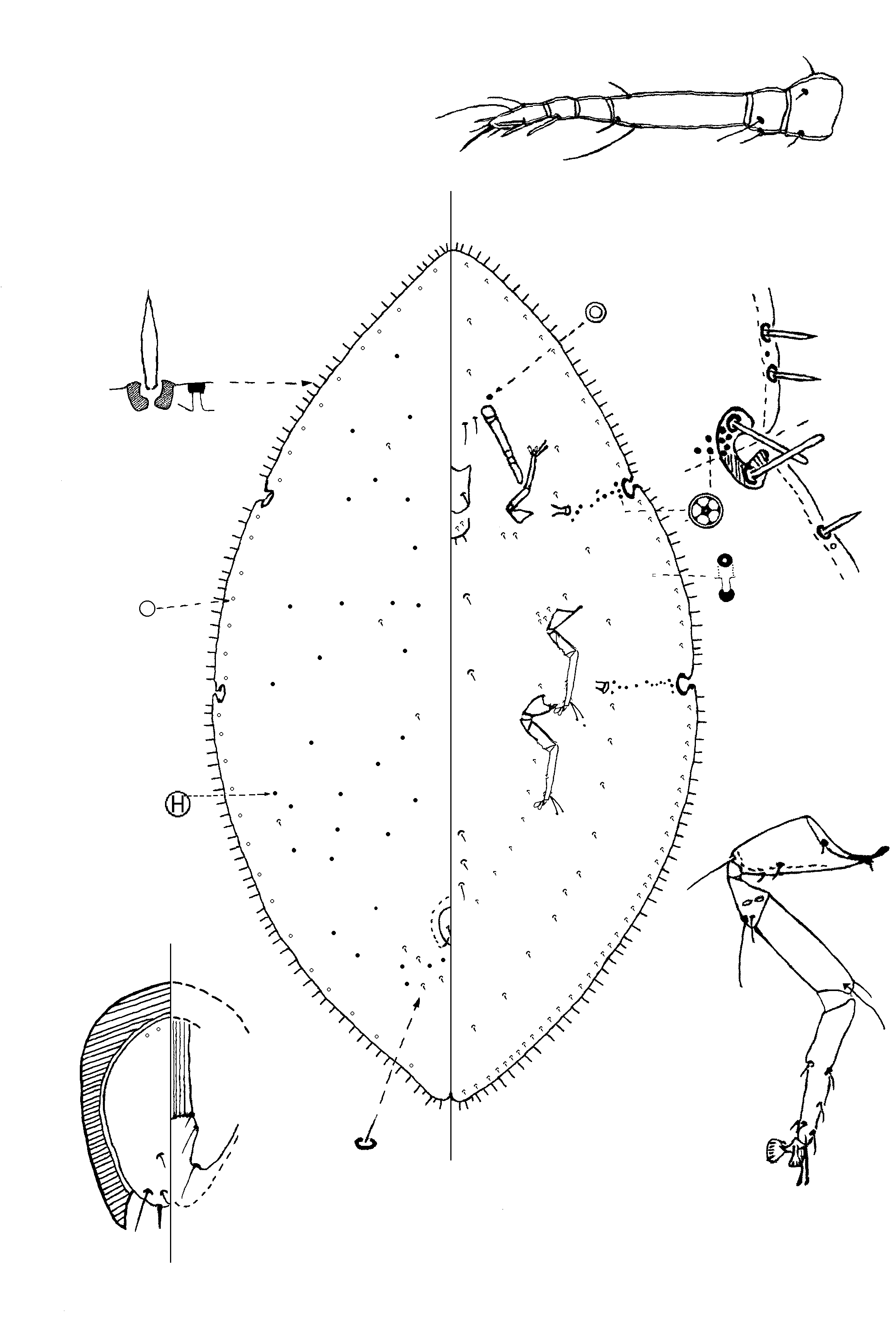
Adult female ( Fig. 1 View FIGURE 1 ). Described from 3 specimens in fair to good condition.
Unmounted material. Reddish brown on the leaf.
Slide-mounted material. Body oval, strongly narrowing at both ends, 6.7–8.4 mm long and 4.0– 4.6 mm wide. Stigmatic clefts distinct and quite deep; anal cleft closely appressed, about 1/5th total body length.
Dorsum. Derm membranous, except for a narrow, heavily sclerotised crescent along inner margins of each stigmatic cleft and around anal plates; derm with a series of irregular-shaped areolations; also with faint radial lines extending medially from the margin as follows: 5 anteriorly on head between clefts; 1 from each cleft; 1 on each side between clefts and 5 on each side of abdomen. Dorsal setae small, each 7–10 µm long, setose, slightly curved, apparently restricted to submedially just posterior to anal plates. Dorsal pores of 2 types, both present throughout dorsum: (i) very small simple pores, each about 2 µm wide, and (ii) larger pores with an inner “H” pattern, each 3– 4 µm wide. Preopecular pores absent. Anal plates together roughly quadrate, with rounded outer angles, each 200– 206 µm long, combined width 149–153 µm, each plate with 2 setae on inner margin near apex, each seta about 8 µm long, and another seta near apex on outer margin, 14–16 µm long; apical seta short, about 4 µm long. Anogenital fold with 1 pair of setae on anterior margin and 2 pairs on lateral margins, one at anterior end and other at posterior end, each 39–43 µm long.
Margin. Marginal setae spear-shaped, each seta 27–44 µm long, sharply pointed, broader medially with a constricted base, with a broad basal socket; setae arranged in a row, abundant, with 44–49 setae between apex and each anterior stigmatic cleft; 23–28 setae on each side between stigmatic clefts; and 7 2–76 setae on each side between posterior stigmatic cleft and anal cleft. A marginal line of sclerotised pores, possibly similar to larger pores on dorsum, often associated with basal sockets of marginal setae, with a pore near every third or fourth seta. Stigmatic clefts deep, with inner margins sclerotised, each with 2 stigmatic spines, all spines with parallel sides, a rounded apex and of similar length, each 30–37 µm long. Eyespots placed well onto dorsum, situated almost dorsad to antennal bases.
Venter. Derm entirely membranous except for sclerotised stigmatic grooves. Pregenital disc pores absent. Spiracular disc pores, each 4–5 µm wide with 5 loculi, present in bands 1–3 pores wide, each band in a sclerotised stigmatic groove near margin; with 57–69 pores in each anterior band and 67–77 in each posterior band. With 1 preantennal pore situated near each scape. Microducts rather few, oval and minute, most frequent in submarginal zone between stigmatic clefts, but probably present throughout. Ventral tubular ducts of 1 type, each without an inner ductule and terminal gland, restricted to a group of 20–24 ducts on either side of genital opening. Ventral setae: frequent submarginally, with about 6–8 setae on each side between stigmatic clefts; and with a pair of moderately long setae on pregenital segment VII, each 65–75 µm long, plus a rather smaller pair on each of proceeding 2 segments; also with a group of 9–13 setae on either side of anterior end of anal cleft; 3 or 4 short setae near each meso- and metacoxa, and 1 short seta near each procoxa. Interantennal setae numbering 2 or 3 pairs, the longest 41–46 µm. Antennae well developed, each 6 segmented; total length 377–435 µm, third segment longest, 166–210 µm long. Setal distribution on antenna: scape 3, pedicel 2, III 3, IV 0, V 1 fleshy seta; apical segment with 3 fleshy setae, 3 stiff setae and 2 hair-like setae; terminal stiff seta 20–24 µm long. Clypeolabral shield 170–200 µm long, with 1 pair of setae, each seta 42–59 µm long; labium with 4 pairs of setae, longest seta 32–39 µm. Spiracles moderately large; width of peritremes: anterior 74–76 µm, posterior 80–83 µm. Legs well developed, segmentation between tibia and tarsus obscure; total lengths of metathoracic legs 740–795 µm (coxa 255–260 µm, trochanter + femur 225–250 µm, tibia + tarsus 240–260 µm, claw 20–25 µm). Setal distribution on leg: coxa with 5 setae, longest about 48 µm long; trochanter with 2, longest about 80 µm long; femur with 2, tibia + tarsus with 6; tarsal digitules similar, both much longer than claw; claw without a denticle, claw digitules dissimilar, one broader than other, each about 39 µm long.
Comment. Of the species currently included in Marsipococcus , adult female of M. ulubendulensis are most similar to those of M. marsupialis , sharing with it the following character states: (i) radial lines extending medially from the margin; (ii) two spines in each stigmatic cleft; (iii) a sclerotised crescent in each stigmatic cleft and also anteriorly around anal plates; (iv) presence of ventral tubular ducts; (v) spiracular disc pores partly in a sclerotised stigmatic groove; (vi) fourth segment of antenna without either normal or fleshy setae (very unusual in Coccidae with well-developed antennae); (vii) marginal setae spear-shaped, each sharply pointed, broader medially with a constricted base; (viii) a similar body shape, being lemon-shaped and slightly pointed at each end; and (ix) dorsal pores that appear bilocular. It differs from M. marsupialis (character states in M. marsupialis in brackets) as follows: (i) multilocular disc pores absent (present in a tight group medially in abdominal segment VII); (ii) preopercular pores absent (present in a narrow band medially); and (iii) dorsal setae restricted to laterad of anal plates (present throughout dorsum). Adult female of M. ulubendulensis differ from those of M. durbanensis and M. proteae (character states in latter two species in brackets) as follows: (i) absence of preopercular pores (present), and (ii) stigmatic spines short, parallel sided and attached to anterior and posterior margins of each cleft (longer, more capitate and present along inner margin of each cleft); M. ulubendulensis also differs from M. durbanensis in having spinose marginal setae (setose), and from M. proteae in having sharply-pointed dorsal setae (blunt).
First-instar nymph ( Fig. 2 View FIGURE 2 ). Described from 3 specimens in good condition and 1 in poorer condition.
Unmounted material. Not seen.
Slide-mounted material. Body elongate oval, about equally rounded at both ends, length 530–1225 µm, width 290–625 µm.
Dorsum. Derm membranous, lacking sclerotisation in stigmatic areas but with a transverse sclerotised band anterior to anal plates. Dorsal setae minute, each about 2 µm long; with 4 pairs submedially: 1 pair nearly dorsad to scape, and perhaps 1 pair on each thoracic segment; also 1 submarginal pair between stigmatic clefts. Dorsal pores of 3 types: (i) minute simple pores in a submarginal line with 2 pairs on head, 2 pairs on thorax (opposite stigmatic clefts) and 7 pairs arranged segmentally on abdomen; (ii) larger pores (possibly microducts), each 3–4 µm wide and appearing to be somewhat trilocular, present as follows: submarginally, with 2 pairs on head; 3 submedially and a submarginal pair between stigmatic clefts; and 1 pair on each of abdominal segments V and VII, and (iii) a pair of small trilocular pores near anterior margin on head. Anal plates elongate, combined width 46–60 µm, with a narrow, sclerotised transverse band on dorsum just anterior to anal plates. Each anal plate 53–60 µm long; with an apical seta, 200–283 µm long; also inner margin of each plate with a long seta near apex, about 30 µm long, and a short seta medially, plus a short seta near apex on outer margin. Anogenital fold with 1 seta on each anterolateral corner, each 10–16 µm long, and another seta on each lateral margin, 25–35 µm long. Anal ring 22–26 µm wide, with 1 row of pores and 6 setae, each seta 60–80 µm long.
Margin. Marginal setae strongly setose, each 10–15 µm long; distribution: 6 setae around head between eyespots; and on each side: 2 setae between eyespots and anterior stigmatic cleft; 2 setae between stigmatic areas and 8 setae from posterior stigmatic cleft to anal cleft. Stigmatic clefts shallow, without any sclerotisation, each with two large stigmatic setae clearly differentiated from marginal setae; each stigmatic seta clavate, anterior seta 38–40 µm long and posterior seta 28–35 µm long, generally pointing somewhat posteriorly. Eyespots situated on margin just above level of antennal scape; width of each lens 9–12 µm.
Venter. Derm membranous. Spiracular disc pores each with 5 loculi, present in a line between spiracle and margin, each anterior band 2 or 3 disc pores wide and each posterior band 3 or 4 pores wide. Microducts present in a submarginal line, with 1 pore on cephalothorax, 1 pore between stigmatic clefts and 3 pores on abdominal segments. Ventral setae: with a pair of long setae medially on abdominal segment VII, each seta 24–29 µm long; a small submarginal and small mediolateral seta on each abdominal segment; 1 pair of interantennal setae, each 18– 23 µm long; a pair of short setae near anterior margin on head, a minute submarginal seta just posterior to each stigmatic cleft and, on each side, with a single seta between stigmatic clefts and on each abdominal segment. Antennae well developed, each 6 segmented, total antennal length 156–165 µm; with third segment longest, about 53–77 µm long. Setal distribution on antenna as follows: scape with 3 hair-like setae; pedicel with 2 long hair-like setae; segment III with 3 setose setae, longest seta 66–99 µm; IV with 1 short setose seta; V with 1 fleshy seta and 1 setose seta; and VI with 3 fleshy setae, 3 long (including apical seta) and 2 short hair-like setae and 1 stiff seta; terminal seta 70–72 µm long. Clypeolabral shield 78–88 µm long, with 1 pair of short clypeal setae. Labium with 4 pairs of slender setae. Spiracles small, each peritreme 7–9 µm wide. Legs 5 segmented, subequal in length, prothoracic leg 220–230 µm long, mesothoracic 205–230 µm long, and metathoracic 220–240 µm long. Length of metathoracic leg segments: coxa 43–60 µm, trochanter + femur 75–82 µm, tibia 46–51 µm, tarsus 38–43 µm, claw 14–16 µm. Setal distribution on metathoracic leg: coxa with 4 setae; trochanter with 2, longest 23–30 µm long; femur with 2 short setae, tibia with 3 setae, and tarsus with 3 or 4 setae. Tarsal digitules dissimilar and offset, with proximal digitule long and stout and more distal digitule on meso- and metathoracic legs much shorter and narrower but both longer than claw; on prothoracic legs, distal digitule short and setose. Claws each with a minute denticle; claw digitules dissimilar, one broader than other, each 14–16 µm long.
Comment. The first-instar nymphs of only a few species within the Paralecaniini have been described ( Austrolecanium cappari (Froggatt) , A. sassafras Gullan & Hodgson , Maacoccus cinnamomicolus (Takahashi) , Paralecanium paradeniyensise (Green) , P. planum (Green) and Xenolecanium takahashii Kondo ) ( Gullan & Hodgson 1998; Hodgson & Martin 2001; Kondo et al. 2005). According to Kondo et al. (2005) and Kondo (2006), the first-instar nymphs of these species share many important features, i.e. a seta next to each mesothoracic and metathoracic coxa, six pairs of ventral submedian setae, and stigmatic setae positioned submarginally on either side of a deep stigmatic cleft. Although the first-instar nymphs of M. ulubendulensis sp. n. lack a seta mesad to each meso- and metacoxa, they otherwise appear morphologically close to those of the above species.
Second-instar female. Described from 1 pharate specimen in good condition. Since not all structures were convincingly visible, it is not illustrated but is basically a smaller version of the third-instar female, described in detail below.
Unmounted material. Not seen.
Slide-mounted material. Body elongate oval, somewhat pointed at both ends; length 1.75 mm, width 0.93 mm.
Dorsum. Derm membranous, except for a narrow, sclerotised crescent around anterior margins of anal plates and sclerotised inner margins to stigmatic clefts. Dorsal setae minute, generally sparse, scattered over dorsum, most abundant posterior to anal plates. Dorsal pores probably of only 2 types: (i) small, round simple pores, each about 1.5 µm wide, present sparsely throughout, and (ii) larger, darker pores with an inner “H” or loculate pattern, scattered over dorsum. Dorsal tubular ducts absent. Anal plates together more or less quadrate but elongate, with a horseshoe-shaped sclerotisation around anterior margins; each plate with rounded outer angles, 96 µm long, combined width 68 µm; each plate with 4 setae near posterior apex, 2 setae near inner margin (one about 1/3rd along inner margin), 1 seta on posterolateral margin, and a stouter apical seta, about 12 µm long. Anogenital fold with seta at each corner of anterior margin plus 2 setae laterally, one placed anteriorly and other posteriorly. Anal ring with 6 setae, each seta about 100 µm long.
Margin. Marginal setae spinose, rather variable in length, each 16–27 µm long, sharply pointed, in a single row; with a total of 33 setae on head between anterior stigmatic clefts, 7–9 setae on each side between stigmatic clefts, and 24 setae on each side between posterior stigmatic cleft and anal cleft. A marginal line of sclerotised pores, similar to larger pores on dorsum, associated with basal sockets of marginal setae, with a pore near every third or fourth seta. Stigmatic clefts distinct, each cleft with a narrow entrance that broadens away from margin, a sclerotised margin and 2 stigmatic spines, one on anterior margin and other on posterior margin; all spines parallelsided, bluntly pointed and of similar length, each about 33–38 µm long. Eyespots not located.
Venter. Derm entirely membranous. Spiracular disc pores, each with 5 loculi, in a narrow band between each spiracle and margin; with 8 or 9 in each anterior band and 10 in each posterior band, but with only a few within each cleft sclerotisation. Ventral microducts not detected for certain. Preantennal pores present, 1 anterior to each antenna. Ventral tubular ducts absent. Ventral setae: with pairs of long setae on abdominal segments V–VII, longest seta about 40 µm long; setae absent medially from segments II–IV; with 2 pairs of interantennal setae; other setae uncertain. Antennae each 6 segmented, 165–170 µm long; apical segment about 35 µm long; setal distribution: scape 3, pedicel 2; segment III 3; IV 0; V with 1 fleshy seta; VI with 3 fleshy setae, 2 flagellate setae and 3 stiff setae; terminal stiff seta about 20 µm long. Clypeolabral shield about 125 µm long, with 1 pair of clypeal setae; labium with 4 pairs of setae. Spiracles relatively small, each peritreme 16–18 µm wide. Legs well developed; lengths of metathoracic leg segments: coxa 83 µm, trochanter + femur 100 µm, tibia + tarsus 112 µm (segmentation indistinct), claw 12 µm; setal distribution: coxa with 6 setae; trochanter with 2; femur with 2; tibia + tarsus with 6 setae. Tarsal digitules similar, both longer than claw; claws each without denticle; claw digitules longer than claw, dissimilar, one broader than other.
Comment. For a comparison with third-instar female nymphs, see Comment under the latter stage below.
Third-instar female ( Fig. 3 View FIGURE 3 ). Described from 1 specimen in very good condition and 1 in poor condition.
Unmounted material. Not seen.
Slide-mounted material. Body elongate oval, somewhat pointed at both ends; length 2.6–3.1 mm, width 1.5– 1.8 mm.
Dorsum. Derm membranous, except for a narrow, sclerotised crescent just anterior to anal plates and sclerotised inner margins of stigmatic clefts. Dorsal setae minute, generally sparse, scattered over dorsum, most abundant posterior to anal plates. Dorsal pores of 3 types: (i) small, round simple pores, each about 1.5–2.0 µm wide, sparsely present throughout and also in a submarginal line; (ii) larger pores with an inner “H” or loculate pattern, scattered over dorsum; and (iii) minute dorsal microducts, each about 1 µm wide, scattered throughout. Dorsal tubular ducts absent. Anal plates together more or less quadrate but elongate, with a horseshoe-shaped sclerotisation around anterior margins; each plate with rounded outer angles, 135–140 µm long, combined width 98–100 µm; each plate with 4 setae near posterior apex: 2 setae on inner margin (one about 1/3rd along inner margin), each 10–15 µm long, 1 seta near apex on outer margin, about 17 µm long, and a stouter apical seta, about 12 µm long. Anogenital fold with a seta at each corner of anterior margin plus 2 setae laterally, one placed anteriorly and other posteriorly, longest about 40 µm long. Anal ring 50–52 µm wide, with 6 setae, each seta 120– 155 µm long.
Margin. Marginal setae spinose, sharply pointed, each 30–39 µm long; distributed in a single row, with 27–29 setae on each side between apex and anterior stigmatic cleft, 15 or 16 setae on each side between stigmatic clefts, and 40–44 setae on each side between posterior stigmatic cleft and anal cleft. Marginal pores, similar to larger dorsal pores, mainly associated with basal sockets of marginal setae, with a pore every second to fourth seta. Stigmatic clefts distinct, with sclerotised inner margins, each with 2 stigmatic spines, one on anterior margin and other on posterior margin, all spines parallel sided, bluntly pointed and of similar length, each about 49 µm long. Eyespots not located.
Venter. Derm entirely membranous. Spiracular disc pores, each with 5 loculi, in a narrow band between each spiracle and margin; with 26–31 in each anterior band and 32–34 in each posterior band; each band with a group of 8–10 pores within cleft sclerotisation. Ventral microducts minute, most frequent submarginally, but probably present throughout. Preantennal pores: 1 pore anterior to each scape. Ventral tubular ducts absent. Ventral setae: with a pair of long setae on each of abdominal segments V–VII, longest seta about 65 µm long; medial setae absent from abdominal segments II–IV, but present medially on thoracic segments (2 pairs) and head (2 pairs); pairs of short setae present in submedial and mediolateral lines on all abdominal segments, with 1 pair per segment; with 2 setae on each side of cephalothorax, between stigmatic clefts, and near each procoxa; also 3 or 4 short setae near each mesocoxa and 1 near each metacoxa; additionally with 2 pairs of interantennal setae, longest seta 25–30 µm long; also with small submarginal setae as follows: 10 or 11 setae between apex and anterior stigmatic clefts, 8 on each side between each lateral stigmatic area and 25 on each side between posterior stigmatic cleft and anal cleft. Antennae each 6 segmented, 250–266 µm long; apical segment about 40 µm long; setal distribution: scape 3, pedicel 2, segment III 3, IV 0, V 1 fleshy seta, VI 3 fleshy setae, 2 flagellate setae and 3 stiff setae, terminal stiff seta about 19 µm long. Clypeolabral shield 160–164 µm long, with 1 pair of clypeal setae; labium with 4 pairs of setae. Spiracles relatively large, each peritreme 33–39 µm wide. Legs well developed; total length of metathoracic leg 457–500 µm, with coxa 125–160 µm, trochanter + femur 150–170 µm, tibia + tarsus 145–155 µm (segmentation indistinct), claw 12–15 µm long. Metathoracic leg setal distribution: coxa with 6 setae; trochanter with 2, longest about 53 µm long; femur with 2; tibia + tarsus with 6 setae. Tarsal digitules similar, both longer than claw; claws each without a denticle; claw digitules dissimilar, one broader than other.
Comment. The second- and third-instar female nymphs of M. ulubendulensis are almost identical morphologically but differ in the size of the appendages and in the frequency of structures such as spiracular disc pores and marginal setae. The third-instar female is also somewhat similar morphologically to the adult female but differs mainly in the absence of a vulva, ventral tubular ducts and in having fewer spiracular disc pores; also in the size of the stigmatic spines, which are shorter and stouter on the adult female. A third-instar female of A. sassafras was briefly described by Gullan & Hodgson (1998), but there is too little detail given for comparison.
Second-instar male ( Fig. 4 View FIGURE 4 ). Described from 3 specimens in good condition.
Unmounted material. Covered in white wax.
Slide-mounted material. Body elongate oval, roundly pointed at both ends; length 1.9–2.6 mm, width 0.9–1.6 mm.
Dorsum. Derm membranous except for a narrow, sclerotised crescent just anterior to anal plates and a sclerotised inner margin to each stigmatic cleft. Dorsal setae minute, sparse, scattered over dorsum. Dorsal pores of 2 types: (i) minute, round simple pores, present sparsely throughout, and also in a submarginal line; and (ii) larger, darker pores, each with an inner trilocular or “H”-shaped pattern, present throughout dorsum, possibly in a sparse reticulate pattern but forming about 7 transverse lines medially on thorax and abdomen. Dorsal tubular ducts of 2 sizes present: (i) long ducts, each 36–42 µm long with a short cup-shaped invagination, a fairly wide inner ductule and a large terminal gland, forming a submarginal band as follows: with 12–20 on head between anterior stigmatic clefts; 6–9 on each side between stigmatic clefts, and 8–15 posterior to posterior stigmatic cleft; and (ii) shorter ducts, each 14–18 µm long, with a wide inner ductule and a large terminal gland, present in fairly broad submedial lines (broadest in about abdominal segments III and IV) running from anal plates anteriorly to dorsad to each scape; with about 80–90 in each line; also with 3 pairs of short radial lines, each formed by 1–7 ducts, with a line extending from anterior margin of head to dorsad to each scape plus lines from approximately each cleft. Anal plates together elongate with rounded outer angles; each plate 90–96 µm long, combined width 62–69 µm; with 3 setae on near apex of each plate plus another about midway along inner margin. Anogenital fold with a seta at each corner on anterior margin and with 2 setae laterally, one located anteriorly and other posteriorly, longest seta about 30 µm long. Anal ring about 39 µm wide, with 6 setae, each seta 90–106 µm long.
Margin. Marginal setae spinose, sharply pointed, mostly with a slight waist basally, in a single row along margin, each 20–37 µm long; distributed as follows: 19 or 20 between apex and anterior stigmatic cleft; 9–11 between stigmatic clefts, and 28–33 between posterior stigmatic cleft and anal cleft. Marginal micropores associated with basal sockets of marginal setae, each about 1.5 µm wide, some with an inner ductule; with a pore associated with every second to fourth seta. Stigmatic clefts distinct, with inner margins sclerotised, each with 2 stigmatic spines, with one on anterior margin of cleft and other on posterior margin, both stout and bluntly pointed, with anterior spine sometimes significantly longer than posterior spine: each anterior spine 40–58 µm long and each posterior spine 28–56 µm long. Eyespots not located.
Venter. Derm membranous. Spiracular disc pores each with 5 loculi, present in narrow bands between each spiracle and margin, with 13–19 in each anterior band and 16–22 in each posterior band. Ventral microducts minute, most frequent submarginally, but probably present throughout. Preantennal pores: singly just anterior to each scape. Ventral tubular ducts present submarginally, each similar to submarginal ducts on dorsum but with a longer sclerotised part to inner ductule, present as follows: 22–28 on head between anterior stigmatic clefts, 6–8 on each side between stigmatic clefts, and 9–16 on each side of anterior abdominal segments. Ventral setae: with pairs of long setae on each of abdominal segments V–VII, longest seta about 27 µm long; medial setae on other abdominal segments absent; with pairs of short submedial and submarginal setae present on each abdominal segment; also with a short seta near each meso- and procoxa; with 2 pairs of interantennal setae, longest seta 26–39 µm long; submarginal setae: about 6 setae between apex and anterior stigmatic clefts, 5 between each stigmatic areas, and 7 between posterior stigmatic cleft and anal cleft. Antennae each 6 segmented, 180–205 µm long, third segment longest, 57–62 µm long; apical segment 35–40 µm long; setal distribution: scape 3, pedicel 2, segment III 3, IV 0, V 1 fleshy seta, VI 3 fleshy setae, 2 flagellate setae and 3 stiff setae, terminal stiff seta about 17–18 µm long. Clypeolabral shield 111–119 µm long; clypeal setae not located; labium with 4 pairs of setae. Spiracles small, each peritreme 23–29 µm wide. Legs well developed; total length of metathoracic leg 320–358 µm, with coxa 90– 110 µm, trochanter + femur 105–120 µm, tibia + tarsus 115–130 µm, claw 12–13 µm; setal distribution: coxa with 5 setae, longest 30–34 µm long; trochanter with 2 setae, longest 40–42 µm long; femur with 2 setae, and tibia + tarsus 6 setae; tarsal digitules similar, each 27–32 µm long; claws without a denticle, claw digitules dissimilar, one broader than other, each 17–22 µm long.
Comment. The only other second-instar male nymph belonging to a species of Paralecaniini that has been described to date is that of Austrolecanium sassafras ( Gullan & Hodgson 1998) . The nymphs of these two species differ significantly in that those of A. sassafras have (character states on M. ulubendulensis in brackets): (i) significantly reduced legs and antennae (normally developed), (ii) apparently only 1 size of dorsal tubular duct (2 sizes), (iii) dorsal tubular ducts arranged in a rather different pattern, with 4 radial lines of ducts and with the marginal ducts tending to be in groups rather than in a line (only 3 radial lines and a marginal band of ducts), and (iv) 3 stigmatic setae in each stigmatic cleft, arranged along base of cleft (only 2 setae, 1 on each side of cleft). The presence of ventral tubular ducts has not been recorded in second-instar male nymphs before but their presence there could be widespread. Location of the dermal openings of these ducts requires extreme care and a good preparation, so they could easily have been missed in the past. In fact, on these specimens, the ventral ducts are more or less out of focus when the dorsal ducts are clear, but this would not be obvious on specimens that had been strongly compressed when slide mounted. In addition, the ventral ducts are situated slightly nearer the margin compared to those on the dorsum. As noted above, although very similar morphologically, the dorsal and ventral ducts differ slightly in the length of the sclerotised tube at the base of the inner ductule, this being longer on the ventral ducts (arrowed in Fig. 4 View FIGURE 4 ).
Male pupa ( Fig. 5). Described from 2 good specimens, 1 poor specimen and 2 quite good pharate specimens.
Unmounted material. Unknown.
Slide-mounted material. Body elongate oval; length 1.7–1.9 mm, head width 397–441 µm. Derm membranous, with small dermal spinules and nodules.
Head. Antennae: total length 662–762 µm (ratio of antennal length to total body length 1:2.5). A sclerotised, transverse yoke-like structure present posteroventrally. Setae: with 3 pairs of minute setae medially, plus a pair anteriorly and 2 pairs ventrally.
Thorax. With a pair of small setae medially on dorsum of all 3 segments, in line with dorsal abdominal setae on abdomen. Without spiracular disc pores associated with either anterior or posterior spiracles. Spiracles: width of anterior peritremes 37–39 µm. With 3 pairs of well-developed legs, each without a small triangular incipient claw on apex; length of metathoracic leg 673–817 µm. Wing-buds: each narrow, length 625–733 µm, width 190–203 µm (ratio length to width 1:0.28).
Abdomen. With 1 pair of small dorsal setae on each of segments V and VI; also with 1 pair of small ventral abdominal setae on each of segments II–VI and 2 pairs on segment VII. Dorsopleural setae, each about 6 µm long, and single minute ventropleural seta present on each side of each of segments II–VI. Lobes on abdominal segment VII moderately developed, rounded and about 2/3rds as long as penial sheath, each lobe with 1 longish seta on apex plus 1 seta subapically. Lobes of segment VIII small, rather rounded and slightly sclerotised, without setae. Penial sheath about 3x longer than lobes of segment VII, 170–177 µm long and about 140–149 µm wide at base (ratio length to width 1:0.8).
Comment. No other pupa of a member of the Paralecaniini has been described.
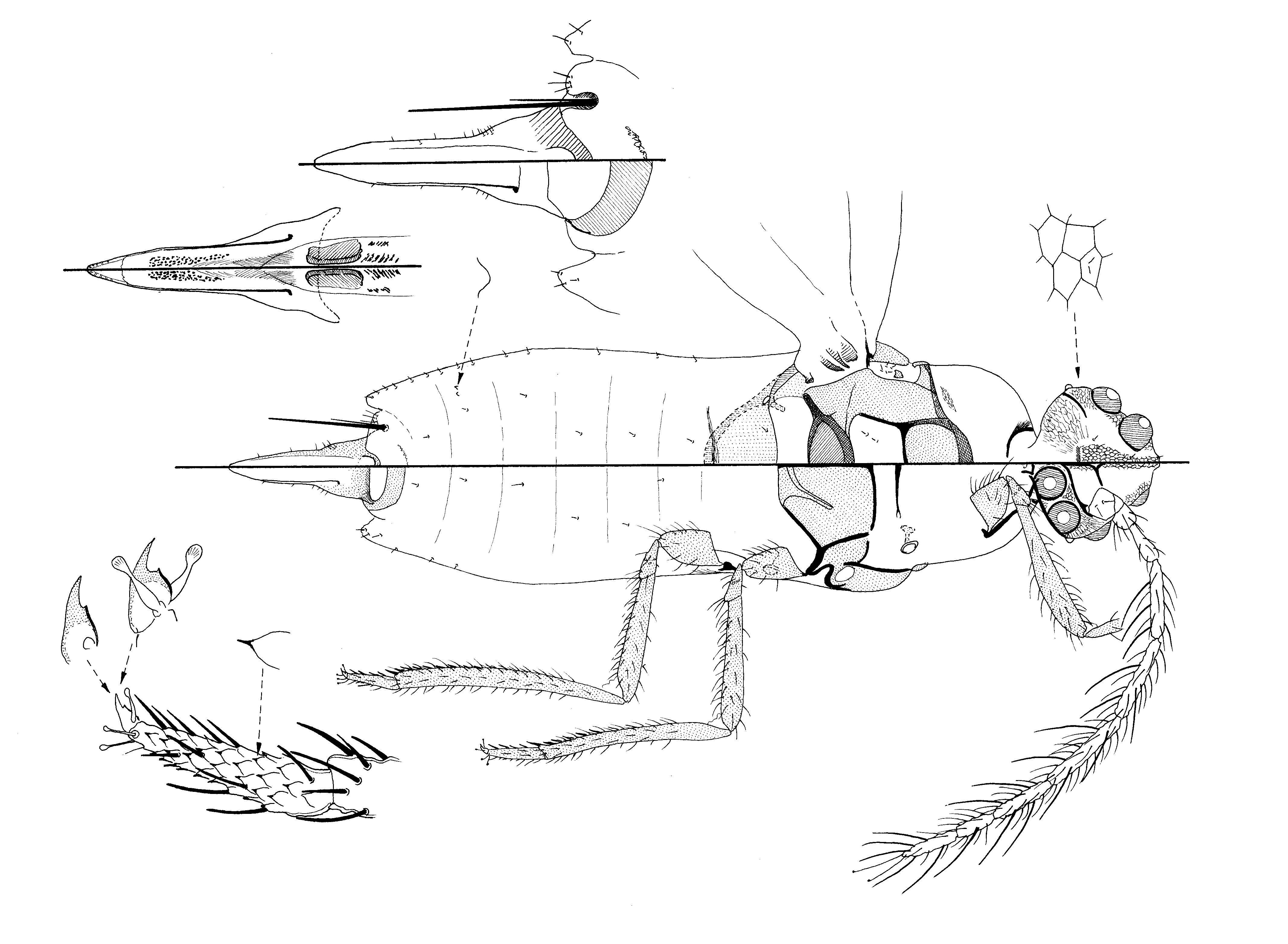
Adult male ( Fig. 6 View FIGURE 6 ). Described from 3 specimens in good condition.
Unmounted material. Unknown.
Slide-mounted material. Body of moderate size; total body length about 1.55–1.60 mm, width across triangular plates about 335–365 µm. Antennae each about half total body length; head with 4 pairs of particularly large simple eyes; head and rest of body with almost no setae, those present all hair-like (hs). Fleshy setae (fs) restricted to appendages and usually easy to separate from hs; procoxae without coxal bristles; trochanterofemur articulation clear. Caudal extensions on abdominal segment VII rounded; glandular pouches present but glandular pouch setae significantly different, one much stouter and longer than other. Antennae each 9 segmented, with only 2 capitate setae on apex. Wings about 7/10th total body length and slightly less than half as wide as long. Hamulohalteres absent.
Head. Roughly diamond-shaped; length about 225–250 µm; width across genae 250–275 µm. Setae few. Median crest well developed, with narrow reticulate striations; postoccipital ridge indicated by a pale area of sclerotisation; with a total of 4 or 5 pairs of hs dorsal head setae. Mid-cranial ridge absent dorsally; ventrally, midcranial ridge narrow but well defined, extending posteriorly as far as ocular sclerite, with a lightly reticulated margin; with 0 or 1 ventral mid-cranial ridge setae present laterad to ridge. Preocular ridge short dorsally but complete ventrally, extending from each scape to ventral mid-cranial ridge. Ocular sclerite strongly polygonally reticulated, each reticulation without inner micro-ridges. Genae large and membranous, without genal setae but with many polygonal reticulations without inner microridges. Eyes: four pairs of particularly large, protuberant, round simple eyes, all subequal in size, each 53–65 µm wide. Ocelli each oval, maximum width about 25–28 µm. Postocular ridge extending only a short distance medially on dorsum. Dorsal ocular setae absent. Ventral head setae absent. Tentorial bridge well developed. Cranial apophysis bifid.
Antennae 9 segmented and filiform. Scape: 45–50 µm long, 45 µm wide, with 0 or 1 fs + 2 or 3 hs. Pedicel: 35– 50 µm long, 45–50 µm wide, apparently without transverse reticulations; with 5 fs + 3 or 4 hs (setae mainly on ventral surface); campaniform pore present. Segments III–IX each 15–20 µm wide; length of fs 28–35 µm (but with several much larger fs on segments IV–VII, each seta up to 60–70 µm long, possibly antennal bristles). Antennal segment lengths (µm): III 80 –90; IV 13 0–140; V 160–165; VI 165–185; VII 110–140; VIII 80 –90; IX 105–115; approximate number of setae per segment: III 7–10 fs + 0–2 hs; IV 16–20 fs + 1 or 2 hs; V & VI 20–35 fs; VII 20–30 fs + 0 or 1 hs + 1 antennal bristle (ab); VIII 15 or 16 fs + 1 ab, and IX 1 1–14 fs + 5 ab and 2 capitate setae (caps).
Thorax. Prothorax: pronotal ridge well developed, probably not fused medially; pronotal sclerite narrow; without lateral pronotal setae. Post-tergites faint; without post-tergital setae. Medial pronotal and other dorsal and pleural setae absent. Proepisternum + cervical sclerite well developed. Sternum with a strong transverse ridge; median ridge poorly developed or obscure; sternite without dermal ridges; prosternal, anteprosternal and antemesospiracular setae absent.
Mesothorax probably strongly convex in life; prescutum wider than long, 105–120 µm long, 145–200 µm wide; prescutal setae absent; prescutal ridge and prescutal suture well developed. Scutum: median membranous area probably about 65–85 µm long, 185–190 µm wide; with 2–5 small hs scutal setae; lateral margins of membranous area sclerotised; scutum without polygonal nodulations laterad to scutellum. Scutellum 50–60 µm long, 155–165 µm wide; with a large foramen; scutellar setae absent. Mesepisternum not nodulated. Postalare without postalare setae. Tegula with 4–6 hs tegular setae. Basisternum large, about 150–165 µm long, 280–290 µm wide; median ridge well developed and complete; bounded anteriorly by strong marginal ridges and posteriorly by strong precoxal ridges; without basisternal setae; lateropleurite extension from marginal ridge laterally poorly defined or absent; furca well developed and extending anteriorly about half-way to anterior margins. Subepisternal ridge well developed. Mesothoracic spiracle: width of peritreme about 36–40 µm; postmesospiracular setae absent. Antemetaspiracular setae absent. Mesopostnotum normally developed.
Metathorax with 0 or 1 pair of metatergal setae. Metapostnotum small and narrow. Ventral part of metapleural ridge well developed, dorsal part absent; metepisternum unsclerotised, without postmetaspiracular setae; metepimeron short, without setae. Metathoracic spiracle: width of peritreme about 38 µm. Dorsospiracular setae absent. Metasternum membranous and not reticulated. Anterior metasternal and posterior metasternal setae absent.
Wings hyaline; of moderate length and width, about 1.1 mm long, 0.47–0.50 mm wide (ratio of length to width 1:0.46). Alar setae, alar lobes and sensoria absent. Hamulohalteres absent.
Legs. Metathoracic legs perhaps marginally longest; all segments with many setae, mainly fs. Coxae: length (µm): I 103–105, II 112, III 112–115; each procoxa without coxal bristles; metacoxa with about 20–30 fs + 5–7 hs. Trochanterofemur with distinct diagonal segmentation; length (µm): I 295–300; II 290–295, III 2 95 –305; each trochanter with 2 pairs of small campaniform sensilla; ‘long’ trochanteral seta barely differentiated from other setae; each femur with many fs + fewer hs. Tibia long: length (µm) I 365–375; II 345–350; III 395–405; metatibia with many fs + a few hs, setae becoming spur-like distally on ventral margin; apical spur (tibs) long and strong, each about 21–25 µm long. Tarsi: lengths (µm): I 85 –100; II 100; III 100; metatarsi with many fs + a few hs, many spur-like, and with a scaly surface; tarsal campaniform pore absent; distal tarsal spur about 18–20 µm long; tarsal digitules shorter than claw. Claws short, each about 17 µm long, shorter than width of tarsi, with a distinct denticle about half way along plantar surface of claw; claw digitules both with small capitate apices, each about as long as claw.
Abdomen. Segments I–VIII: tergites absent; sternites only apparent on segment VIII. Caudal extensions of segment VII distinct, rounded and as long as or longer than those of VIII; those on segment VIII short and rounded, with 3 or 4 shortish hs pleural setae. Dorsal abdominal setae few, with 2 on each of segments II–VII; with 0 or 1 ante-anal seta on segment VIII. Pleural setae all hs; dorsal and ventral pleural setae few. Ventral abdominal setae all hs, number of pairs: VIII 0; VII–II with 1 pair on each segment. Glandular pouches deep, each divided into an inner and an outer section; glandular pouch setae very different, with one very long and stout, about 190–200 µm long, and other seta less than half as long and thin, about 75 µm long.
Genital segment. Penial sheath rather atypical of male Coccidae : quite short, broad basally and gradually narrowing to a blunt apex; length 275–280 µm, about 1/5th total body length (ratio of total body length to penial sheath length 1:0.17); width at base about 125–135 µm; with strongly sclerotised margins. Basal rod absent; aedeagus very broad (70–75 µm wide at base) and long (about 150–155 µm long), remaining subequal in width throughout its length, ending in what appears to be an inverted eversible endophallus, (possibly with two sperm bundles inside). Apex of penial sheath with a cluster of small sensillae and with about 6–10 small setae along each margin.
Comment. The adult male of M. ulubendulensis sp. n. is very unusual for adult male Coccidae in having the following characteristics: (i) 9-segmented antennae (normally 10 segmented); (ii) what might be considered to be antennal bristles on antennal segments IV–VI (otherwise unknown); (iii) tarsi with a scaly appearance (absent); (iv) glandular pouch setae of very different lengths (normally subequal in length), and (v) possible presence of an eversible endophallus. The adult male of Chloropulvinaria tailungensis Hodgson & Martin ( Hodgson & Martin 2001) from Hong Kong also has what might be an eversible endophallus but is otherwise very different. Other features which are significant are: (i) only 2 capitate setae on apex of each antenna (normally 3), and (ii) 4 pairs of very convex large simple eyes (present on a few other species, e.g., Inglisia theobromae (Newst.) ( Giliomee 1967) — which is actually not a species of Inglisia and not a member of the Paralecaniini). These character states are significantly different from those of the few other males of Paralecaniini that have been studied (Hodgson unpublished).


No known copyright restrictions apply. See Agosti, D., Egloff, W., 2009. Taxonomic information exchange and copyright: the Plazi approach. BMC Research Notes 2009, 2:53 for further explanation.