
Marilia discaulis, Morse, John C., 2017
|
publication ID |
https://doi.org/ 10.11646/zootaxa.4320.1.5 |
|
publication LSID |
lsid:zoobank.org:pub:C6Aa0B10-E6Cb-4B49-8Efb-Bc183B69354B |
|
DOI |
https://doi.org/10.5281/zenodo.6032273 |
|
persistent identifier |
https://treatment.plazi.org/id/03C787EF-751C-FFC6-FF39-CD35FBD8D8CB |
|
treatment provided by |
Plazi |
|
scientific name |
Marilia discaulis |
| status |
sp. nov. |
Marilia discaulis , n. sp.
( Figs. 8 View FIGURES 7 – 8 , 10 View FIGURE 10 )
Diagnosis. The new species is similar to Marilia ceylanica from Sri Lanka, Marilia enikiana from Laos, and Marila megalopos sp. n. from Gui-zhou ( China) by having (1) forewings each convex subapically, crossvein r-m absent with (2) M fused basally with R4+5, and (3) R5+MA branching from MP at mid length; (4) distal segment (harpago) of each male inferior appendage paler and (5) much narrower than apical end of basal segment (coxopodite) in lateral view, somewhat water-drop-shaped in ventral view. Additionally, it is similar to Marilia enikiana and Marilia megalopos in that (6) Cu1 stem is aligned with Cu1+MP in the hind wings and (7) male segment IX has dorsal and ventral margins shorter than the median lateral length, such that the dorsal lateroapical corners of tergum IX are not developed in lateral view. It differs from Marilia ceylanica in having MA branching from R5+MA beyond the nygma in each forewing and Cu1 stem is aligned with Cu1+MP in each hind wing, whereas MA branches from R5+MA before the nygma in each forewing and the Cu1 stem is not aligned with Cu1+MP in each hind wing of Marilia ceylanica . The phallobase forms a 120° angle between the basal tube-like portion and the distal portion, differing from Marilia enikiana (which is without a distinct basal tube and is gradually curved about 90°) and Marilia megalopos (angled about 140°). Furthermore, it differs from Marilia enikiana by male segment IX of this species being short, 7 times as broad as its average length in dorsal view, whereas segment IX is long, about 3 times as broad as its length in Marilia enikiana . It differs from Marilia megalopos by the smaller compound eyes separated from each other and by male segment IX having only one longitudinal groove, whereas the latter has large compound eyes appressed against each other and segment IX has 2 longitudinal grooves.
The new species is also very similar to Marilia aerope Malicky & Chantaramongkol 1996 from Thailand in male genitalia in that (1) the dorsal and ventral margins of segment IX are much shorter than the median lateral length, such that the lateroapical corners of tergum IX are not developed in lateral view and (2) the phallobase forms a 120° angle between the basal tube-like portion and the distal enlarged, backward-directed portion. However, the new species differs from it in that segment IX has only one longitudinal groove on each pleural region, the subapical margins of the forewings are convex and MA branches from R5+MA, whereas there are 2 longitudinal grooves on segment IX, the forewing subapical margins are concave, and MA does not branch from R5+MA in Marilia aerope .
Marilia discaulis is possibly also very close to Marilia simulans from Si-chuan, especially in wing venation and the generally shape of the male genitalia. However, the original description and drawings by Forsslund (1935) were very simple. See Remarks for the latter species below.
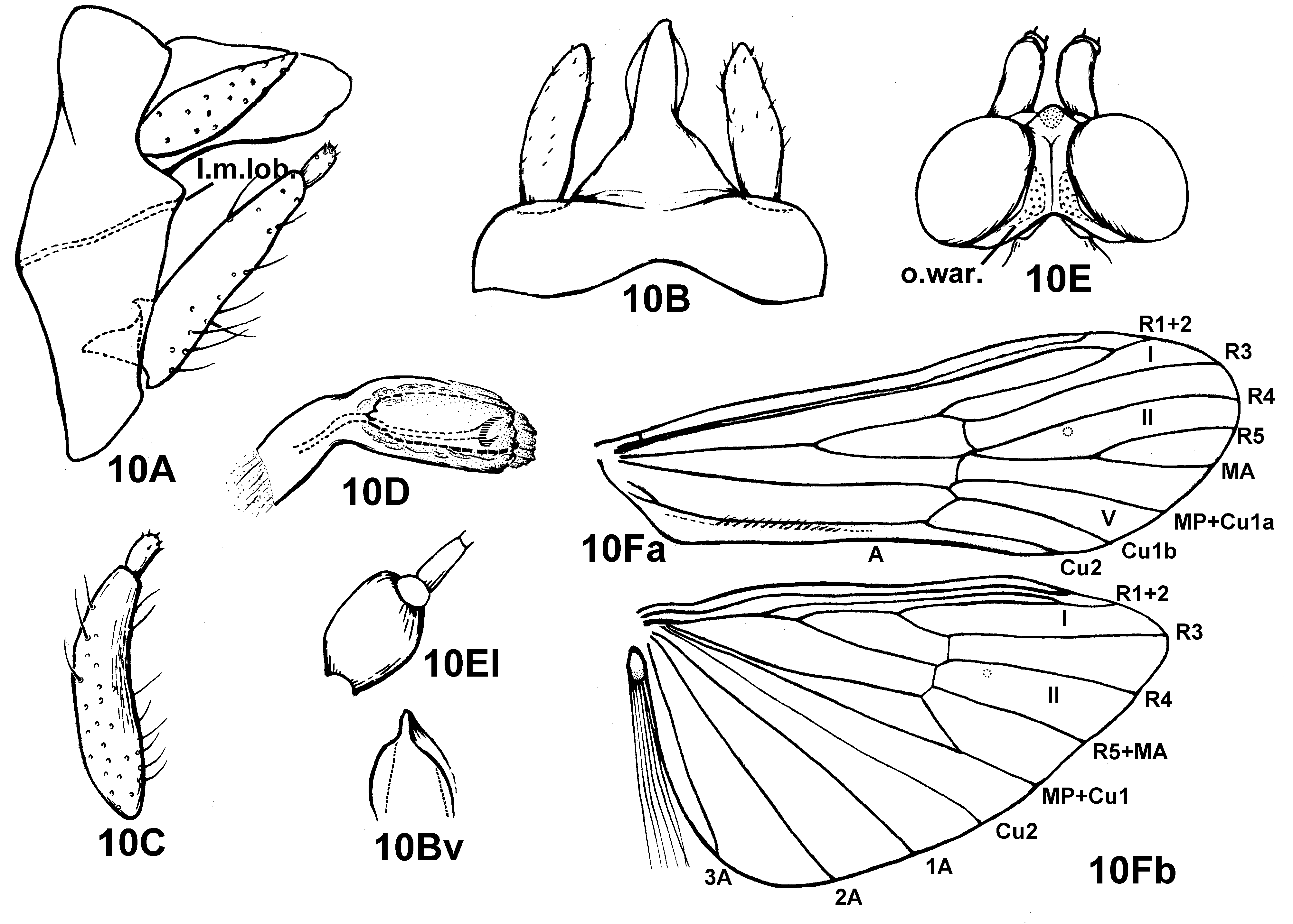
Male (in alcohol). Body small, head and thorax dark brown; palps dark brown and densely covered with dark brown hairs, sometimes first segment of maxillary palps dark, gradually paler to tip; cephalic and thoracic setal warts are concolorous with head and thorax; thoracic legs brown, forelegs slightly darker than mid- and hind legs. Forewings with convex termen, membranes dark brown, covered with dark brown hairs, hind wings pale. Vertex constricted; interocular distance about 3 times as long as broad with posterior margins widely divergent. Vertexal mediantennal compact setose wart single. Occipital compact setose warts (o.war.) large, elongate triangular, located at posterior half of compound eyes along their interior margins ( Fig. 10E View FIGURE 10 ).
Forewings ( Fig. 10 View FIGURE 10 Fa) each with length 6.5–7.5 mm (n = 6); R1 confluent with R2 shortly beyond end of Sc; forks I, II, and V present, all forks sessile, MA branching from R5+MA beyond nygma. Hind wings ( Fig. 10 View FIGURE 10 Fp) similar to those of most species of this genus, R2 very long, running into R1 at same level as end of Sc; Cu1 stem aligned with Cu1+MP, forming very acute angle (about 35°) at the confluence of MP and Cu1. A long basal brush is present on a small anal lobe at the base of each hind wing, this brush composed of about a dozen, very fine setae, with the same color and thickness as the marginal hairs.
Male genitalia. Tergum IX short, narrowly transverse, 6 times as broad as its median length in dorsal view ( Fig. 10B View FIGURE 10 ); in lateral view on each side, broadest at median portion at ends of diagonally longitudinal groove and nearly 2 times as long as dorsal or ventral margins, height of segment IX 5 times length of dorsal or ventral margins ( Fig. 10A View FIGURE 10 ); anterolateral margin protruded in 140° angle, posterior lateroapical corners not well developed, the lateromedian apical lobe (l.m.lob.) forming 110° angle ( Fig. 10A View FIGURE 10 ); dorsolateral longitudinal grooves absent. Segment X forming long hood, about 2 times as long as tall, with ventral margins slightly sinuous, almost straight in lateral view; in dorsal view, basal half of segment X broadly triangular, distal half quite compressed with only lower portion slightly extended laterad and with narrow apex not divided ( Fig. 10B View FIGURE 10 ), dorsomesal portion of apex highly elevated in ventrolateral view ( Fig. 10 View FIGURE 10 Bv). Preanal appendages slender, elongate, foliaceous, about 2 times as long as tergum IX, each broadest in basal 1/3, tapering to acute apex in lateral view ( Fig. 10A View FIGURE 10 ), elongate elliptical ( Fig. 10B View FIGURE 10 ) or slender (as in Fig. 9 View FIGURE 9 BI) in dorsal view. Inferior appendages much longer than segment X and preanal appendages, directed caudodorsad to apex of segment X; each with basal segment (coxopodite) dark and straight, not curved in lateral view ( Fig. 10A View FIGURE 10 ), almost straight with distal 1/3 gradually curved slightly inwards in ventral view, apical segment pale, short and small, obviously narrower than distal end of coxopodite, somewhat water-drop shaped, narrowed at base with outer margin convex in ventral view (Fig, 10C). Phallobase constricted at 1/3 distance from base and forming about 120° angle between tube-like basal portion and enlarged distal portion directed caudad; phallicata retracted within phallobase in our specimens ( Fig. 10D View FIGURE 10 ); phallotremal sclerite lightly sclerotized, U-shaped in ventral view.
Female genitalia. Segments IX and X and gonopods VIII and IX firmly united with each other. In dorsal view, terga IX+X forming short, transverse plate, roundly tapered posteriorly; anterior parts of tergite IX ( Fig. 8B, t View FIGURES 7 – 8 .IXa+IXb) weakly distinguishable posteriorly from posterior part of tergite IX and tergum X ( Fig. 8B, t View FIGURES 7 – 8 .IXc+X), terga IXc+X divided by median indentation into pair of setose apical lobes with rounded apices, bottom of indentation slightly produced at center, but never forming triangular projection; area between apical lobes small, triangular, and slightly depressed. In lateral view, outline of segments IX+X generally triangular, dorsal margin of fused IX+X ( Fig. 8A, t View FIGURES 7 – 8 .IXa+IXb, t.IXc+X) sloping downward in straight line without intervening declivity ( Fig. 8A View FIGURES 7 – 8 ); on each side, outlines of pleura IXa+IXb poorly discernible, small triangular projection ( Fig. 8A View FIGURES 7 – 8 , arrow) located on anterior margin nearly 1/3 distance below dorsal margin; subgenital plate ( Fig. 8C View FIGURES 7 – 8 , subg.pl. = fused external gonopods VIII, or e.gon.VIIIa+VIIIb) terminating in posterior wall of genitalic segments and separated from ventral lobes ( v.lob . = e.gon. IX) by deep concavity ( Fig. 8A View FIGURES 7 – 8 ). In ventral view, anal opening (a.) close to end of segments, below setose apical lobes (t.IXc+X), ventral lobes of anus ( v.lob .) small, with transverse posterior margins much shorter than longitudinal median margins, each ventral lobe bearing small patch of sensory pores ( Fig. 8C View FIGURES 7 – 8 ); weakly striate areas not well defined, pair of anterolateral sclerotized ridges (scl.rid.) each gently curved caudad, much longer than posterior margins of ventral lobes, this pair of ridges together forming semicircle ( Fig. 8C View FIGURES 7 – 8 ). Subgenital plate ( Fig. 8C View FIGURES 7 – 8 , subg.pl.) projecting posterad, subtriangular, about 2.5 times as broad as long, anterior margin of subgenital plate excised at mesal 1/3, with large patch of sensory pores ( Fig. 8C View FIGURES 7 – 8 , arrow) on each anterolateral corner of plate, with posterior margin terminating in single triangular projection between pair of oblique ridges of subgenital plate separating it from ventral lobes. Spermathecal sclerite (sper.scl.) having long triangular outline in ventral view, broadest near anterior end, and with 2 pairs of small ear-like subapical lobes ( Figs. 8A, 8C View FIGURES 7 – 8 ); in lateral view, dorsal margins of spermathecal sclerite nearly horizontal, its ventrolateral edges each with 2 small, lobe-like projections ( Fig. 8A View FIGURES 7 – 8 ).
Holotype male. PR CHINA: Jiang-xi Province , Gui-xi County, Xi-qi River , 59 km south east of Gui-xi, N28.3°, E117.2°, alt. 210 m, 5 June 1990, Coll. Yang L-f. GoogleMaps
Paratypes. Same data as holotype, 7 males and 8 females ( NAU); 3 males and 3 females ( CUAC); An-hui Province, Huang-shan City, Tang-kou Town, Fu-xi Village, Fu-xi stream, N30.085°, E118.142°, alt. 639 m, 10 July 2014, Coll. Xu J-h., 21 males and 3 females ( NAU).
Etymology. Latin, discaulis = without stem, with reference to the sessil fork II of each forewing.
Distribution. Oriental Region, southeastern China (Jiang-xi and An-hui).


No known copyright restrictions apply. See Agosti, D., Egloff, W., 2009. Taxonomic information exchange and copyright: the Plazi approach. BMC Research Notes 2009, 2:53 for further explanation.
|
Kingdom |
|
|
Phylum |
|
|
Class |
|
|
Order |
|
|
Family |
|
|
Genus |