
Halictophagus urucui, Kogan, Marcos, 2012
|
publication ID |
https://doi.org/ 10.5281/zenodo.282617 |
|
DOI |
https://doi.org/10.5281/zenodo.6174176 |
|
persistent identifier |
https://treatment.plazi.org/id/03DD3830-FF92-FFB0-FF54-FC9DFF77AB97 |
|
treatment provided by |
Plazi |
|
scientific name |
Halictophagus urucui |
| status |
sp. nov. |
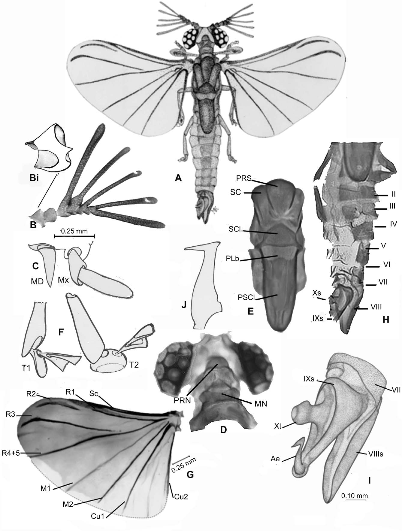
Halictophagus urucui sp. n.
( Figures 1 View FIGURE 1 A–J)
Holotype. Male collected in light trap at Coari, Rio Urucú, Amazonas, Brazil, 4°51’56.5” S, 65°0.4’56.6” W; Paulo Bührheim, N. O. Aguiar, and F.A. Penta, coll., 11–18 May, 1991. Specimen dissected and mounted in Hoyer’s, on three slides: slide 1 – right wing, left antenna, tarsus of leg 2; slide 2 – left wing, aedeagus; slide 3– head, thorax and abdomen (see note at end of paper). Set of original digital microphotographs copied onto a DVD, all deposited in the Strepsiptera Collection, INPA, Manaus, Brazil.
Description. Large species, length from tip of frons to edge of 8th abdominal sternite 3.49 mm ( Fig. 1 View FIGURE 1 A). Head ( Fig. 1 View FIGURE 1 D) 0.85 mm wide measured between edges of eye globes; eyes with 9 large, round eyelets occupying ca. 2/3 of the head’s width dorsally. Margin of frons concave between insertions of antennae. Antenna ( Fig. 1 View FIGURE 1 B) sevensegmented; segments III–VI extended laterally with slender flabella; segment I ( Fig. 1 View FIGURE 1 Bi) with pointed projection on distal margin; base of segment IV enlarged towards extension of flabellum; segments III–VII, including all flabella, covered with sensorial pits of various shapes and areas. Length of antennal segments (in μm): I = 92; II = 68; III + flabellum = 660; IV + flabellum = 561; V + flabellum = 470; VI + flabellum = 473; VII = 470. Mandible 59.5 μm long, cone-shaped ( Fig. 1 View FIGURE 1 C–MD), slightly sinuous; base of maxilla 51.0 μm; palp 77.5 μm; base and palp covered with sensorial trichomes ( Fig. 1 View FIGURE 1 C– MX).
Thorax ( Figs. 1 View FIGURE 1 D, 1E): pronotum much reduced, totally enclosed within the head space between posterior extension of eyes ( Fig. 1 View FIGURE 1 D–PN); mesonotum angular-shaped anteriorly, expanding laterally to connect to metanotum ( Fig. 1 View FIGURE 1 D–MN). Metanotum ( Fig. 1 View FIGURE 1 E) 1.37 mm long, 3.4 times longer than pro- and mesonotum combined; praescutum (PRS) broadly rounded anteriorly, moderately protruded from scuti (SC); scutellum (SCl) pentagon-shaped with anterior edge pointed and posterior edge slightly rounded; postlumbium broad, mildly constricted in center by inward curvature of both anterior and posterior margins; postscutellum (= postnotum) about as long as the other metanotal components together, broadly rounded posteriorly. Hind wing ( Fig. 1 View FIGURE 1 G) span from axillary joint to edge beyond end of R3 = 2.23 mm; R1 converging with Sc at distal tip within a diffusely pigmented area, R2 curved and about half the length of R3; R4 and R5 fused, M1 present, M2 and Cu1 reaching edge of wing; Cu2 developed. Tarsi three-segmented, basal segment elongated in mid- and hind legs ( Fig. 1 View FIGURE 1 F–T2); first segment of front tarsi rounded and pad-like ( Fig. 1 View FIGURE 1 F–T1), segments II and III of all pairs with well-developed paddle-like, fairly profusely pubescent pads.
Abdominal segments ( Figs. 1 View FIGURE 1 H–J) partly sclerotized ( Fig. 1 View FIGURE 1 H). The mounted specimen when photographed had terminal segments right rotated about 45o. Sternite VIII long, tapered sharply posteriorly ( Fig. 1 View FIGURE 1 I); sternite IX inverted sub-conically shaped with one edge stretched posteriorly and with a dorsal concavity, from center of which protrudes a clubbed cylindrical structure, possibly a greatly modified tergite X; aedeagus ( Fig. 1 View FIGURE 1 J) toothed distally, with distal branch at ca. 77o angle with shaft; shaft stout.
Discussion. Halictophagus urucui n. sp. differs from most other species within its zoological region by the shape of the aedeagus, which is similar to that of H. besucheti Luna de Carvalho, but the shaft in H. urucui is stouter and the apical process longer and at a sharper angle with the shaft. In addition, the postlumbium is wider and medially compressed in both front and hind margins. The wings have R4 and R5 fused, M1 present, and C2 well defined; base of front tarsi pad-like; base of antennal segment III with a pointed projection; and base of segment IV swollen towards projection of flabellum. The terminalia, with the long and cupped sternite VIII, the unusual shape of sternite IX and projection of tergite X ( Fig. 1 View FIGURE 1 I–Xt), does not seem to have similarity to the terminalia of any other described species.
Etymology. The name urucui relates to Rio Urucú, a tributary of the River Solimões, the type locality of the species.
| INPA |
Instituto Nacional de Pesquisas da Amazonia |
No known copyright restrictions apply. See Agosti, D., Egloff, W., 2009. Taxonomic information exchange and copyright: the Plazi approach. BMC Research Notes 2009, 2:53 for further explanation.