
Faunus ater, (LINNE, 1758)
|
publication ID |
https://doi.org/ 10.1111/j.1096-3642.2010.00687.x |
|
persistent identifier |
https://treatment.plazi.org/id/039687CF-FFD4-AB74-FC3D-9522FBA5DCF2 |
|
treatment provided by |
Valdenar |
|
scientific name |
Faunus ater |
| status |
|
FAUNUS ATER (LINNÉ, 1758) View in CoL
Material examined
Philippines: Bohol Island, Abatan River , 9°43.9′N, 123°53.4′E ( USNM 1143354 View Materials ) GoogleMaps .
Description
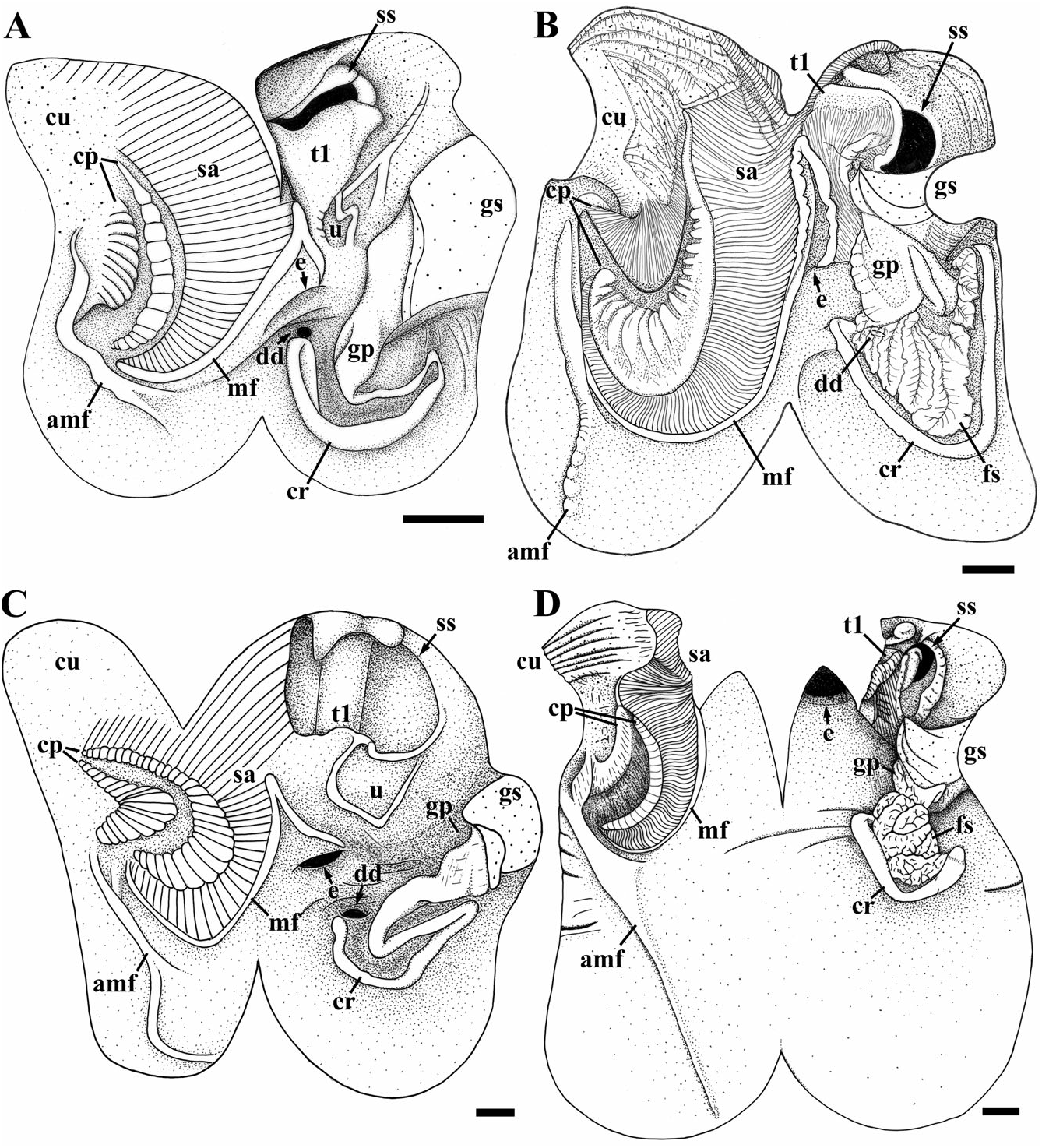
Marginal fold S-shaped ( Fig. 13B View Figure 13 ), terminating at posterior tip of sorting area; oesophageal aperture slightly behind tip of marginal fold recurved segment; sorting area highly curving, broad anteriorly, tapering rapidly to blunt tip; left margin of sorting area even, lacking conspicuous bulge; paired crescent-shaped pads bordering left margin of sorting area; inner crescentic pad broad, flat with thick, rounded transverse ridges along left margin; outer crescentic pad with fine, parallel grooves; accessory marginal fold extending posteriorly from tip of outer crescentic pad, bordering posterior margin of sorting area, with bifurcate posterior tip; sorting area pad lacking; proximal glandular pad tall, broad, longitudinally ridged, extending well posterior to gastric shield; distal glandular pad modified into crescent-shaped series of branched leaflets; foliate structure extending in broad arc across midgut floor towards right, embracing proximal glandular pad, to shallow depression behind gastric shield; single digestive gland duct opening at left of glandular pad; crescentic ridge long, extending from digestive gland duct, curving around foliate structure, separated by broad, shallow crescentic groove, joining distal tip of foliate structure behind gastric shield; accessory pads, caecum, and caecal folds lacking; U-shaped fold present below style sac aperture; typhlosoles unfused.
Remarks
Previously described by Houbrick (1991a: fig. 24), showing the large sorting area, inner crescentic pad (= ‘ribbed ridge’), the ‘platform-shaped’ proximal glandular pad, and the most intriguing aspect of Faunus ’ midgut anatomy – the modification of the distal glandular pad into a foliate structure. An identical modification of the distal glandular pad is present in Potadoma cf. freethi ( Fig. 14B View Figure 14 ; see below).
The distribution of the foliate structure among other cerithioideans has been misinterpreted, and hence its phylogenetic significance has been misunderstood. Houbrick (1991a) stated that a similar structure is present in Madagasikara spinosa (Lamarck, 1822) (as Melanatria fluminea ), citing Starmühlner (1969) as the source, and in Melanopsis species citing personal communication from Altaba. Examination of Starmühlner’s (1969: fig. 214) illustration of the midgut for Madagasikara spinosa confirms only the presence of the paired crescentic pads, which were evidently mistaken by Houbrick (1991a) for the foliate structure; absence of the structure in this species has been confirmed by personal examination and by Köhler & Glaubrecht (2010). Examination of several Melanopsis species (see Morton, 1952; Bilgin, 1973; present study) similarly reveals no foliate structure; Altaba undoubtedly confused the sorting area for a ‘foliate’ structure.
Houbrick (1991a) also likened the foliate structure to a system of leaflets in Campanile ( Houbrick, 1981a, 1989) and Gourmya ( Houbrick, 1981c) . Although there is insufficient information available to assess homology of the leaflets in Gourmya , the system of leaflets in Campanile occurs in a different position at the base of the major typhlosole, and is not considered here to be homologous with the foliate structure in Faunus . Instead, it is hypothesized to represent a modification of a flap at the base of the major typhlosole present in vermetids and strombids (see character 24, below).
Thus, the foliate structure – documented thus far only in Faunus and Potadoma – is apparently unique to pachychilids. Although autapomorphic and not coded in the analysis of Strong et al. (in press), the presence of this feature may support a close relationship between these two taxa; this hypothesis can only be evaluated in a more comprehensive phylogenetic analysis of the family. The distal glandular pad in several South American species of Doryssa (Pachychilidae) has a surface covered with U-shaped acini ( Simone, 2001: figs 230, 254). Like the foliate structure, these probably represent modifications for increasing surface area for absorption, and perhaps are homologous. Insufficient ecological information is available to speculate further what selective pressures may be driving these modifications uniquely among Pachychilidae .

No known copyright restrictions apply. See Agosti, D., Egloff, W., 2009. Taxonomic information exchange and copyright: the Plazi approach. BMC Research Notes 2009, 2:53 for further explanation.