
Euophrys flordellago, Barry J. Richardson, 2010
|
publication ID |
https://doi.org/ 10.5281/zenodo.194411 |
|
DOI |
https://doi.org/10.5281/zenodo.6205395 |
|
persistent identifier |
https://treatment.plazi.org/id/911E87A7-E418-FFD3-FF4B-F8EA38818DFD |
|
treatment provided by |
Plazi |
|
scientific name |
Euophrys flordellago |
| status |
sp. nov. |
Euophrys flordellago View in CoL n. sp.
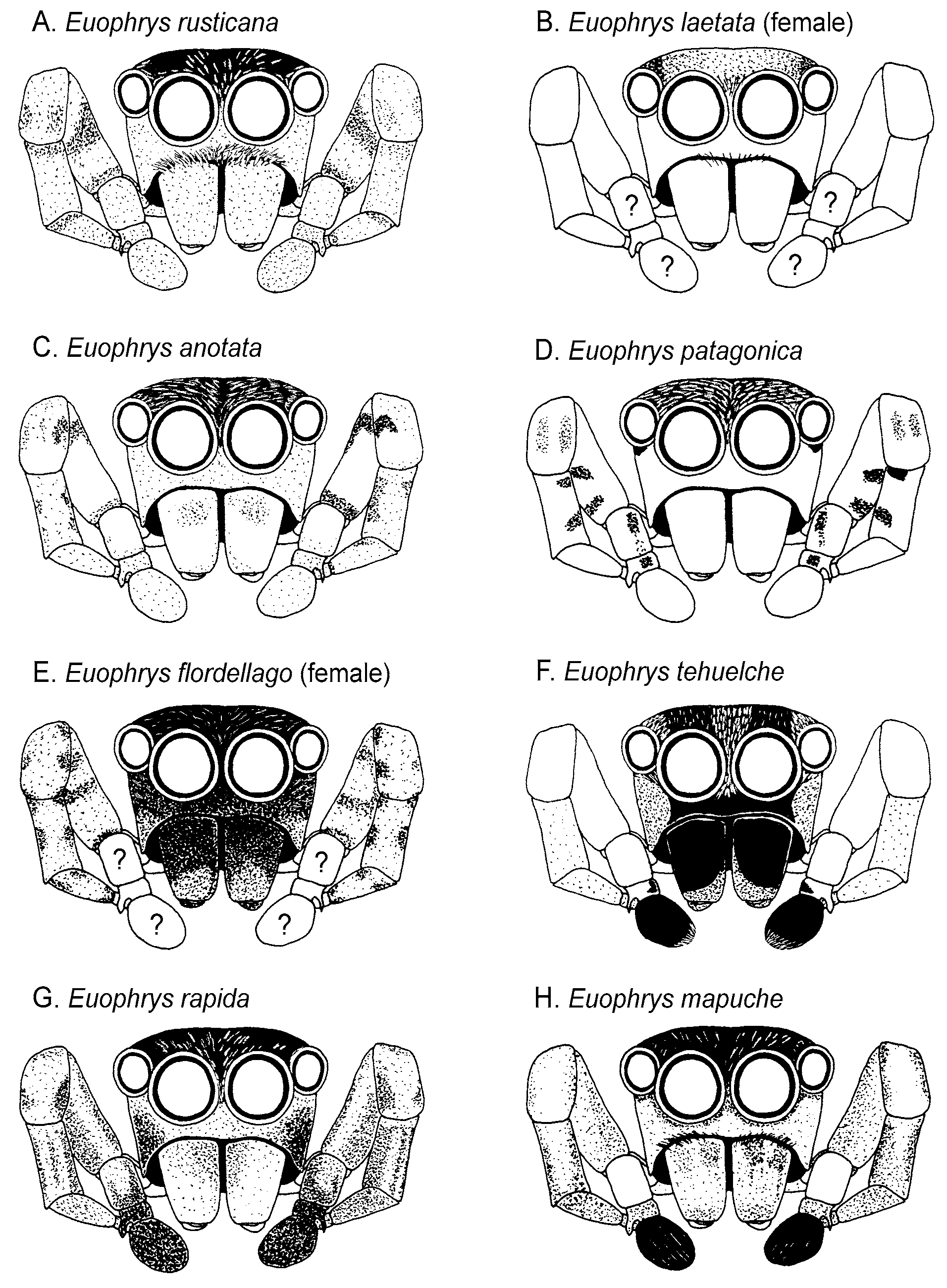
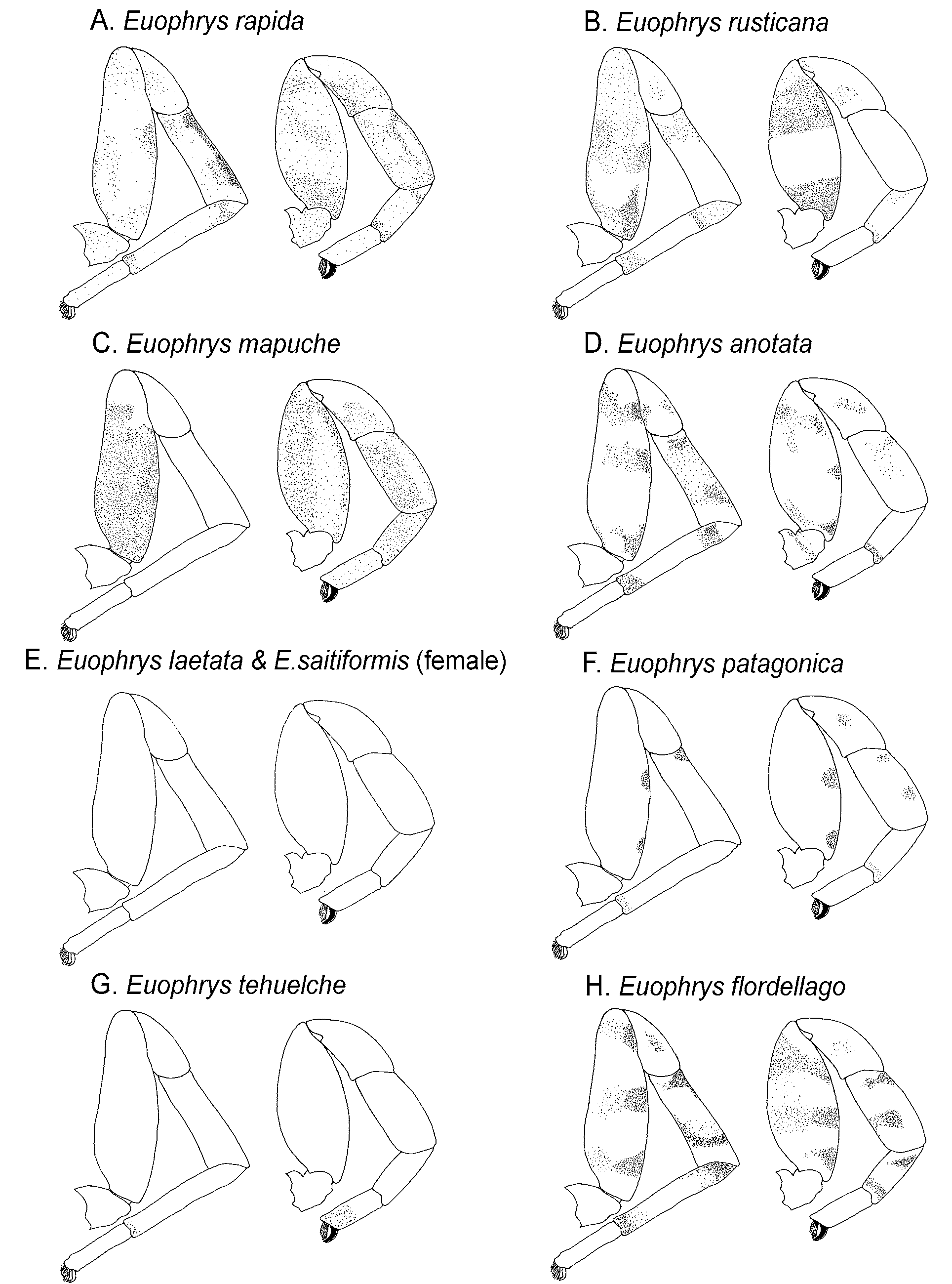
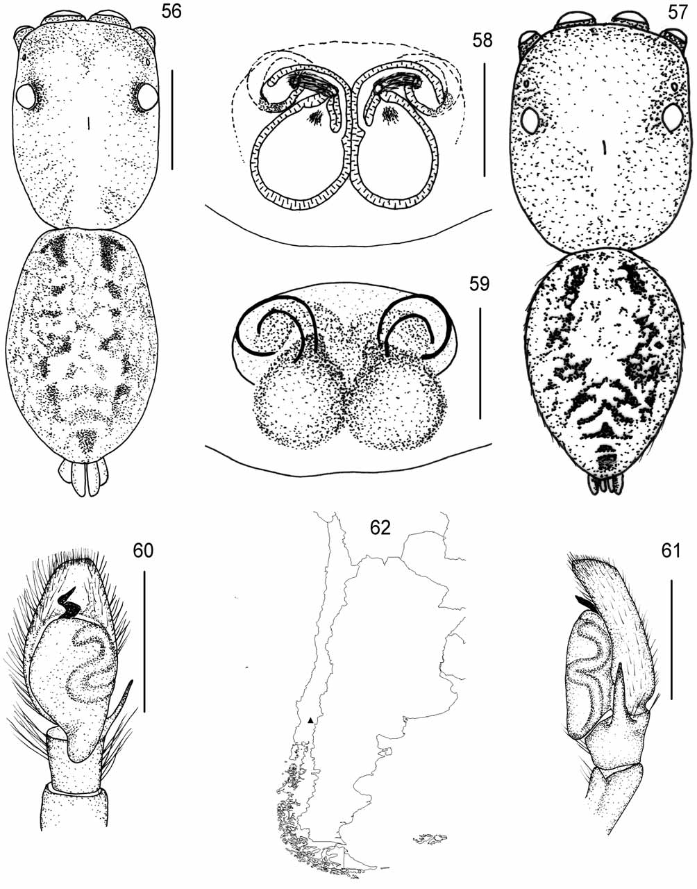
Figs 2 View FIGURE 2 E, 3H, 56–62
E. saitiformis: Braul et al. 1997: 146 View in CoL –147, figs 7–9. (male misidentified).
Etymology. A combination of letters, to be treated as a noun in apposition.
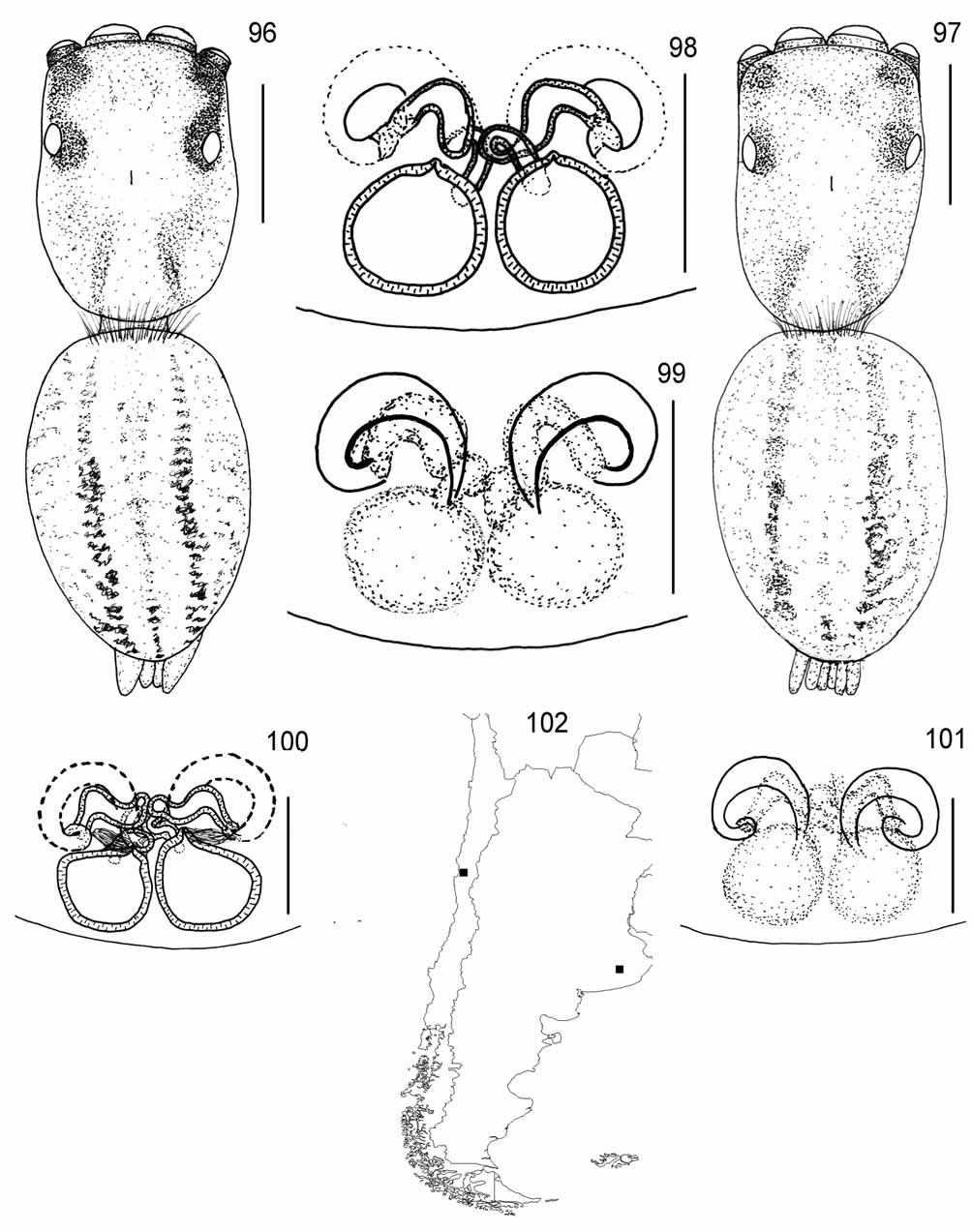
Remarks. The illustration of the male of E. saitiformis by Braul et al. (1997) based on material from Brazil, matches the abdominal colour pattern of this species but not that of E. saitiformis ( Fig. 96 View FIGURES 96 – 102 ). The fact that the external female genitalia of the two species are indistinguishable and different from that of other species may have misled these workers.
Type material: Holotype: ♀, Villarrica , Flor del Lago Ranch , Chile, 72.13°W, 39.20°S, 11 December 2003, I. Avila et al, hand ( BJR974 , MNNC 6805 ) GoogleMaps .
Paratype: 1♀, Villarrica , Flor del Lago Ranch , Chile, 72.13°W, 39.20°S, 12 December 2001, E. Arias et al. ( BJR1001 , CAS 9029849 ) GoogleMaps .
Diagnosis. Females of the species are distinguishable from the other species of Euophrys in Chile, except E. saitiformis , by the external morphology of the epigynum. The spermatheca is larger than the fossa and lies immediately posterior to it. It is distinguishable from E. saitiformis by the internal anatomy of the insemination duct which is uncoiled ( Fig. 58 View FIGURES 56 – 62 ), unlike that of E. saitiformis ( Fig. 98 View FIGURES 96 – 102 ). The animals also have black rather than yellow chelicerae and patterned rather than the plain yellow legs seen in E. saitiformis ( Figs 3 View FIGURE 3 H vs. 3E). The males of the species, though not seen, should be distinguishable from the other species of Euophrys in Chile, except E. tehuelche , by the presence of black chelae and by the pattern of black markings on the legs ( Fig. 3 View FIGURE 3 H) not seen on E. tehuelche ( Fig. 3 View FIGURE 3 G).
Description. Male: If it follows the pattern of the other species in this genus in Chile, the male will be similar in colouring to the female and therefore the Braul et al. (1997) description is of this species, then: Cephalothorax dark brown, a little lighter in the thoracic region with a lighter area around the fovea, white hairs on the posterior region. Yellowish bristles around the eyes. Chelicera with two promarginal teeth arising from a single base and a single retromarginal tooth. Clypeus narrow. Endites and labium dark brown. Sternum orange. Dorsal abdomen beige with darker patterning as in Fig. 57 View FIGURES 56 – 62 , ventral abdomen with scattered spots. Legs beige with brown areas. Palp ( Figs 60–61 View FIGURES 56 – 62 ): tibia with a thin and very long apophysis. Tegulum large and tapering, embolus sinuous, arising from the margin of the tegulum. Dimensions (after Braul et al. 1997): CL 2.26, EFL 1.58, CW 1.64, AL 2.17, SL 0.86, L1 5.0 0 (1.42+0.86+1.14+1.0 2+0.56), L 2 3.88 (1.20+0.64+0.80+0.76+0.48), L3 4.50 (1.30+0.64+0.80+1.26+0.50), L4 4.70 (1.40+0.64+0.90+1.18+0.58).
Female (holotype): Cephalothorax; pars cephalica orange, PLE surroundings black, pars thoracica pale yellow, with pale orange markings, not darker around lower margin. Clypeus narrow, pale-yellow. Chelicerae vertical, yellow, with two promarginal teeth and one retromarginal tooth. Endites, labium, sternum and ventral abdomen yellow. Abdomen pale yellow, elliptical with pair of parallel light brown bands leading to black patterning posteriorly ( Fig. 56 View FIGURES 56 – 62 ). Spinnerets yellow. All legs yellow. L1, femur with three dorsal and two distal dorsolateral spines. Patella with one pair of dorsolateral spines. Tibia and metatarsus with three and two pairs of ventrolateral spines, respectively. Similar spination on other legs to L1, except all have three distal spines rather than two on the femur and L3 tibia has four pairs of ventrolateral spines. Epigynum ( Figs 58–59 View FIGURES 56 – 62 ): poorly sclerotised, with two anterior fossae directed posteriorly and with distinctive, well separated copulatory openings. Insemination ducts relatively short and direct, spermathecae placed directly posterior to the fossae and much larger than the fossae, pear-shaped, with pointed fertilization ducts on anterior dorsal edge. Dimensions (holotype): CL 2.29, EFL 0.93, CW 1.55, AEW 1.49, AMEW 0.93, PEW 1.42, SL 0.93, L1 3. 5 4 (1. 1 8+ 0. 6 8+ 0. 6 8+ 0. 5 0+ 0. 5 0), L 2 3. 6 6 (1. 1 8 + 0. 6 8+ 0. 6 8+ 0. 6 2+ 0. 5 0), L 3 4. 7 7 (1.49+0.68+0.93+0.80+0.87), L4 4.83 (1.49+0.68+0.93+1.11+0.62).
Distribution. Known only from type locality, and southeastern Brazil (Braul et al. l997).
No known copyright restrictions apply. See Agosti, D., Egloff, W., 2009. Taxonomic information exchange and copyright: the Plazi approach. BMC Research Notes 2009, 2:53 for further explanation.
|
Kingdom |
|
|
Phylum |
|
|
Class |
|
|
Order |
|
|
Family |
|
|
Genus |
Euophrys flordellago
| Barry J. Richardson 2010 |
E. saitiformis: Braul et al. 1997 : 146
| Braul 1997: 146 |