
Dinelytron museunacional, Heleodoro & Rafael, 2020
|
publication ID |
https://doi.org/ 10.1016/j.jcz.2020.01.005 |
|
DOI |
https://doi.org/10.5281/zenodo.3716936 |
|
persistent identifier |
https://treatment.plazi.org/id/9D0A8794-FFDA-0668-0668-671CB887E821 |
|
treatment provided by |
Plazi |
|
scientific name |
Dinelytron museunacional |
| status |
sp. nov. |
Dinelytron museunacional View in CoL sp. nov.
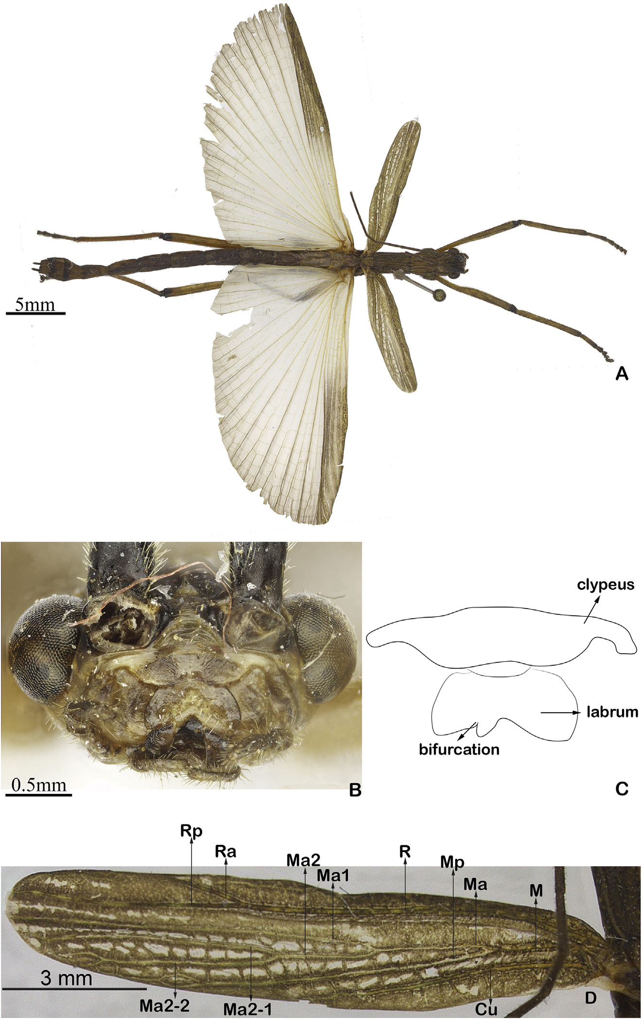
Fig. 17A-B View Fig .
Examined material. Holotype _: “ Parque Sooretama, Sede, Sooretama , Espírito Santo, 20.ii.1959, Martins Gomes de Amorim leg.” ( MNRJ LOST IN THE BURN).
Paratype _: “ Itatiaia , Rio de Janeiro, 10.xi.1961, na luz do lampi~ ao [collected on lamp], coleta manual [manual collecting], 19-23 horas” ( MNRJ LOST IN THE BURN) .
Etymology. The species name is dedicated to the Museu Nacional do Rio de Janeiro to solidarize with its burning.
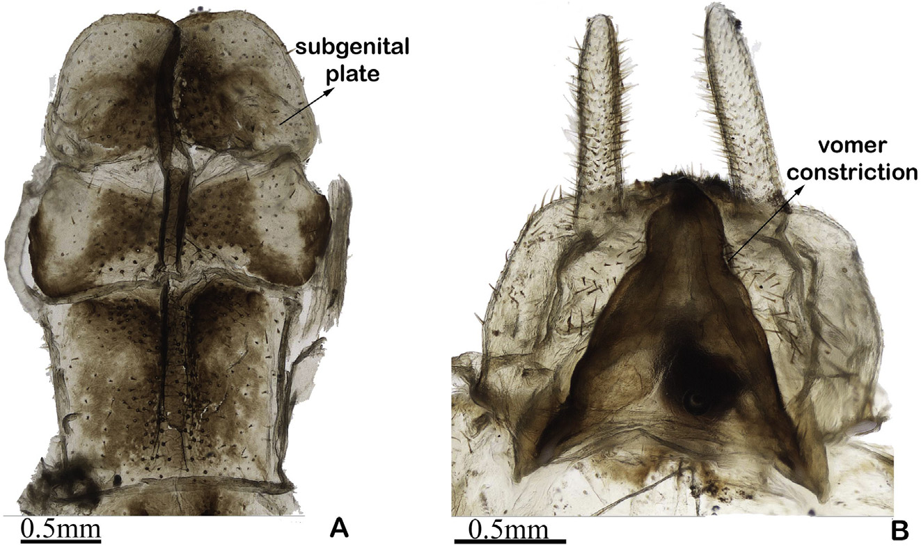
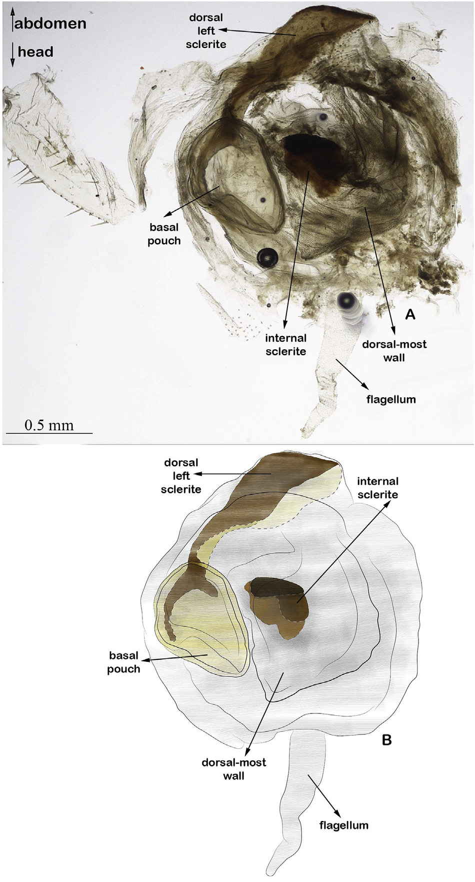
Diagnosis. Labrum asymmetrical, apically bifurcated at right half ( Fig. 17B, C View Fig ). Tegmina mud green with white spots; radial vein light brown, remaining veins translucid ( Fig. 17A, D View Fig ). Radial vein subdivided in Radial anterior and posterior at apical third; Radial anterior straight, approximately four times shorter than Radial posterior; Radial posterior straight, reaching tegmina apex ( Fig. 17D View Fig ). Medial bifurcated in Medial anterior and posterior at the basal third; Medial anterior bifurcated in Medial anterior 1 and 2 at approximately half the tegmina length; Medial anterior 1 straight, reaching tegmina apex; Medial anterior 2 bifurcated in Medial anterior 2-1 and 2-2, with both bifurcations straight and not reaching tegmina apex ( Fig. 17D View Fig ). Subgenital plate with basal margin conspicuously concave, apical margin with slight emargination medially ( Fig. 18A View Fig ). Vomer with deep constriction in apical third ( Fig. 18B View Fig ). In dorsal view ( Fig. 19 View Fig ): basal pouch egg-shaped; dorsal left sclerite with acute apex. Internal sclerite reniform, 3.5 times shorter than basal pouch.
Description. Head. General coloration light brown ( Fig. 17A View Fig ). Frontal suture conspicuous, forming an inconspicuous dark triangular yellow sulcus ( Fig. 17B View Fig ); coronal suture inconspicuous. Clypeus light yellow, medially with dark brown spot; basal margin medially emarginated, lateral margin convex, apical margin slightly sinuous, four times wider than high ( Fig. 17B, C View Fig ). Area between clypeus and labrum ellipsoid, conspicuous ( Fig. 17B, C View Fig ). Labrum asymmetrical, apically bifurcated at right half, laterally light yellow with dark brown spots ( Fig. 17B, C View Fig ). Antenna with flagellum dark brown ( Fig. 17A View Fig ); flagellomere 1 rectangular, longer than wide, 2.2 times longer than flagellomere 2; flagellomere 2 subtriangular; flagellomere 3 rectangular, longer than wide, 1.2 times longer than flagellomere 2. Compound eye globose, light brown with dark brown spots ( Fig. 17A, B View Fig ).
Thorax. General coloration light brown ( Fig. 17A View Fig ). Pronotum with two longitudinal parallel-arched sulci; posterior margin with horizontal black spot ( Fig. 17A View Fig ). Mesonotum 1.6 times longer than pronotum, with conspicuous longitudinal medial carina ( Fig. 17A View Fig ). Coxopleurite smooth, concolor with pronotum. Mesothoracic epimeron subtriangular, with straight margins, rugose, concolor with pronotum. Mesothoracic episternum idem with epimeron. Metapleural region dark brown smooth, shiny. Thoracic sterna dark brown, smooth. Mesobasisternum with medial circular sclerite concolor with sterna.
Legs. All legs: dorsally and ventrally light brown; anteriorly and posteriorly covered by setae. Anterior femur dorsally rugose, with dark brown spots, longitudinal medial carina; ventrally with small setae. Mid leg dorso-posteriorly with three rhomboid projections. Posterior femur with anterior and posterior margins straight.
Wings. Tegmina mud green with white spots; radial vein light brown, remaining veins translucid ( Fig. 17A, D View Fig ). Radial vein subdivided in Radial anterior and posterior at apical third; Radial anterior straight, approximately four times shorter than Radial posterior; Radial posterior straight, reaching tegmina apex ( Fig. 17D View Fig ). Medial bifurcated in Medial anterior and posterior at the basal third; Medial anterior bifurcated in Medial anterior 1 and 2 at approximately half the tegmina length; Medial anterior 1 straight, reaching tegmina apex; Medial anterior 2 bifurcated in Medial anterior 2-1 and 2-2, with both bifurcations straight and not reaching tegmina apex ( Fig. 17D View Fig ). Cubital vein long, straight ( Fig. 17D View Fig ) Posterior wing with veins light yellow.
Abdomen. Abdominal terga dark brown, opaque ( Fig.17A View Fig ). Terga 1-6 dorsally rectangular, longer than wide with conspicuous longitudinal carina ( Fig. 17A View Fig ). Tergum 7 dorsally rectangular, longer than wide, with longitudinal medial carina. Tergum 8 dorsally trapezoidal, as long as wide, 1.4 times longer than Tergum 9. Tergum 9 rectangular, approximately 1.2 times wider than tergum 10. Tergum 10 with straight base, arched, and convex lateral and apical margin; 1.5 times shorter than tergum 9. Cercus cylindrical, with small setae, and oblong apex ( Fig. 18B View Fig ). Sterna 1-7 rectangular, longer than wide, gradually shortening in length. Sternum 7 rectangular, 1.3 times longer than wide, with basal and apical margin slightly convex; lateral margin straight (18A). Sternum 8 rectangular, 1.8 times wider than long, with slightly concave basal margin; lateral margin convex; apical margin conspicuously concave ( Fig. 18A View Fig ). Subgenital plate with basal margin conspicuously concave, apical margin medially with slight emargination ( Fig.18A View Fig ). Vomer dark brown, with deep constriction at apical third, apically acute ( Fig. 18B View Fig ).
Genitalia ( Fig. 19 View Fig ). Basal pouch egg-shaped in dorsal view. Dorsal left sclerite with acute apex. Internal sclerite reniform, 3.5 times shorter than basal pouch. Ventral lobe having small scattered spines. Flagellum present.
Measurements. Body length 37.2-38.4; dorsal head length 2.5-2.7; pronotum 2.4-2.8; mesonotum 2.7-2.8; anterior femur 7.4-7.5; anterior tibia 5.5-5.8 mid femur 5.3; mid tibia 5.0-5.1; posterior femur 9.9; posterior tibia 7.0-7.2.
Type condition. All lost in September 2018 burning of the MNRJ.
Geographical records. Brazil, Espírito Santo: Sooretama ; Rio de Janeiro: Itatiaia.
Remarks. Di. museunacional sp. nov. has the longest body of the Dinelytron species. It is morphologically similar to Di. ramusculus sp. nov. but it can be differentiated by the subgenital plate, which is concave at the basal margin (versus convex in Di. ramusculus sp. nov.). It is the only species with a bifurcation in the right side of the labrum, which is present in both holotype and paratype, which come from different populations. Therefore, the possibility of this character being a populational abnormality is excluded.
| MNRJ |
Museu Nacional/Universidade Federal de Rio de Janeiro |
No known copyright restrictions apply. See Agosti, D., Egloff, W., 2009. Taxonomic information exchange and copyright: the Plazi approach. BMC Research Notes 2009, 2:53 for further explanation.