
Danionella priapus, Britz, Ralf, 2009
|
publication ID |
https://doi.org/ 10.5281/zenodo.191105 |
|
DOI |
https://doi.org/10.5281/zenodo.6213021 |
|
persistent identifier |
https://treatment.plazi.org/id/03B3E452-FFFC-FFC3-BF92-2F82A465BB66 |
|
treatment provided by |
Plazi |
|
scientific name |
Danionella priapus |
| status |
sp. nov. |
Danionella priapus View in CoL , sp. nov.
( Figs. 1–4 View FIGURE 1 View FIGURE 2 View FIGURE 3 View FIGURE 4 )
Danionella sp.: Jorai River, Britz et al., 2009
Type material. Holotype: BMNH 2009.9.9.1, male, 14.4 mm SL; India, West Bengal, Jalpaiguri District, Brahmaputra drainage, Jorai River, a tributary of the Sankosh at Laskarpara, outskirts of Barobisha town. 26° 28' 52.3''N, 89° 49' 29.8''E; M. Das, 2 Apr 2008.
Paratypes: BMNH 2009.9.9.2-37 (36), 11.8–16.0 mm SL; data as for holotype. BMNH 2009. 9 .9.38–43 (6), cleared and double stained, 13.6–15.9 mm SL; data as for holotype.
Diagnosis. Danionella priapus is distinguished from its congeners, D. dracula , D. translucida and D. mirifica , by the presence in adult males of a conical projection of the genital papilla situated between the pelvic fins, which form a funnel-like structure (vs. genital papilla not developed as a conical projection and pelvic fins not funnel-shaped), by possessing 8 pectoral-fin rays (vs. 6–7) and 20–21 anal–fin rays (vs. 12–14 in D. dracula , 12–16 in D. translucida , and 17–19, rarely 20 in D. mirifica ), and by having the last anal-fin pterygiophore inserted in front of haemal spine of vertebra 27 or 28 (vs. 21 or 22 in D. translucida , 23–25 in D. mirifica , and 22–24 in D. dracula ). It differs further from D. translucida and D. mirifica in the presence of a median and two paramedian rows of pigment cells on the dorsal side of the body (vs. pigment rows absent). It is further distinguished from D. dracula and D. mirifica by 7–8 dorsal caudal procurrent rays (vs. 5–6) and 6–8 ventral procurrent rays (vs. 4–5). In addition it differs from D. dracula in having two upper jaw bones (vs. one); edges of jaw bones entire, without processes (vs. dorsal face of dentary and ventral face of upper jaw in males each with a single row of 6–13 odontoid processes, anterior most large and canine-like); presence of maxillary-mandibular cartilage (vs. absence); absence of membrane bone flanges on basipterygium (vs. presence); and 9+9 principal caudal fin rays (vs. 7–8 + 7–8). It differs also from D. translucida in having 15– 16 abdominal (vs. 13–14) and 22–23 caudal (vs. 19–20) vertebrae and a well developed lateral stripe extending from ear capsule to caudal peduncle (vs. a row of few melanophores midlaterally, restricted to posterior part of body).
Description. Morphometric information, based on 15 specimens, is presented in Table 1. Known maximum size 16.0 mm SL. Head and eye large, mouth supraterminal ( Fig. 1 View FIGURE 1 ). Body laterally compressed, elongate; dorsal fin short, situated opposite posterior half of long anal fin. Caudal fin furcate, with remnants of larval fin fold in front of its dorsal and ventral margins. Remnant of pre-anal larval fin fold present in females only ( Fig. 1 View FIGURE 1 b). Anus and genital papilla of adult males located between pelvic fins, in females at normal position in front of anal fin. Genital opening of adult males located at tip of short, conical projection at proximal end of funnel-like structure formed by pelvic fins being fused along their midline ( Figs. 1 View FIGURE 1 a, 2). Pseudotypanum present in body musculature at lateral side of anterior gas-bladder chamber, rendering its pigmented surface visible ( Fig. 1 View FIGURE 1 ). Body muscles greatly thinned out at lateral side of posterior gas bladder chamber. Scales absent. Lateral-line canals and pores on head and body absent.
Skull, hyopalatine arch, gill arches, and endoskeletal shoulder girdle mostly cartilaginous with perichondral ossifications giving skeleton overall larval appearance. Numerous bones and cartilages absent: kinethmoid (and kinethmoid cartilage), preethmoid, vomer, dermethmoid, nasal, parietal, intercalar, extrascapular, infraorbitals 2–5, ectopterygoid, metapterygoid, urohyal, hypobranchials 1–3, sclerotic bones, posttemporal, postcleithrum, mesocoracoid, pectoral radials 3–4 (and pectoral radial cartilages 3–4), pelvic radials 1–3 (and pelvic radial cartilages 2–3), supraneurals 2 and 5–9 (and supraneural cartilages 5–9), middle and distal radials in dorsal and anal fins, epineurals, epipleurals, uroneural 2, and scales. Ceratobranchial 5 heavily ossified, bearing 4–5 tri- to quadricuspid teeth plus the tips of one or two replacement teeth. Outer arm of os suspensorium sexually dimorphic, being enlarged and hyperossified in males and in confluence with lateral process of second vertebra via anterior process and with inner arm of os suspensorium below tripus via broad median flange. Males with a relatively large conical nodule of cartilage between fifth rib and outer arm of suspensorium, its proximal surface approaching swimbladder wall.
Vertebrae totalling 37 (2) or 38 (4), abdominal vertebrae 15 (3) or 16 (3); caudal vertebrae 22 (5) or 23 (1). Ribs present on vertebrae 5 to 14. Dorsal-fin rays ii,6,i (5) or ii,7,i (1). First dorsal-fin ray pterygiophore inserted behind neural spine of vertebra 21 (3) or 22 (3), and last in front of neural spine of vertebra 25 (2) or 26 (4). Anal-fin rays ii,17,i (2) or ii,18,i (4). Number of anal-fin pterygiophores in front of first hemal spine: 0 (2), 1 (2) or 2 (2). Last anal-fin pterygiphore inserted in front of neural spine of vertebra 27 (3) or 28 (3). Principal caudal-fin rays 9+9 (6) plus 7 (5) or 8 (1) dorsal and 6 (2), 7 (1) or 8 (3) ventral procurrent rays. Pectoral-fin rays 8 (6) and pelvic-fin rays 5 (6).
Colouration. Pigmentation in alcohol specimens restricted to several rows of melanophores ( Figs. 1 View FIGURE 1 , 3 View FIGURE 3 ): mid-lateral row along horizontal septum from shoulder girdle to hypural plate; ventral row from in front of and slightly above anal-fin base along ventral larval fin fold to end of hypural plate; row along anal-fin base; mid-dorsal row starting behind head with several enlarged melanophores forming small blotch and extending usually halfway between head and anterior base of dorsal fin, rarely to anterior dorsal-fin base; paired dorsal paramedian rows from head to base of caudal fin; dorsal-fin base row; abdominal mid-ventral row from ventral tip of cleithrum to anus; several melanophores on otic capsule; melanophores capping dorsal and dorso-lateral face of gasbladder chambers and their connecting duct and lining peritoneum covering gonads; streaks of melanophores lining anterior edge of lower cleithrum, ventral edge of opercle and preopercle, branchiostegal rays and fin rays in dorsal, anal and caudal fins and anterior two or three rays of pectoral fin; and several larger melanophores near symphysis of lower jaw. Head with supraoccipital blotch formed by several large contiguous melanophores, with numerous large scattered melanophores between supraoccipital blotch and epiphyseal bar, with a preepiphyseal blotch, and a few prenasal melanophores.
In life, body colourless and largely translucent ( Fig. 4 View FIGURE 4 ), except for melanophore patterns described above, a thin yellowish-greenish line running along body at level of neural tube; golden chromatophores forming ring around pupil, capping swimbladder chambers and connecting duct.
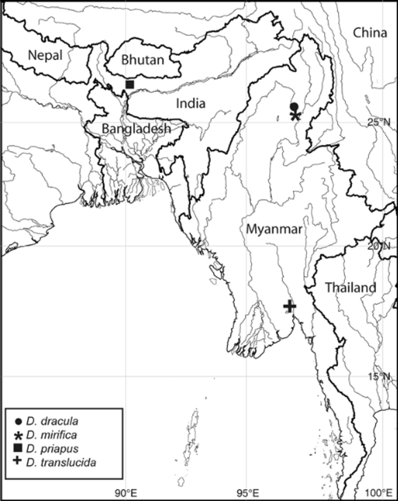
Distribution. The species is known only from the type locality, Jorai River in West Bengal, India ( Fig. 5 View FIGURE 5 ).
Etymology. The species name priapus is derived from the greek ρίαπος, the god of fertility of Greek mythology. It was inspired by the conical projection of the genital papilla in males, which—at a superficial level—is reminiscent of the penis of mammals. A noun in apposition.
Holotype Paratypes
Male Males Females
Range Mean St. Dev Range Mean St. Dev
Standard length (SL) 14.4 12.5–14.1 13.7–15.0
In % of SL
Head length 17.4 17.4–20.0 18.3 0.9 19.0–20.7 19.8 0.7 Predorsal-fin length 70.1 68.7–70.4 69.4 0.7 68.7–71.0 70.1 0.8 Prepelvic-fin length 30.6 29.8–32.8 30.8 1.1 34.0–35.6 34.8 0.5 Preanal-fin length 52.1 51.1–52.1 51.5 0.4 53.3–54.8 54.2 0.6 Snout to anus 29.9 29.1–32.0 30.1 1.0 51.4–52.7 51.9 0.4 Body depth at dorsal-fin origin 13.2 13.1–15.2 14.1 0.7 14.4–16.0 15.4 0.5 Caudal-peduncle depth 7.6 7.5–8.5 8.0 0.3 7.5–8.9 8.0 0.5 Caudal-peduncle length 18.8 17.9–21.6 19.6 1.4 18.1–21.3 19.6 1.1 Length of dorsal-fin base 9.7 9.7–11.3 10.7 0.5 10.3–11.7 10.9 0.5 Length of anal-fin base 30.6 28.4–30.7 29.5 0.9 26.0–28.3 27.4 0.7 Length of pelvic fin 8.7 7.2–8.7 7.4 0.3 6.3–8.3 7.0 0.6
In % of HL
Eye diameter 44.0 40.0–44.0 40.8 1.5 35.0–39.3 37.3 1.3 Snout length 16.0 16.0–16.7 16.1 0.3 17.9–21.4 20.0 1.2


No known copyright restrictions apply. See Agosti, D., Egloff, W., 2009. Taxonomic information exchange and copyright: the Plazi approach. BMC Research Notes 2009, 2:53 for further explanation.