
Cratera joia (Froehlich, 1956)
|
publication ID |
https://doi.org/ 10.11646/zootaxa.4500.4.3 |
|
publication LSID |
lsid:zoobank.org:pub:70672C0A-EC78-40BA-85EE-6206184CE0F0 |
|
persistent identifier |
https://treatment.plazi.org/id/4F7187CF-C55D-FFE5-D5A8-F498FD1CFF6E |
|
treatment provided by |
Felipe |
|
scientific name |
Cratera joia (Froehlich, 1956) |
| status |
|
Cratera joia (Froehlich, 1956) View in CoL
Synonymy
Material examined. Syntypes. Parque Estadual Serra do Mar (between Curitiba and Paranaguá), State of Paraná, Brazil, July 1953, Froehlich and Froehlich, coll. Syntype A ( MZUSP PL 2121 ): sagittal sections of copulatory apparatus on 3 slides (S444-S446) . Syntype B ( MZUSP PL 2122 ): sagittal of copulatory apparatus on 4 slides (S447-S450) . Syntype A or B: transverse sections of pre-pharyngeal region on 2 slides (S440-S441); sagittal sections of pharynx on 2 slides (S442-S443). Syntype C (laboratory number: F7239; MZUSP PL 2120 ): transverse sections of cephalic region on 13 slides; horizontal sections of ovaries region on 5 slides; transverse sections of prepharyngeal region on 13 slides; sagittal sections of pharynx and copulatory apparatus on 15 slides .
Distribution. Only known from the type locality, Parque Estadual da Serra do Mar, State of São Paulo, Brazil.
Diagnosis. Species of Cratera with some 40 mm in maximum length. Anterior tip, ventrally and dorsally, rust colored, same as a median and a pair of marginal lines. Rest of dorsum dark. Epidermis ciliated dorsally and ventrally. Eyes over entire dorsum. Sensory pits pear-shaped. Glandular margin unsharpened. Proximal portion of prostatic vesicle bifurcated, extrabulbar, and attached to penis bulb. Penis bulb extends 0.6 millimeters anteriorly to penis papilla. Ejaculatory duct may be slightly dilated distally. Dorsal insertion of penis papilla slightly anterior to the ventral. Penis papilla projects into female atrium. Distal dorsal section of penis papilla lined with an epithelium higher and sinuous; this epithelium pierced by numerous cyanophil glands. Common glandular duct present.
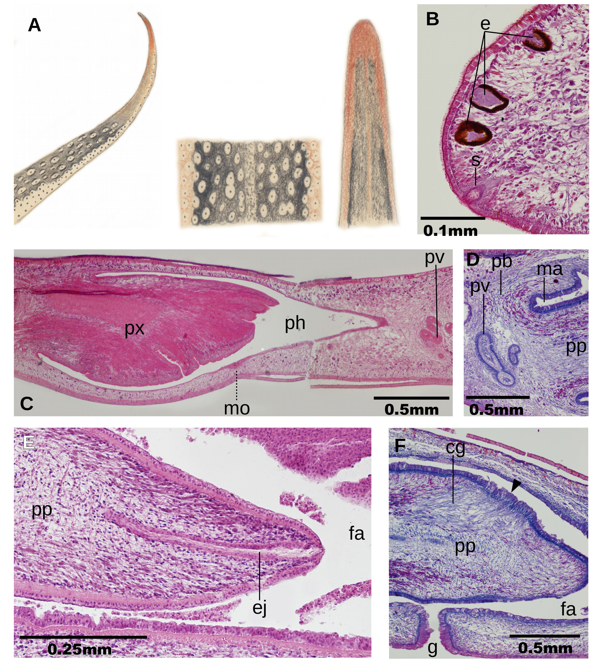
Description. External aspect. Preserved animals up to 40 mm long, and 5 mm wide. Body margins nearly parallel; anterior and posterior end pointed ( Fig. 5A View FIGURE 5 ). Dorsum slightly convex. From the original description, eyes spread over entire dorsal surface. Sensory pits are invaginations 50 µm deep; this invagination is slightly different from the straight shape in most geoplaninids, since it widens slightly at half its depth ( Fig. 5B View FIGURE 5 ). Sensory pits are arranged in a single row. A ~10 µm long section of anterior apex of the body was lost; it is assumed sensory pits contour anterior end. Behind, sensory pits extend backwards to at least a length equal to 35% of body length. Relative mouth:body length, 70% and relative gonopore:body length, 80%, in syntype C.
Internal morphology. Creeping sole occupies 90% ventral body width. Externally to the creeping sole, there is a 200 µm wide band of nonciliated epidermis; remaining epidermis ciliated, even dorsally ( Fig. 5B View FIGURE 5 ). Erythrophil glands producing erythrophil granules pierce dorsal epidermis. A second type of gland cell discharges its granules through the dorsal epidermis; these granules stained brownish black in syntypes A and B; and orangish in syntype C. The two types of glands present a progressive increase in number from the mid region to the body margins, where suddenly they cease, so the glandular margin is indistinct. Ventral epidermis pierced by two types of glands producing erythrophil and cyanophil granules, respectively.
Cutaneous musculature with the usual three layers present in the subfamily Geoplaninae : subepithelial circular (one-fiber thick), diagonal with decussate bundles (7 µm thick) and an innermost longitudinal layer (25 µm thick), the latter is arranged in bundles. Cutaneous musculature as thick as 8% of body height. Parenchymal musculature composed of three muscular layers: a weak dorsal layer with diagonal decussate fibers (10-12 µm thick) and a supraintestinal (25 µm thick) loose layers and a subintestinal transverse layer (40 µm thick). Ventral nerve plate present.
Relative position mouth:pharyngeal pouch length, of 73%. ( Fig. 5C View FIGURE 5 ). Pharynx cylindrical, with its dorsal insertion slightly placed backwards. Esophagus present, with 30% of pharynx length. Outer pharyngeal musculature consisting of a subepithelial layer (3 µm thick) of longitudinal muscle, followed by a layer (5 µm thick) of circular fibers. Inner pharynx musculature consisting of a subepithelial layer (60 µm thick) of circular musculature with some longitudinal fibers interspersed.
Testes located between supraintestinal parenchymal muscle layer and intestine. Testes are arranged in singleto-double row on each side of the body. They extend from the level of the ovaries (equal to 35% of the body length in syntype C) to closely the root of the pharynx (equal to 61% of the body length in syntype C). Penis bulb consisting of packed muscle fibers variously oriented. This bulb extends from 0.6 millimeters anterior to penis papilla to envelope the anterior half of the male atrium approximately. Sperm ducts run laterally to open into short paired branches of the extra-bulbar portion of the prostatic vesicle ( Fig. 5D View FIGURE 5 ). Extra-bulbar portion of the prostatic vesicle is C-shaped in lateral view and is attached to the penis bulb. Subsequently, the prostatic vesicle penetrates the anterior section of the penis bulb to continue as a sinuous tube until it continues as the ejaculatory duct inside the penis papilla. The prostatic vesicle is lined by a columnar, ciliated epithelium. This epithelium is surrounded by a 15 µm thick circular muscle. Glands producing very weakly stained reddish (syntypes A and B) or bluish (syntype C) secretion open into prostatic vesicle. Ejaculatory duct runs straight through the penis papilla. The ejaculatory duct is lined with a cuboidal, ciliated epithelium. It is surrounded by a 5 µm thick circular muscle. The lumen of ejaculatory duct is 12.5 µm in diameter proximally, subsequently it narrows to 7.5 µm and it widens slightly to 17.5 µm distally before opening at the tip of the penis papilla (syntype A, Fig. 5F View FIGURE 5 ). There is no distal widening of the ejaculatory duct in syntypes B and C.
Penis papilla relatively long, cylindrical, pointed distally, and with its dorsal insertion slightly anterior than the ventral. It exceeds the gonopore level to occupy anterior half of the female atrium. This papilla is lined with an epithelium which is columnar anteriorly, and cuboidal distally and is underlain by a circular muscle (5 µm thick), followed by a longitudinal muscle (12 µm thick). Most epithelium of penis papilla is pierced by erythrophil glands. These glands are absent in distal dorsal section of the penis papilla, which in turn is pierced by numerous glands producing fine cyanophil granules ( Fig. 5F View FIGURE 5 ). The lining epithelium of this section of the penis papilla is sinuous and two times higher; underlying muscle is also thicker. The male atrium is not folded. It is lined with a cuboidal, nonciliated epithelium, and is underlain by an 8 µm thick circular muscle.
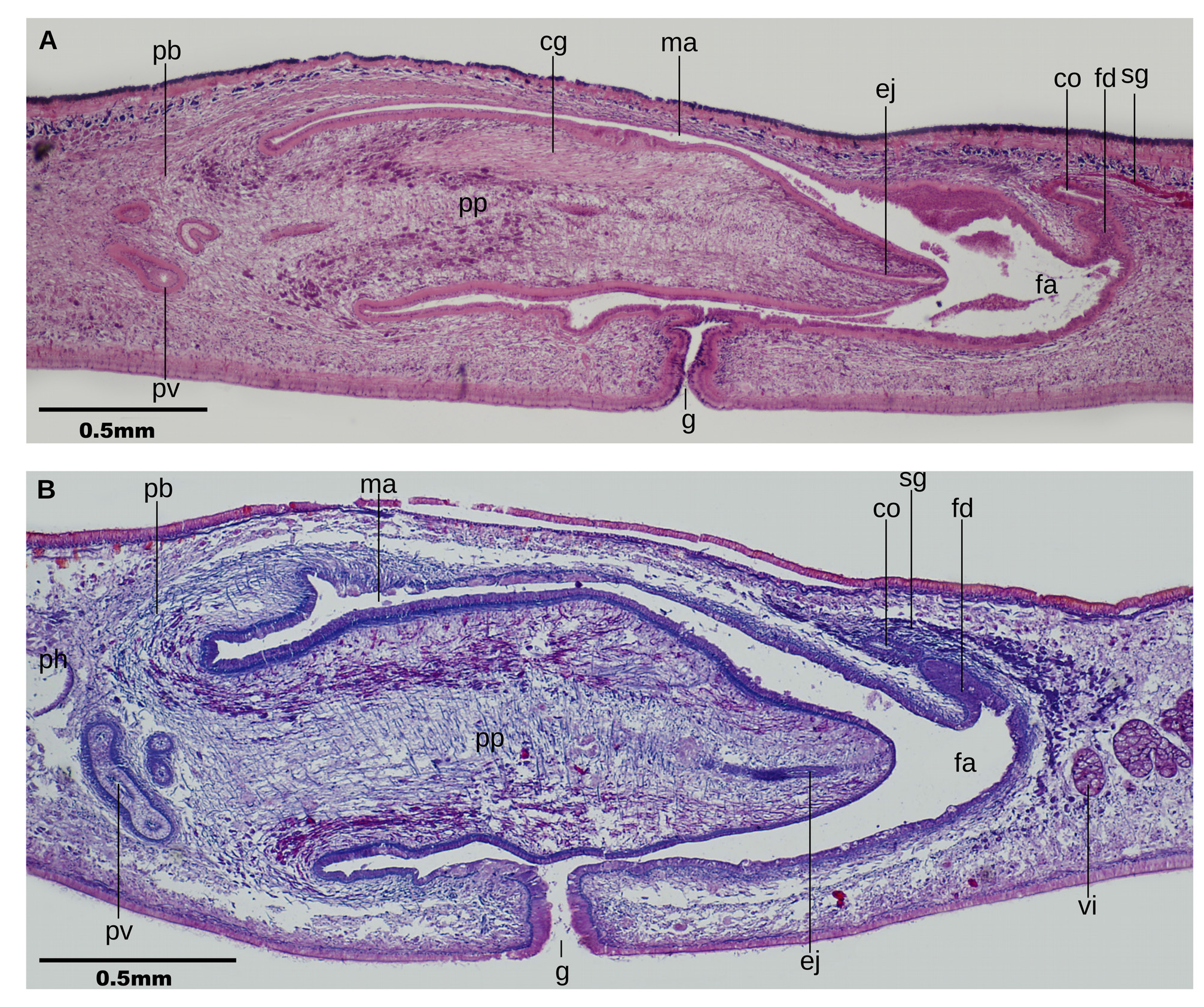
The ovaries are rounded, 300 µm in diameter (syntype C). They are located at a distance from anterior end equal to 35% of body length. The ovovitelline ducts arise from the dorso-lateral aspect of the ovaries. Ovovitelline ducts 50 µm in diameter along most of its length. They run laterally, rise laterally to the female atrium, and join to form the common glandular ovovitelline duct above female atrium ( Figs. 6A, B View FIGURE 6 ). The distal section of the ovovitelline ducts widens to a thickness of 80 µm in syntype A. Numerous shell glands discharge into final portion of ovovitelline ducts. The common glandular ovovitelline duct runs posteriorly to open to the female genital duct. The female genital duct is an anteriorly directed projection from the posterior region of female atrium.
Female atrium is ample, and not folded. It is not separated from the male atrium; the female:male atrial length is 2:1.5. This female atrium is lined with a nonciliated epithelium, which is 38 µm high posteriorly, and 20 µm high anteriorly. This epithelium has a multilayered aspect and presents scattered gaps containing fine erythrophil granules. The female lining epithelium is surrounded by a thin longitudinal muscle with circular fibers intermingled ectally.
Remarks. Froehlich did not designate explicitly holotype and paratypes among the eight specimens he mentioned in the original description. He sectioned two specimens, the ones we here named syntype A and B, respectively. A diagrammatic representation of the copulatory apparatus of syntype A was illustrated in the original description of the species (Fig. 20 in Froehlich, 1956). A diagrammatic representation of the pharynx of either syntype A or B was illustrated in the original description of the species (Fig. 21 in Froehlich, 1956). We sectioned a third type specimen, here named syntype C, from Eudóxia’s wet collection.
We noted some minor differences with respect to original description, namely (a) the mouth is not placed at the end of the pharyngeal pouch, but in its second third; (b) proximal portion of the prostatic vesicle is not intrabulbar but extrabulbar, albeit attached to it; and (c) Froehlich describes erythrophil glands discharging through the dorsal surface of the male atrium. However, we could not distinguish muscle tissue from any other structure such as glands surrounding dorsal surface of the male atrium in syntypes A and B. In syntype C, the dorsal surface of the male atrium is missing these glands as well.
| MZUSP |
Museu de Zoologia da Universidade de Sao Paulo |
No known copyright restrictions apply. See Agosti, D., Egloff, W., 2009. Taxonomic information exchange and copyright: the Plazi approach. BMC Research Notes 2009, 2:53 for further explanation.
|
Kingdom |
|
|
Phylum |
|
|
Order |
|
|
Family |
|
|
Genus |