
Coarazuphium formoso, Pellegrini, Thaís Giovannini & Ferreira, Rodrigo Lopes, 2011
|
publication ID |
https://doi.org/ 10.5281/zenodo.206526 |
|
DOI |
https://doi.org/10.5281/zenodo.6189721 |
|
persistent identifier |
https://treatment.plazi.org/id/039E87B8-FFBA-0125-FF4C-BC6646F55C48 |
|
treatment provided by |
Plazi |
|
scientific name |
Coarazuphium formoso |
| status |
sp. nov. |
Coarazuphium formoso View in CoL sp. n.
( Fig. 1–11 View FIGURE 1 View FIGURE 2. A View FIGURE 3 View FIGURE 4 View FIGURE 5 View FIGURE 6 View FIGURE 7 View FIGURE 8 View FIGURE 9 View FIGURE 10 View FIGURE 11 ).
Description. Morphometric data from paratypes are given in parenthesis.
Holotype male ( Fig. 1 View FIGURE 1 A). Total length, from the apex of the mandible to the apex of the elytra: 5.41 mm (5.16– 5.53), width, from at the widest region of the elytra: 1.51 mm (1.32–1.45). Body pale reddish brown, two paratypes are yellowish to pale brown, dorsal integument of the elytra covered with short recumbent hairs.
Head. Subtrapezoidal ( Figs. 1 View FIGURE 1 A–B) with similar width and length, width/length ratio: 1.06 (1.06–1.13). Maximum width of head at its base, 1.07 (1.07–1.13). Head slightly narrower than pronotum. Dorsal surface with one pair of setae internal to the ocular area, one pair of lateral setae located immediately behind ocular area, one pair at the widest region of the head, and two pairs close to posterior margin of head (both more internally). Ventral surface with one pair of anterior setae close to median line of head; another pair of setae almost medially, close to gula and a pair close to the posterior margin of the gular region ( Fig. 2A View FIGURE 2. A ). Eyes reduced, which are laterally positioned at the end of antennal impression of the head ( Fig. 4 View FIGURE 4 ). Ocular area smooth with sparse fine hairs, no ocelliform spots detected. Ocular margin present.
Antennae. Antennae filiform and flagellar, ( Fig. 1 View FIGURE 1 ) 4.03 mm (4.03–4.28), 4.27 (4.53–4.57) times longer than pronotum; first antennomere 0.83 times as long as 2nd to 4th together (0.69–0.74). First antennomere with a long bristle close to the middle and a few shorter setae. The antennae consists of 11 antennomeres, including the scape and pedicel with an attachment on the head that provides a remarkable mobility to antennae in every direction ( Fig. 5 View FIGURE 5 A–B); and nine antennomeres of similar shape ( Fig. 5 View FIGURE 5 C–D). Antennomeres are almost round in cross-section, except for the tip of the terminal, which is laterally flattened ( Fig. 5 View FIGURE 5 D).
Based on external morphology and their attachment to the antennal surface, long hair-like sensilla were divided into chaetoid and trichoid sensilla (s.ch. and s.t. respectively). The sensilla chaetoid (sensory bristles or spines) are distinguished by standing in a wide articulary socket at the base and the shaft is smooth; they are present in all antennomeres, and are the most abundant ( Fig. 5 View FIGURE 5 B). Long hairs, without any specialized basal cuticular ring, inflexible in their sockets, were classified as trichoid sensilla (sensory hairs) also distinguished by the relatively greater length; they can be found on the 5th to 11th antennomeres.
Short pegs and cones, which reach above the socket, were classified as basiconic sensilla (s.b.) (sensory pegs or cones) irrespective of whether they sit in a wide or tight socket; they are present on the 4th to 11th antennomeres. Some BÖhm sensilla (B.s.) (sensory pit-pegs) typical bristle are also present found in areas opposite the intersegmental membrane between head and scape, as well as between scape and pedicel on the scape and pedicel bases, respectively ( Fig. 5 View FIGURE 5 B). Coeloconic sensilla (s.co.), (sensory pit-pegs) are small pit organs, on the floor of depressions in the antennal cuticle, these organs ending at the very tip of the cone; they can be found on the 4th to 11th antennomers ( Fig. 5 View FIGURE 5 C–D). Appendages of cuticular plates (ACP), which are small cuticular process, are abundant at the bases of all antennomeres, close to the intersegmental joints ( Fig. 5 View FIGURE 5 C).
Mouthparts. Sensilla on the mandible, maxilla, labial palpus, labrum, and clypeus of the paratype 1 were examined. The mandible is acutely bent inwardly at its tip. On the ventral side, longitudinal rows of setae are present ( Fig. 6 View FIGURE 6 A–B). On the dorsal surface, a series of hair sensilla projects from the submolar region to near the cuticular processes ( Fig. 6 View FIGURE 6 C–D).
The maxilla basically consists of the lacinia, maxillary palp, and galea ( Fig. 7 View FIGURE 7 A). The lacinia is shorter than the galea, with an acute and curved end, with rows of long setae and cuticular process. The four-segmented maxillary palp is long and filiform with spaced basiconic sensilla present on the surfaces of the segments. Trichoid sensilla are distributed along the maxillary palp, and they become more abundant and smaller on the last segment ( Fig. 7 View FIGURE 7 C). There are also grooves in this segment that may indicate a sensory organ or gustatory receptors ( Fig. 7 View FIGURE 7 B).
The galea is biarticulated, composed of 2 segments, with different types of basiconic sensilla. These sensilla become more abundant near the apex of the last segment ( Fig. 7 View FIGURE 7 D–E).
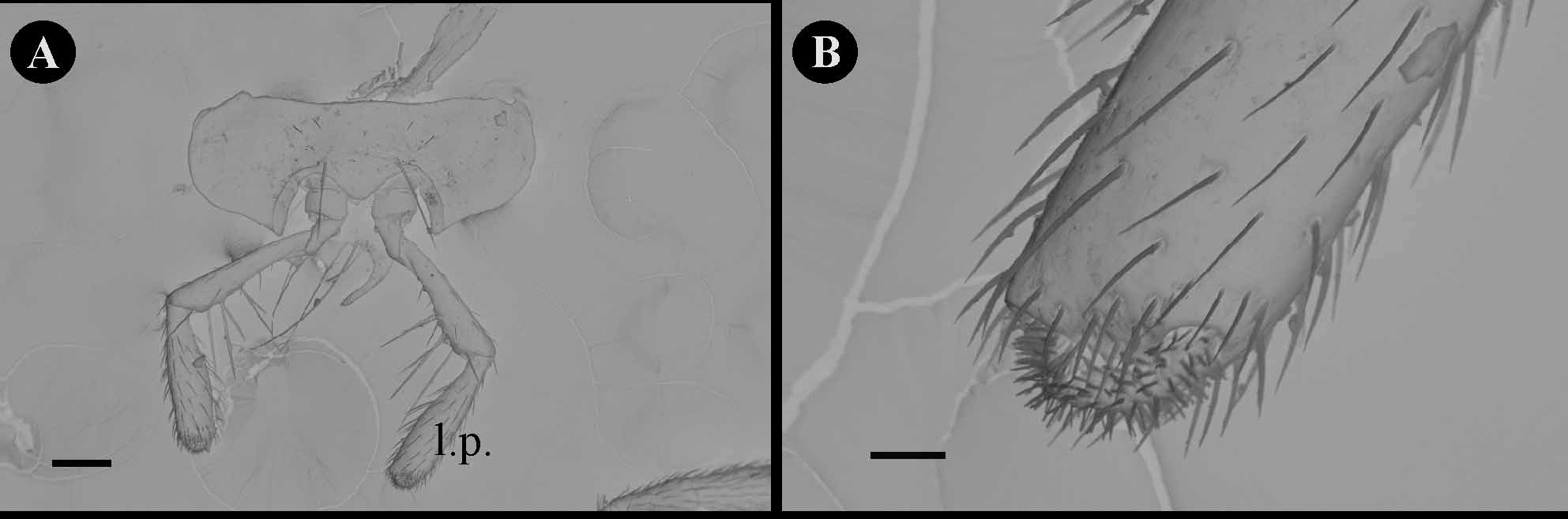
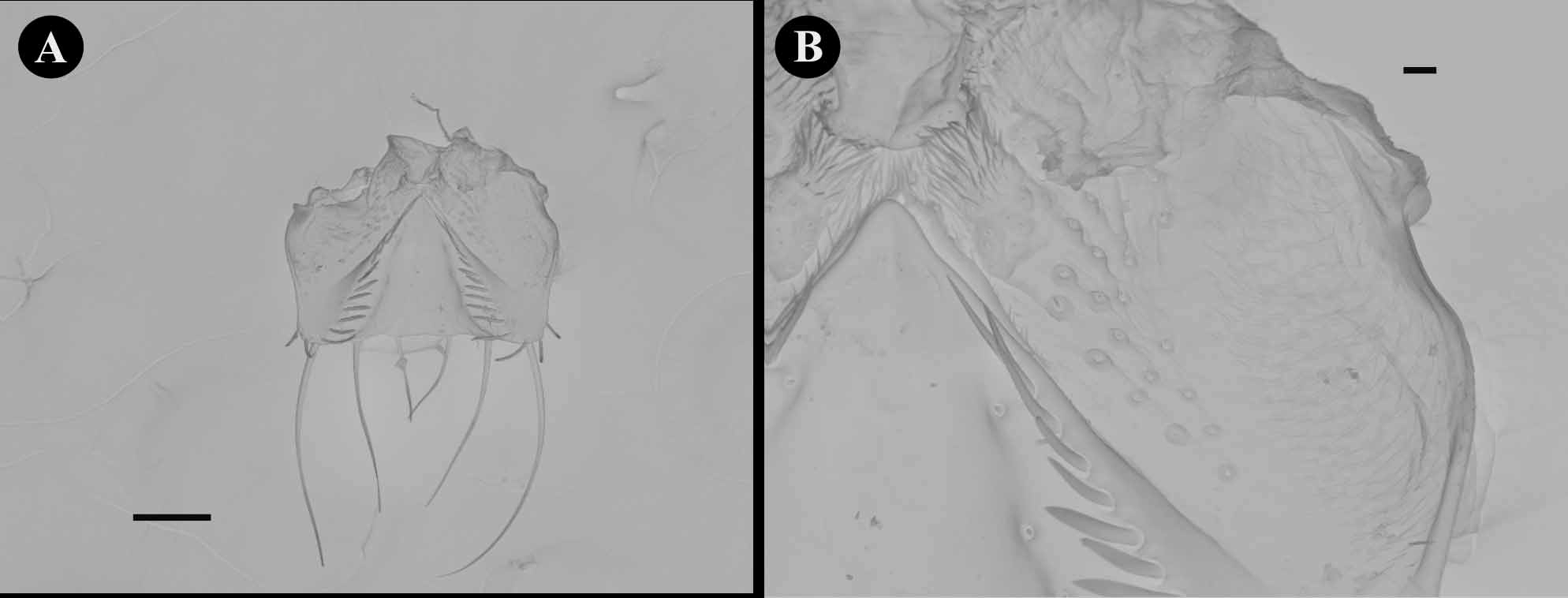
The labium has two pairs of small setae at its base and one pair of long setae near the labial palpi ( Fig. 8 View FIGURE 8 A), which has some long hairs within. The types of sensilla on the three-segmented labial palpomeres are the same as those on the maxillary palpomeres ( Fig. 8 View FIGURE 8 B). The labrum is quadrangular ( Fig. 9 View FIGURE 9 ). On the ventral side confluent rows of long setae are present.
Pronotum. Shape trapezoidal, 1.17 (1.13–1.29) times wider than long ( Figs. 1 View FIGURE 1 , 2 View FIGURE 2. A B). Maximum width close to anterior angle and as wide as head. Anterior and posterior angles are acute. Dorsal surface ( Figs. 1 View FIGURE 1 A–B) with two pairs of erect setae: one close to the anterior angle of the pronotum and the other, shorter, close to the posterior angle. Ventral surface with one pair of anterior setae medially located ( Fig. 2A View FIGURE 2. A ).
Elytra. Elytra are free ( Fig. 1 View FIGURE 1 ), together 1.78 (1.67–1.86) times longer than wide. Maximum width nearly one third the distance from the apex and 2.73 (2.60–2.79) times longer than pronotum. Apex of elytra sinuous. Seven large setae in each elytron: 3 close to the anterior angle, 2 marginal in posterior half, and 2 on posterior margin. There is also one smaller seta on the posterior internal margin of each elytra. Wings absent. Abdominal sterna 1–5, glabrous, sixth sternum with a small pair of setae close to its posterior margin.
Legs. Procoxa with one pair of setae at posterior margin; meso and metacoxa with one pair of setae close to the anterior margin ( Fig. 1 View FIGURE 1 ). Pro- and mesotrochanters bear one medial setae at posterior margin; metatrochanter lacking setae. Profemur with some long and short setae. Profemur 1.26 (1–1.05) mm, as long as the mesofemur and 0.88 (0.75–0.82) times the length of metafemur. Protibia 1.06 (1–1.06) mm as long as the mesotibia and 0.72 (0.68–0.79) times the length of metatibia. Protibia 1.21 (0.86–1.36) times longer than protarsus. Mesotibia 0.97 (0.69–0.98) times the length of mesotarsus and metatibia 1.02 (0.76–1.05) times longer than the metatarsus. First tarsomere almost equal to tarsomeres 2–4 together. Length of protibia and tarsus together 1.95 (0.76–1.05) times the length of the pronotum. Mesotibia and tarsus length 2.17 (2.27–2.36) times, and metatibia and tarsus length 3.04 (3.13–3.25) times the length of pronotum.
The ultrastructural analysis showed that the coxal segment has 2 types of sensilla: sharply pointed long trichoid sensilla (s.t.) and small peg sensilla, which are similar to the antennal basiconic sensilla (s.b.). Appendages of cuticular plates (ACP) are abundant on the procoxae, covering all the tissue ( Fig. 10 View FIGURE 10 A). On the femur, trichoid sensilla are regularly distributed throughout all the tissue ( Fig. 10 View FIGURE 10 C). ACP are abundant at the bases of protibia, close to the intersegmental joint, where there is also an aggregate of basiconic sensilla ( Fig. 10 View FIGURE 10 B). Protibia also have a row of trichoid sensilla, which became more abundant at tibial apex, and spaced basiconic sensilla occur at its bor- der ( Fig. 10 View FIGURE 10 D–E). The tarsus has abundant trichoid sensilla ( Fig. 10 View FIGURE 10 F).
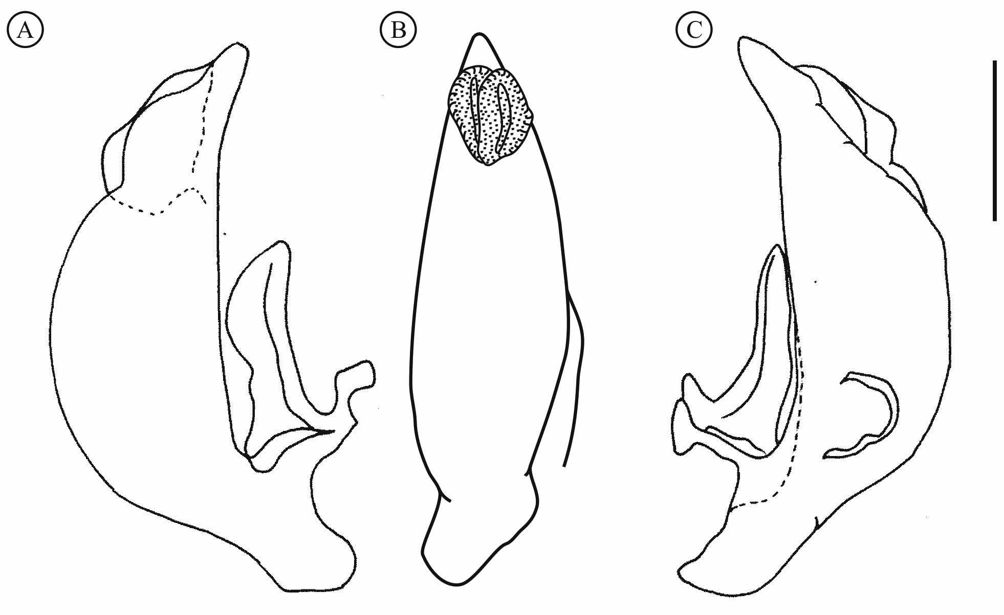
Aedeagus. Dorsally curved and elongate, with some round (ovoid) protuberance at the apices ( Figs. 3 View FIGURE 3 A–C). Left paramere about two times longer than wide, with an irregular inner margin in dorsal view; right paramere slighter curved and elongate.
Etymology. The epithet is given in apposition as a toponymic for the name of the municipality (Campo Formoso ) where the species was collected.
Differential diagnosis. All the characteristics of C. formoso are consistent with the description of the genus Coarazuphium . This new troglobitic species of Coarazuphium differs from the others in the genus by having three pairs of setae on the dorsal surface of the head close to the posterior margin, while the other species have only one or two pair of setae. The aedeagus differs from other Coarazuphium species; it has some singular round (ovoid) protuberance at the apices, and the left paramere bears some irregular margin in dorsal view.


No known copyright restrictions apply. See Agosti, D., Egloff, W., 2009. Taxonomic information exchange and copyright: the Plazi approach. BMC Research Notes 2009, 2:53 for further explanation.