
Charonina elephanti, Gürelli, 2019
|
publication ID |
https://doi.org/ 10.11646/zootaxa.4545.3.6 |
|
publication LSID |
lsid:zoobank.org:pub:5B9851C2-AF9C-4D74-85CB-20E1BDBD98D6 |
|
DOI |
https://doi.org/10.5281/zenodo.5933389 |
|
persistent identifier |
https://treatment.plazi.org/id/F6268789-FF9D-FFDE-FF61-CE4EC06D7FDE |
|
treatment provided by |
Plazi |
|
scientific name |
Charonina elephanti |
| status |
sp. nov. |
Charonina elephanti n. sp.
urn:lsid:zoobank.org:act:25421C9C-4771-4929-BCA6-C2B9F0ED706B
Description. The body is ovoid in shape with rounded anterior and posterior ends. The vestibular opening is at the
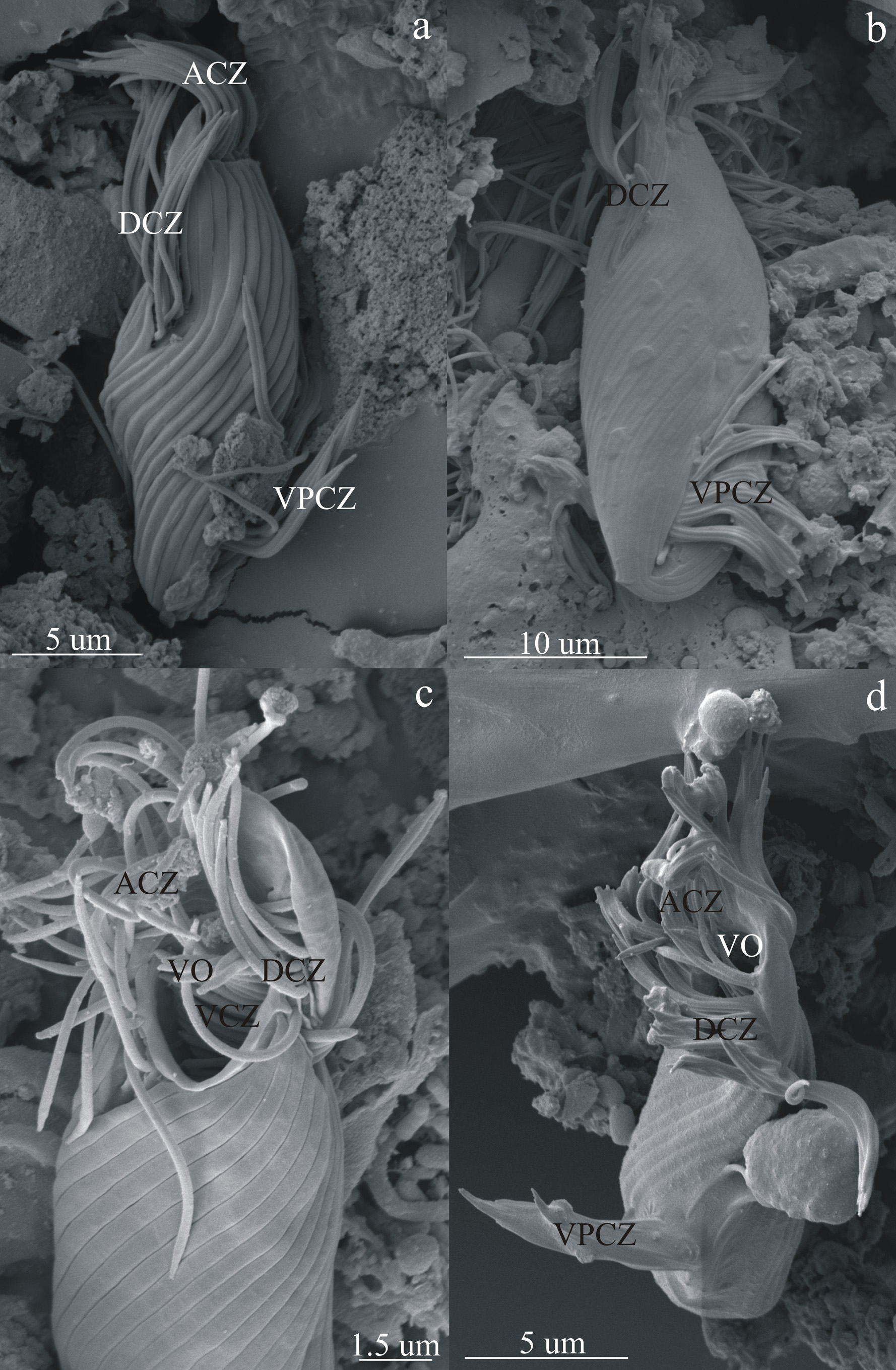
left side of the anterior end of the body and leads to a long, funnel-shaped vestibulum, which extends nearly to the midpoint of the body. The macronucleus and micronucleus are ovoid in shape, and the micronucleus adheres to various sides of the macronucleus. The position of the macronucleus is not constant in the body. A contractile vacuole is present near postero-ventral part of the body. The cytoproct is at the posterior end of the body. The surface of the body has long, spiral pellicular striations. Two buccal ciliary zones are present. The adoral ciliary zone extends along the vestibular opening. The vestibular ciliary zone is in the vestibulum and extends to the midline of the body. Three somatic ciliary zones are present. The dorsal ciliary zone is at the dorsal side of the vestibular opening and is oblique from the left to the right surface. Two posterior ciliary zones, dorsal and ventral, are near the posterior end of the body and are oblique from the right to the left surface. All ciliary zones are nonretractable (Table 3, Figures 7a View FIGURE 7 , 8 View FIGURE 8 , 9 View FIGURE 9 ).
Habitat, type host, and locality. The hindgut of Asian elephants ( Elephas maximus ) in Gaziantep Zoo, Gaziantep, Turkey .
Etymology. The name elephanti refers to elephants because the new species is detected from elephants.
Type material. The specimens are held in the endocommensal ciliates of herbivores collection in the Department of Biology, Faculty of Sciences and Arts, Kastamonu University, Kastamonu, Turkey. Holotype and paratypes are on the slide numbered as EIC-5.
Remarks. C. elephanti n. sp. is distinguishable from other Charonina species by its ovoid body shape with rounded anterior and posterior ends, and ovoid macronucleus shape and position.
Infraciliature of C. elephanti n. sp. The buccal infraciliature of C. elephanti n. sp. is composed of adoral and vestibular polybrachykinety. The adoral polybrachykinety is crescent-shaped and extends along the vestibular opening. It is composed of short, oblique kineties. The vestibular polybrachykinety is Y-shaped and composed of short, oblique, and transverse kineties extending to the dorsal and ventral walls of the vestibulum. The dorsal part of the vestibular polybrachykinety begins near the level of the dorsal polybrachykinety and extends to the midpoint of the body. The ventral part of the vestibular polybrachykinety is oblique from ventral to dorsal and begins near the posterior end of the adoral polybrachykinety. The ventral part of the vestibular polybrachykinety is composed of oblique kineties and connects the dorsal part of the vestibular polybrachykinety at its midpoint. The dorsal vestibular kineties are oblique in the anterior part and transverse in the posterior part of the vestibulum. Vestibular fibrils are transverse, parallel, and long and arise from the dorsal posterior wall of the vestibulum. Paralabial kineties were not impregnated. The somatic infraciliature of C. elephanti n. sp. is composed of three polybrachykineties. The dorsal polybrachykinety is ribbon shaped, located at the dorsal side of the vestibular opening, extends obliquely from the left to the right surface, and is composed of short, oblique kineties. The dorsal and ventral posterior polybrachykineties are ribbon shaped, composed of short oblique kineties, and extend obliquely from the right to the left surface ( Figures 7b View FIGURE 7 , 8 View FIGURE 8 , 9 View FIGURE 9 ).


No known copyright restrictions apply. See Agosti, D., Egloff, W., 2009. Taxonomic information exchange and copyright: the Plazi approach. BMC Research Notes 2009, 2:53 for further explanation.