
Paracanthopoma capeta, Pinna & Dagosta, 2022
|
publication ID |
https://doi.org/ 10.11606/1807-0205/2022.62.072 |
|
publication LSID |
lsid:zoobank.org:pub:A32FD3AF-C87F-4C75-9100-D695C3578283 |
|
DOI |
https://doi.org/10.5281/zenodo.10839404 |
|
persistent identifier |
https://treatment.plazi.org/id/A81A87C0-FFCC-FC4A-FC3F-1189209DAB74 |
|
treatment provided by |
Felipe |
|
scientific name |
Paracanthopoma capeta |
| status |
sp. nov. |
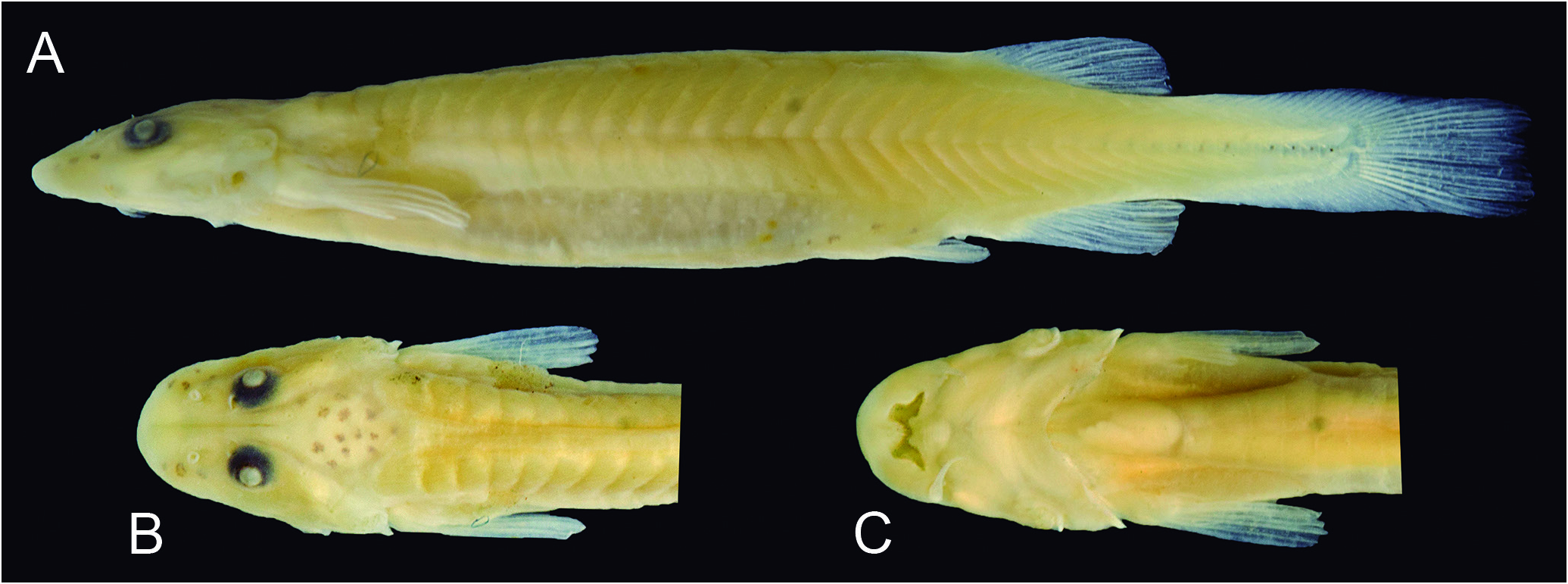
Paracanthopoma capeta , new species ( Fig. 13 View Figure 13 )
Holotype: MZUSP 29154 View Materials , 14.5 mm SL, Brazil, Amazonas , rio Negro , praia Mari-Mari , upstream from Barcelos and slightly above mouth of rio Cuiuni (approximately 00°32′S, 63°24′W), col., M. Goulding, 30 May 1979. GoogleMaps
Paratypes: MZUSP 100144,6 ex (2 c&s), 14.1-15.6 mm SL, collected with holotype.
Diagnosis: Distinguished from all congeners by the supraoccipital well-developed, its anterior margin extending transversely across the skull (vs. supraoccipital receded into deep concavity or produced anteriorly as median spike); by the extremely long, thread-like maxilla (vs. maxilla not thread-like); and by the single-row dentition disposed in a v-shape, with lateral teeth gradually more anteriorly and the central tooth inserted most posteriorly (vs. row or rows of teeth approximately aligned transversely). Distinguished from all congeners except Pc. ahriman , Pc. cangussu , and Pc. irritans by the presence of five median premaxillary teeth (one or two often in replacement) (vs. either three or 9 to19 in total). Distinguished from Pc. ahriman by the narrower head (head width 68.0-72.0% HL; vs. 80.7-87.6). Distinguished from Pc. cangussu by the shorter caudal peduncle (18.0-20.4% SL; vs. 21.8-24.0); by the less deep caudal peduncle (7.0-8.8% SL; vs. 10.8-13.0); by the longer predorsal and preanal lengths (72.2-74.1 and 71.8-75.9% SL; vs. 66.7-71.3 and 68.6-70.3, respectively); by the narrower head (68.0-72.0% SL; vs. 75.7-83.3); by the larger eye (16.7-21.7% SL; vs. 14.7-16.2). Distinguished from Pc. irritans by the narrower head (68.0-72.0% HL; vs. 73.3-76.9); by the mouth cleft directed more strongly laterally than posteriorly (vs. opposite); and by the median premaxilla trapezoidal with nearly straight anterior margin (vs. roundish).
Description: Morphometric data for the holotype and paratypes are provided in Table 4 View Table 4 . Body short (HL 16.5-21.1% SL). Cross-section of body approximately as deep as broad at pectoral-fin insertion and increasingly compressed posterior to that point, tapering to caudal fin. Dorsal profile of body gently convex or straight from head to origin of dorsal fin ( Fig. 13 View Figure 13 ). Dorsal and ventral profiles of caudal peduncle gently convex posterior to dorsal and anal fins, moderately spatulate, expanded by procurrent caudal-fin rays ( Fig. 13 View Figure 13 ). Ventral profile of body straight at pectoral-fin base and then gently convex until pelvic-fin origin, with some specimens with greatly distented abdomens. Myotomes and longitudinal skeletogenous septum clearly visible through thin integument along whole body. Axillary gland very large, elongate in shape, protruding markedly on surface of body when full with secretion, extending along limit between hypaxial musculature and abdominal cavity. Anterior end of gland surrounding dorsal, ventral and posterior margins of muscular pectoral-fin base, as thick corselet, extending posteriorly to beyond margin of adpressed pectoral fin. Posterior end of gland blunt and round, its large round or oval pore opening dorsally at its middle portion, approximately at vertical through half of pectoral- fin length. Condition of gland posterior to pore evidently related to amount of secretion stored.
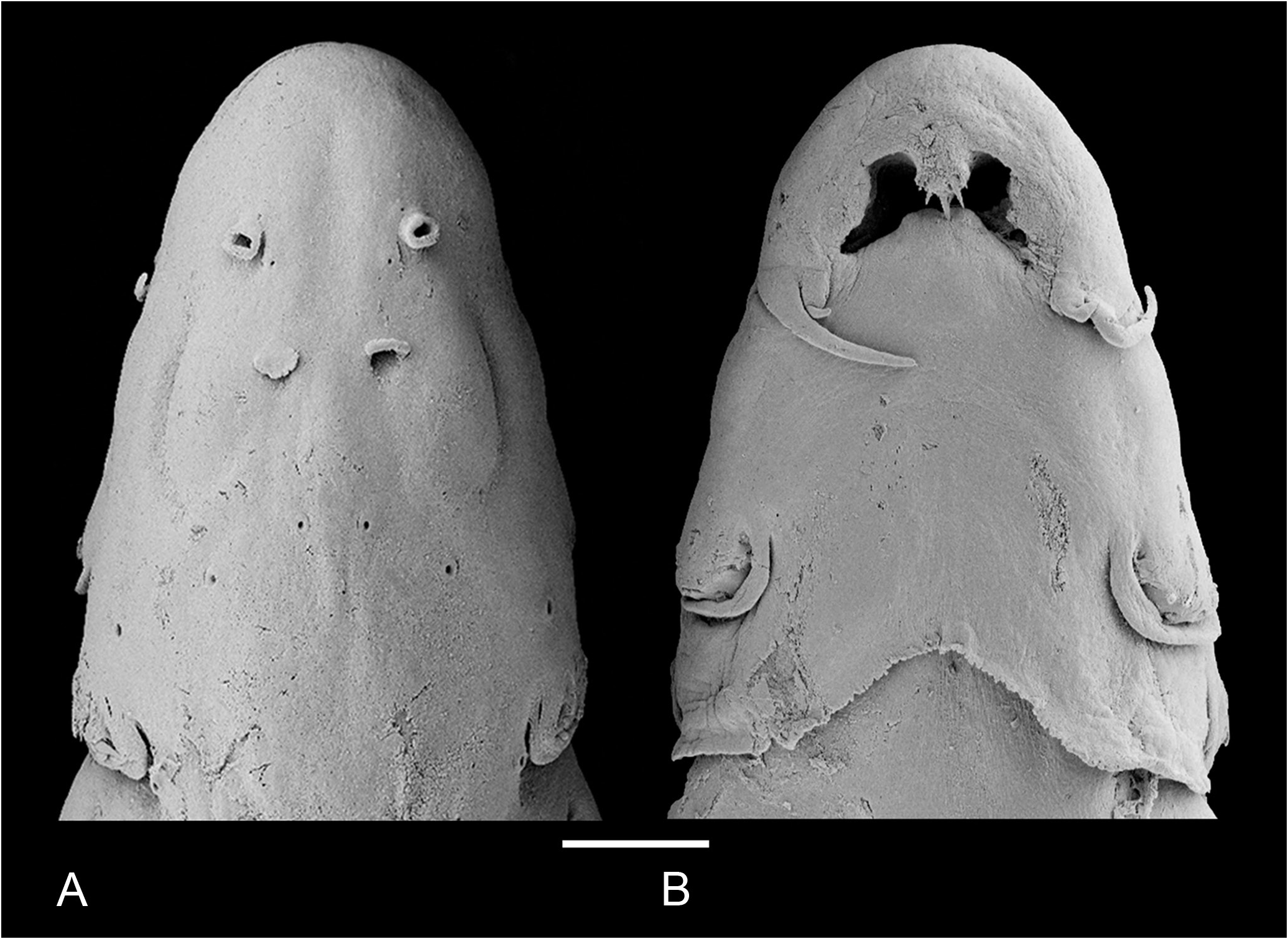
Dorsal profile of head continuous with that of dorsum ( Fig. 13 View Figure 13 ), its origin sometimes indicated by slight constriction of anterior end of epaxial musculature. Head longer than broad (head width 68.0-72.0% HL), snout broad, parabolic with a continuous round anterior margin. Head muscles not entering skull roof. Head moderately depressed (head depth 41.2-63.9% HL), with dorsal profile in lateral view straight until eye, then bending ventrally and straight again to tip of snout. Ventral profile of head straight or slightly convex. Eye large (16.7-21.7% HL), without free orbital rim, located dorsolaterally on head and directed dorsolaterally, with pronounced lateral component ( Fig. 13 View Figure 13 ). Integument over eye thin and transparent. Middle of eye approximately at middle of HL, interorbital width almost equal to longitudinal diameter of eye. Eyelens constricted by iris only marginally, with large round or oval pupil in specimens examined. Anterior nostril small, surrounded by short tubule of integument produced posteriorly into small pointed process ( Figs. 13 View Figure 13 , 14 View Figure 14 ), with double elastin cores. Conspicuous recess-like elongate depression immediately posterior to base of anterior nostril, with plicate inner surfaces. Anterior internarial width slightly larger than interorbital. Posterior naris small, slightly larger than anterior ones, roundish or triangular in shape, adjacent to mesial margin of eye and partly occluded by anterior flap of integument ( Figs. 13 View Figure 13 , 14 View Figure 14 ). Anterior margin of posterior naris posterior to transverse line through anterior margin of eye. Posterior internarial width narrower than interorbital and 2-2.5 times diameter of one nostril.
Opercular odontodophore medium-sized and elongate, dorsolaterally located on head, on dorsal half of head depth in lateral view, anterodorsally to pectoral-fin base. Opercular odontodes 6 or 7, irregularly positioned with larger ones posteriorly. Main axis of opercular odontodes oriented horizontally in lateral view, with distal portions of larger posterior ones curved mediodorsally. Two or three caps of replacement odontodes interspersed with mature ones. Opercular periodontodal fold well-differentiated, extending well beyond tips of odontodes. Interopercular odontodophore larger than, or as large as, opercular one, located ventrolaterally on head, at horizontal through origin of pectoral fin, with 6 or 7 odontodes closely positioned in one main posterior row with five odontodes, plus one or two smaler ones anteriorly. Interopercular odontodes progressiely larger posteriorly, with largest ones strongly compressed at base. Interopercular odontodophore slightly closer to opercular one than to eye. Interopercular periodontodal fold of integument well-developed but narrow, roundish, extending shortly beyond tips of odontodes. Epiodontodeal velum poorly-differentiated, thin and transparent, irregularly covering most of odontodes.
Mouth inferior (ventral), flattened ventrally ( Fig. 13 View Figure 13 ). Each premaxilla with 1 or 2 large scalpelloid teeth (always two tooth sockets) attached to its distal tip disposed in parallel ( Figs. 4D View Figure 4 , 15 View Figure 15 ). Scalpelloid teeth deeply hidden in labial tissue and difficult to expose in preserved specimens without damage to soft tissue. No conical teeth on premaxilla. Upper lip very broad, continuous with ventral surface of snout. Median premaxilla small, occupying only central portion of upper jaw, with 5 teeth with insertions disposed in single v-shaped row ( Figs.4D View Figure 4 , 15 View Figure 15 ). Bases of teeth strongly off-set,with those of lateral-most teeth most anterior, with following teeth inserted half-way to posterior margin of median premaxilla and central (largest) tooth posterior-most, inserted close to posterior margin of bone. All teeth posteriorly oblique to ventral surface of median premaxilla, with anterior one on each side also strongly inclined mesially. Distal pungent portion of lateral teeth curved posterolaterally, and of median one curved posteriorly. Basal portion of all median premaxillary teeth strongly compressed laterally. Two or three replacement tooth caps interspersed with mature dentition. Median premaxillary velum small, but covering most of tooth surace. Hypodontal pad of median premaxilla small, roundish, occupying small area proportional to small median premaxillary dentition. Lower jaw narrow, composed mostly of small knob-like dentary lobes, largely confluent basally, round anteriorly and, continuous with mental region posteriorly ( Fig. 13 View Figure 13 ). Jaw cleft short and oriented obliquely to longitudinal axis. Dentary diastema angulate. Dentary teeth 3 or 4 (when three, one obviously missing, in process of replacement), closely set at mesial end of dentary and disposed as two ventral and two dorsal ones, not exactly aligned ( Figs. 4D View Figure 4 , 15 View Figure 15 ). Dentary teeth very long, their main axis sloped medially in ventral view, and their distal portions strongly curved dorsally, hooked.
Branchiostegal velum forming large, continuous, hyperbolic and posteriorly concave, free fold across whole of mental region, its lateral portion plicate near margin ( Figs. 13 View Figure 13 , 14 View Figure 14 ). Dorsal portion of velum reaching, but not covering, anterior margin of pectoral-fin base. Branchial openings medium-sized, spanning approximately area between ventral margin of opercular odontodophore and ventral margin of interopercular odontodophore, anteriorly to base of pectoral fin. Maxillary barbel long and thin, not reaching base of interopercular odontodophore (extending approximatelly three-fourths of distance to it). Posterior point of its base anterior to vertical through anterior margin of eye in lateral view. Mesial (or ventral) part of maxillary-barbel base inserting directly onto corner of mouth without intervening membranous outgrowth. Rictal barbel small but well-differentiated, attached mesially to base of maxillary one and approximately one-fifth of its length.Nasal barbel vestigially represented by posterior elongated portion of fold around anterior naris described above ( Figs. 13 View Figure 13 , 14 View Figure 14 ), with double internal elastin core.
Lateral line short and straight, extending alongside dorsal margin of anterior portion of axillary gland. Terminal lateral-line pore immediately dorsal to axillary gland opening.Very short secondary branch splitting off ventrally (or laterally and immediately curving ventrally) from proximal portion of main canal, with corresponding pore opening anteriorly to midlength of main canal. Single lateral-line tubule straight, extending for half of main canal posterior to bifurcation.
Pectoral fin short (62.5-70.0% HL), elongate with gently convex or truncate margin. Margin of fin irregular at close range. Pectoral-fin rays i + 5, its base on ventral side of body. Pelvic fin small, well-separated at base, with i + 4 rays. Pelvic splint present. Origin of pelvics close to origin of anal fin, well anterior to vertical through origin of dorsal-fin, entirely covering anus and urogenital papilla and extending posteriorly to origin of anal fin. Posterior margin of pelvic fin gently convex or truncate. Dorsal fin elongate, roughly rectangular, with roundish edge and gently convex distal margin. Dorsalfin rays ii + 5 or i + 6, plus 3 to 6 procurrent ones. Anal fin similar in shape to dorsal fin, with ii + 5 rays, plus 3 or 4 procurrent ones. Origin of anal fin at or slightly posterior to vertical through origin of dorsal-fin. Anal fin with same size, slightly smaller or slightly larger than dorsal one. Caudal fin truncate or slightly concave, its maximum depth when expanded deeper than maximum depth of caudal peduncle. Principal caudal-fin rays 6 + 6. Procurrent caudal-fin rays 16 to 21 dorsally and 19 or 20 ventrally.
Vertebrae 36 (n = 1) or 37 (n = 1). First dorsal-fin pterygiophore subsequent to neural spine of vertebra 20 (n = 1). First anal-fin pterygiophore subsequent to haemal spine of vertebra 20 (n = 1) or 21 (n = 1). Dorsalfin pterygiophores 7 (n = 2). Anal-fin pterygiophores 6 (n = 2). Branchiostegal rays 3.
Pigmentation in preservative: Body almost entirely white. Few scattered dark chromatophores on posterior part of sides of abdominal wall, more evident in specimens with distended abdomens. Small black dot at middle of each vertebra along posterior part of caudal peduncle, formed by internal chromatophores. Posterior half of neurocranium with irregular dark brain pigment seen by transparency. Field of dark chromatophores anteriorly to eyes, at lateral part of snout.
Etymology: From the Portuguese vernacular term capeta (probably from a combination of capa, meaning cape, and -eta, diminutive suffix), meaning the devil.
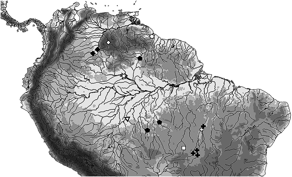
Geographical distribution: Paracanthopoma capeta has been recorded from a single locality in the middle rio Negro, Northern Brazil ( Fig. 20 View Figure 20 ).
Biology: The type series was collected from the gill chamber of a specimen of Phractocephalus hemioliopterus (Pimelodidae) , 1.08 m in length (label does not specify if SL or TL) and 22 kg. Three paratypes have distended abdomens, apparently full with discolored blood.
| MZUSP |
Museu de Zoologia da Universidade de Sao Paulo |
No known copyright restrictions apply. See Agosti, D., Egloff, W., 2009. Taxonomic information exchange and copyright: the Plazi approach. BMC Research Notes 2009, 2:53 for further explanation.
|
Kingdom |
|
|
Phylum |
|
|
Class |
|
|
Order |
|
|
Family |
|
|
Genus |