
Austrodecus taylorae, Staples, 2019
|
publication ID |
https://doi.org/ 10.11646/zootaxa.4567.3.1 |
|
publication LSID |
lsid:zoobank.org:pub:0AEFAF80-B001-4A18-88AC-5B6A189F6DCD |
|
DOI |
https://doi.org/10.5281/zenodo.5944894 |
|
persistent identifier |
https://treatment.plazi.org/id/03895C33-2902-4F12-FF01-FD61FD55FD0D |
|
treatment provided by |
Plazi |
|
scientific name |
Austrodecus taylorae |
| status |
sp. nov. |
Austrodecus taylorae View in CoL sp. nov.
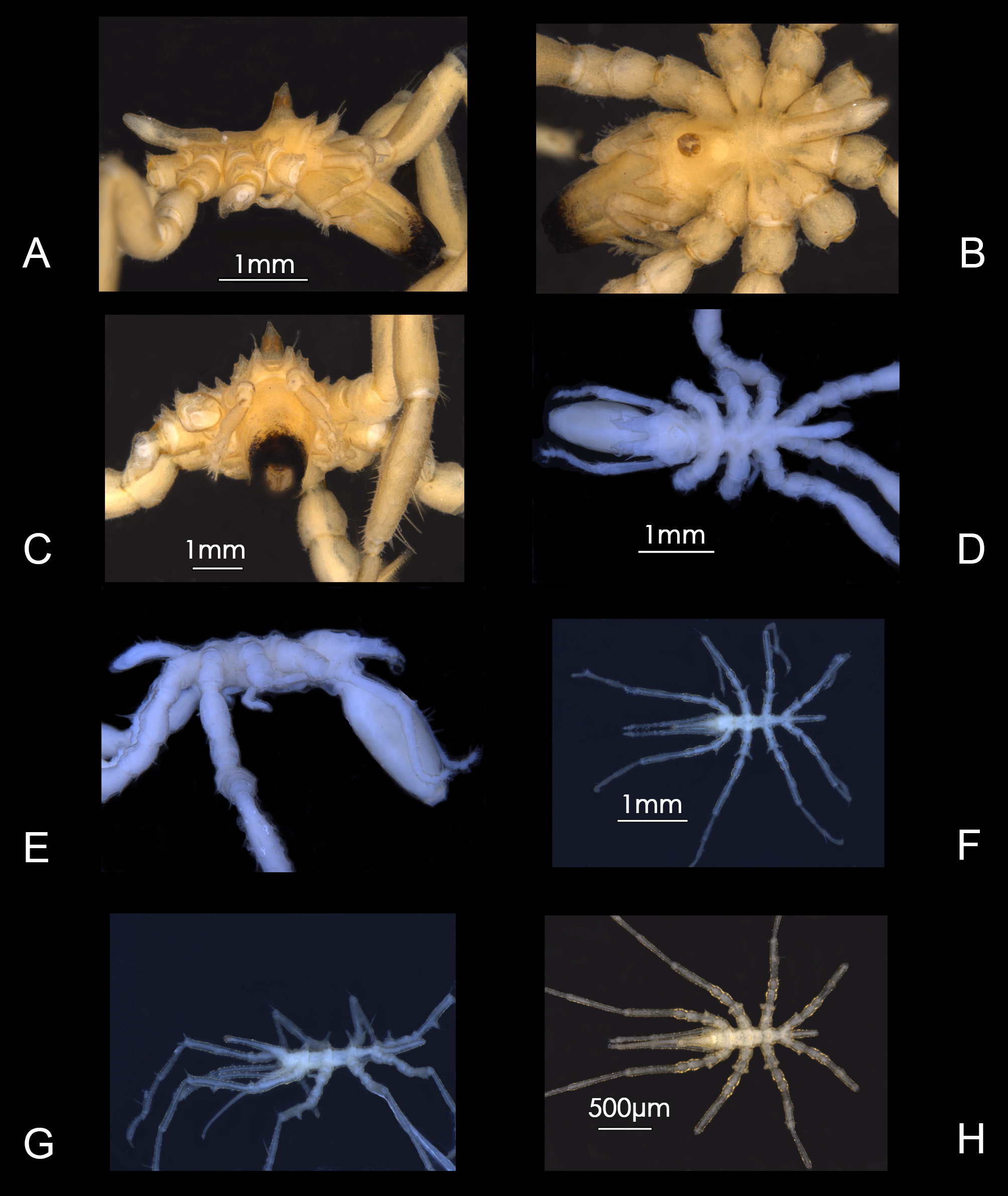
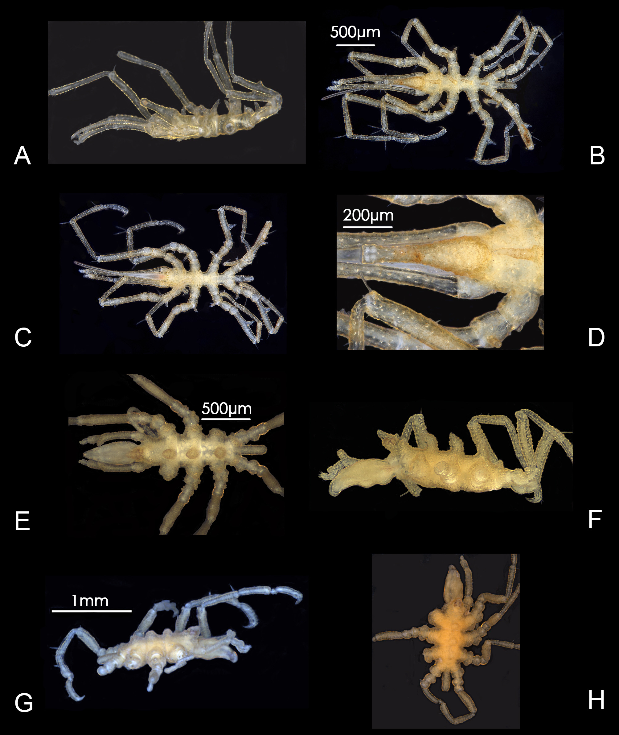
Figure 11 View FIGURE 11 A–I; Plates 3H View PLATE 3 ; 4A View PLATE 4
Material examined. Holotype, male ( NHMUK 2018.24 View Materials ), Southwest Indian Ocean, Atlantis Bank , 32° 42.86´S, 57° 16.34´E, ROV, 750 m, specimen JC066-4213C, stn 8.29, mooring site, on net containing Mango wood, 14 December 2011. GoogleMaps
Paratype. One male ( NHMUK 2018.25 View Materials ), same collection details as for holotype. Two males ( NHMUK 2018.26 View Materials ), Southwest Indian Ocean, Atlantis Bank , 32° 42.86´S, 57° 16.34´E, ROV, 750 m, specimen JC066- 4214C, stn 8.29, mooring site, on net containing Mango wood, 14 December 2011 GoogleMaps .
Description. Male holotype. Trunk ( Fig.11A, B View FIGURE 11 . Plates 3H View PLATE 3 , 4B View PLATE 4 ) papillose, quite transparent, segments 1 to 4 with dorsomedian tubercles, height each little more than half depth of trunk, those on segments 2 and 4 slightly shorter than those on segments 1 and 3, margins of tubercles uneven; lateral processes smooth, separated by about half basal width, diverging distally. Ocular tubercle tall, height almost equal to 60% of trunk length, inclined forward, over-reaching base of proboscis, tapering from broad base to narrow truncate tip, surface of basal part coarsely rugose; four eyes, unpigmented.
Proboscis 30% longer than trunk, typical of genus, down-curved, annulated distally, basal part inflated, smooth, jaws vertically bilateral.
Abdomen ( Fig. 11C View FIGURE 11 ) unarticulated at base, surface coarsely rugose, length about 35% of trunk length, carried horizontally, tapering slightly distally.
Chelifores absent.
Palp ( Fig. 11E View FIGURE 11 ) five-segmented, attached to lateral extension of cephalon, segment 1 longest segment, with 1 or 2 strong inward-facing curved spines, segment 3 next longest, greater than half length segment 1, with six inwardfacing curved spines of similar shape and size.
Oviger ( Fig. 11F View FIGURE 11 ) four-segmented, attached to ventral extension of cephalon, second segment with strong recurved spine on distal margin, segment 4 longest, constricted at tip and accompanied by several spines.
Third leg: ( Fig. 11D View FIGURE 11 ) femur widest and longest segment, single mid-ventral femoral cement gland duct legs 3 and 4 only, conical, height little more than one-third width of femur, tibia 1 longer than tibia 2, tibia 2 more setose ventrally than other segments, femur with single long dorsodistal hair-like spine on low process, first coxae with prominent dorsodistal digitiform tubercles in the order of 1:2:2:1, coxa 2 longer that coxae 1 or 3, third coxa with a distinctly bifurcate tubercle about one-third the height of those on coxa 1 ( Fig. 11I View FIGURE 11 ), tarsus short, with several median spines and two longer dorsolateral spines, propodus gently curved, heel absent, sole lined with about ten short spines, without major heel spines, terminal claw about 40 percent of propodus length, auxiliary claws almost half length main claw, flared outward. Gonopores not evident.
Measurements of holotype (mm). Trunk length (frontal margin of cephalic segment to tip of 4 th lateral process), 0.784; width across 2 nd lateral processes, 0.400; proboscis length (dorsal), 1.016; abdomen length (lateral), 0.280; height ocular tubercle (lateral), 0.456. Third leg: coxa 1, 0.120; coxa 2, 0.128; coxa 3, 0.088; femur, 0.480; tibia 1, 0.440; tibia 2, 0.368; tarsus, 0.048; propodus, 0.280; claw, 0.112; auxiliary claws, 0.048. Palp: seg. 1, 0.576; seg 2, 0.080; seg. 3, 0.304; seg. 4, 0.040; seg. 5, 0.032. Oviger: seg. 1, 0.032; seg. 2, 0.040; seg. 3, 0.032; seg. 4, 0.088.
Etymology. Named for Dr Michelle Taylor, whose drive, direction and organization of all biological aspects contributed significantly to the efficient operation of the voyage.
Remarks. The basal part of ocular tubercle surface is particularly rugose with tiny pyramid-shaped projections. There is variability in the shape of oviger segment 4 which may be age dependent. In the holotype there is a strong constriction near the tip accompanied by one or two small spines ( Fig. 11F View FIGURE 11 ) whereas oviger segment 4 of one paratype is without apparent constriction ( Fig. 11G View FIGURE 11 ) but spination is similar. The fourth oviger segment of one smaller (less mature?) male is shorter, slightly ovate and without distal constriction ( Fig. 11H View FIGURE 11 ).
Austrodecus taylorae View in CoL sp. nov. is one of seven species in which males possess cement glands on legs 3 and 4 only. The other species are A. acone Hedgpeth and McCain, 1971 , A. bathyale Stock, 1991 View in CoL , A. excelcum Stock, 1991 , Austrodecus View in CoL sp. C (below), A. latum Stock, 1991 View in CoL and A. tuberculatum Stock, 1991 View in CoL . The cement gland in all these species except A. acone opens through a long tube on the posterior surface on the distal end of the femur; in A. acone the gland opens through a basal cone with apical tube on the ventral surface of the femur. Austrodecus taylorae View in CoL and the unnamed species ‘C’ described herein are the only species in which the cement gland opens through a ventral cone lacking an apical tube. Three species are known from female specimens only and accordingly the shape, number and position of the cement glands are unknown. These species are A. elegans Stock, 1957 View in CoL , A. frigorifugum Stock, 1954 View in CoL and A. macrum Child, 1994b View in CoL . Austrodecus elegans View in CoL differs primarily from the new species in having very low mid-dorsal tubercles and tiny auxiliary claws. Austrodecus frigorifugum View in CoL differs in possessing very tall tubercles on the mid-dorsal surface of the trunk, no auxiliary claws and a very much shorter proboscis. Austrodecus macrum View in CoL is a far more elongate species with mid-dorsal tubercles low to absent. Cement gland ducts have also not been recorded in A. varum Child, 1994b View in CoL and A. palauense Child, 1983 View in CoL however the descriptions of both species were based on juveniles. Austrodecus varum View in CoL is distinguished from the new species by low or non-existent mid-dorsal tubercles and an exceptionally long dorsodistal process on coxa 1. Austrodecus palauense View in CoL can be distinguished from A. taylorae View in CoL by more widely spaced lateral processes and the absence of auxiliary claws.
Compared to A. bamberi View in CoL , A. taylorae View in CoL is more compact and differs most noticeably in the shorter ocular tubercle and in the lesser number of femoral cement glands.

No known copyright restrictions apply. See Agosti, D., Egloff, W., 2009. Taxonomic information exchange and copyright: the Plazi approach. BMC Research Notes 2009, 2:53 for further explanation.
|
Kingdom |
|
|
Phylum |
|
|
Class |
|
|
Order |
|
|
Family |
|
|
Genus |
Austrodecus taylorae
| Staples, David A. 2019 |
Austrodecus taylorae
| Staples 2019 |
Austrodecus taylorae
| Staples 2019 |
A. taylorae
| Staples 2019 |
A. taylorae
| Staples 2019 |
A. macrum
| Child 1994 |
Austrodecus macrum
| Child 1994 |
A. varum
| Child 1994 |
Austrodecus varum
| Child 1994 |
A. bathyale
| Stock 1991 |
A. excelcum
| Stock 1991 |
A. latum
| Stock 1991 |
A. tuberculatum
| Stock 1991 |
A. palauense
| Child 1983 |
Austrodecus palauense
| Child 1983 |
A. acone
| Hedgpeth and McCain 1971 |
A. acone
| Hedgpeth and McCain 1971 |
A. acone
| Hedgpeth and McCain 1971 |
A. elegans
| Stock 1957 |
Austrodecus elegans
| Stock 1957 |
A. frigorifugum
| Stock 1954 |
Austrodecus frigorifugum
| Stock 1954 |
Austrodecus
| Hodgson 1907 |