
Arumatia motenata Ghirotto, 2022
|
publication ID |
https://doi.org/ 10.5852/ejt.2022.827.1849 |
|
publication LSID |
lsid:zoobank.org:pub:8B6F1573-B627-4C62-94CA-DB0F1146ED2C |
|
DOI |
https://doi.org/10.5281/zenodo.6798881 |
|
persistent identifier |
https://treatment.plazi.org/id/935C1661-AF01-473F-A0B3-884C968257DF |
|
taxon LSID |
lsid:zoobank.org:act:935C1661-AF01-473F-A0B3-884C968257DF |
|
treatment provided by |
Felipe |
|
scientific name |
Arumatia motenata Ghirotto |
| status |
gen. et sp. nov. |
Arumatia motenata Ghirotto gen. et sp. nov.
urn:lsid:zoobank.org:act:935C1661-AF01-473F-A0B3-884C968257DF
Figs 34–46 View Fig View Fig View Fig View Fig View Fig View Fig View Fig View Fig View Fig View Fig View Fig View Fig View Fig , 53G–H View Fig
Diagnosis
Females
Differs from all other known females of Arumatia Ghirotto gen. nov., except those of A. diamante gen. et sp. nov., by the very elongate and subrectangular head, larger microtrichia on the galea, the presence of an apical sessile spine on the apex of ventral, antero- and posteroventral carinae of the tibiae, the very elongate epiproct, the shorter subgenital plate and the wider gonoplac in lateral view and from all other known species of the genus by the lobed, well-developed praeopercular organ. It also differs from all other species of the genus except A. diamante and A. anyami gen. et sp. nov. by the absence of stronger spiniform setae on the carinae of the tarsi and on the apex of the mid and hind tibiae. It further differs from A. dubia gen. et comb. nov. and A. fulgens gen. et comb. nov. by the shorter median segment in relation to the metanotum, the longer segment VII in relation to VIII, the slightly shorter cerci and the longer gonapophyses VIII. From A. anyami it further differs by the less emarginate posterior margin of tergum X. From A. crassicercata gen. et sp. nov. it further differs by the shorter median segment in relation to the metanotum and the longer cerci. From A. aramatia gen. et sp. nov. it further differs by the shorter median segment in relation to the metanotum, the shorter cerci and the longer gonapophyses VIII. From A. diamante it further differs by the less elongate head, the relatively longer cerci, the shorter epiproct, and paraprocts and the lack of tubercles on tergum VI.
Males
From Arumatia aramatia gen. et sp. nov., the only other species with known male, it differs by the head with parallel to concave sides slightly widening towards the anterior in dorsal view rather than convex and not widening, the absence of stouter spiniform setae at the posterior region of all carinae of the tibiae, the presence of an apical sessile spine on the apex of the ventral, antero- and posteroventral carinae of the tibiae, tergum IX as long as X, the poculum shortened and round in lateral view, tergum X with lateral flat expansions with thorn pads further extending to the ventral area of the lateral expansions.
Eggs
The egg of Arumatia motenata gen. et sp. nov. differs from the eggs of all other Arumatia gen. nov. whose eggs are known by the not sinuous, somewhat radial elevations of the capitulum and from eggs of A. diamante gen. et sp. nov. it further differs by the constricted opercular collar, not enlarged.
Etymology
This species is named after its unusual male-female attaching mechanism consisting of the posterior margin of the tergum X of the male bearing lateral expansions that fold around the well-developed and prominent female praeopercular organ. The name is a Latinized adjective formed by the old Tupi word ‘ motena ’ (= ‘to attach, to fit’) with the Latin suffix ‘-ata’ meaning ‘shaped to attach’. The old Tupi language was spoken by the indigenous Tupi people of Brazil and is now extinct. However, the word survives with the same meaning in the more recently developed Nheengatu language of the TupiGuarani family which is spoken by some peoples of the Amazon.
Material examined
Holotype BRAZIL • ♀; Minas Gerais, Santana do Riacho, side of highway MG-010, next to Sítio Velozias; 19°16’51.7″ S, 43°34′52.6″ W; 1100–1200 m a.s.l.; 31 Oct. 2021; V.M. Ghirotto leg.; Serra do Cipó plateau , in campo rupestre vegetation; MZUSP V0603 . GoogleMaps
Paratypes BRAZIL • 5 ♀♀; same collection data as for holotype; MZUSP V0626 , V0659 , V0662 , V0663 , V0675 GoogleMaps • 6 ♂♂; same collection data as for holotype; MZUSP V0620 to V0625 GoogleMaps • eggs; same collection data as for holotype; MZUSP GoogleMaps .
Other material
BRAZIL • 3 ♀♀; same collection data as for holotype; MZUSP V0660 , V0661 , V0664 GoogleMaps .
Description
Female holotype (MZUSP V0603)
MEASUREMENTS (in mm). Body (without cerci) 100.1, head 6.5, antennae 57.0, pronotum 4.5, mesonotum 26.3, metanotum 10.0, median segment 6.7, abdomen (excluding median segment) 57.1, cercus 6.0, profemur 26.5, protibia 27.0, mesofemur 19.5, mesotibia 20.0, metafemur 24.0, metatibia 26.0.
COLOUR ( Figs 35–37 View Fig View Fig View Fig ). Entirely yellowish beige with irregular stains of different tones.
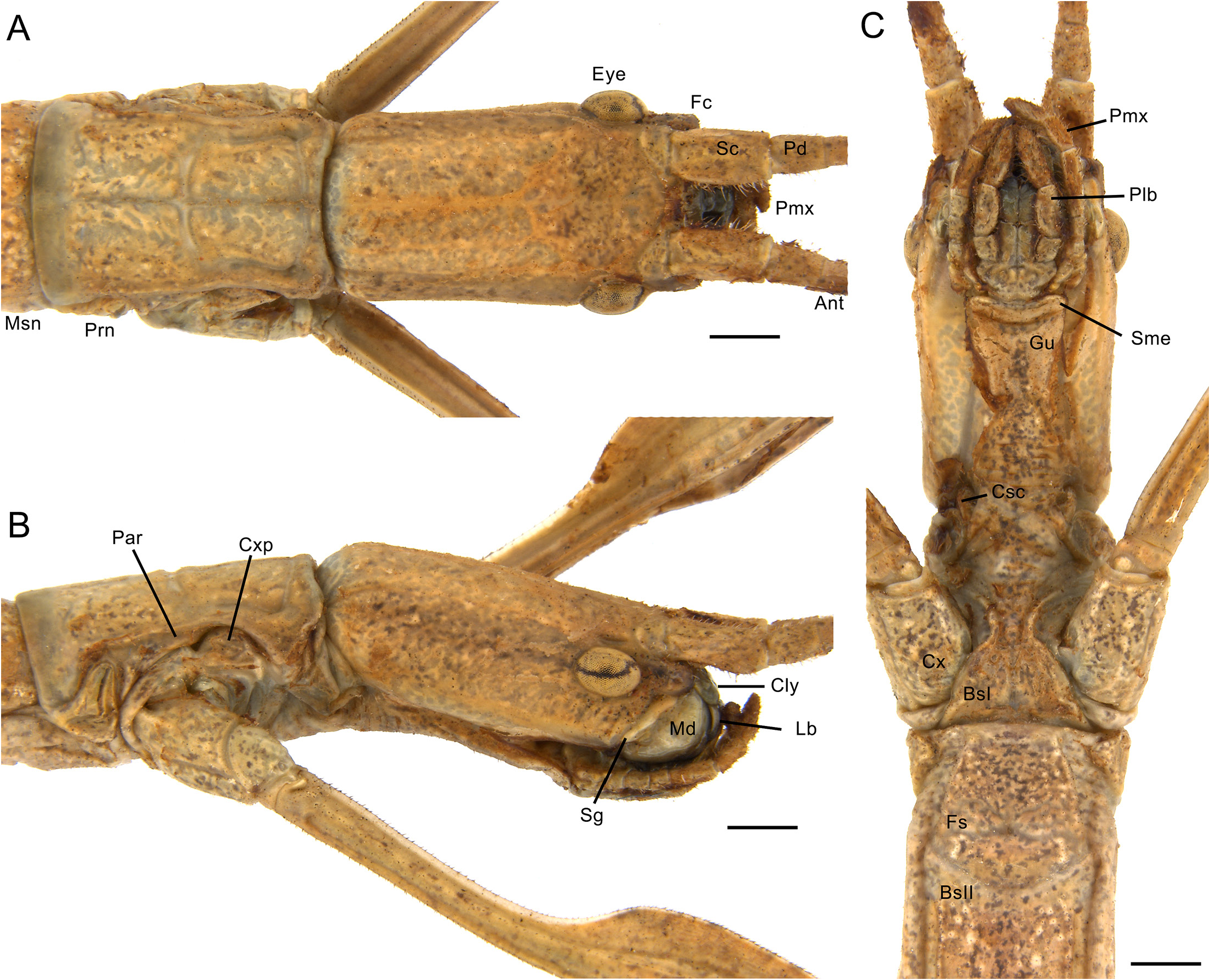
HEAD ( Fig. 35 View Fig ). Very elongate, smooth, with sparse small setae, vertex flat but very gently convex at posterior margin to fit underneath the pronotum. Frontal convexity developed, frontal suture round ( Fig. 35A–B View Fig ). Eyes small and slightly elongate, approximately 0.15 × as long as head. Cervix covering slightly more than half head, cervical sclerites developed, gula bearing setae and covering slightly less than half of cervix ( Fig. 35C View Fig ). Subgena fairly narrow, with posterior projection ca height of middle of eye. Submentum narrow and curved towards posterior, mentum simple, prementum somewhat wide. Glossa elongate, rounded, paraglossa roundly falcate, with round lateral margins and curved inner margins, reaching middle of labrum. Lacinia with three distal teeth, one large medially and two smaller laterally, all prominent, mesal edge bearing bundles of ca 11 large setae from base to sclerotized portion. Galea bearing long setae, slightly elongate, slightly widened before apical portion, apex round. Inner edge of galea apically with dense round tuft of hairy, long, large microtrichia and a few distinct circular granules dorsally and posteriad to this patch. Galealobulus present, small, round and basally fused to galea. Maxillary palpal segments cylindrical and labial slightly widened. Clypeus wide, subrectangular. Labrum strongly notched anteromedially, delimiting two round lobes, asymmetric with right lobe larger. Antennae filiform, reaching at least tergum IV. Scapus ca 2 × as long as wide, slightly compressed dorsoventrally. Pedicellus subglobose, large, slightly more than half as long as scapus ( Fig. 35A–B View Fig ). Antennomeres bear three types of setae as in A. dubia .
THORAX ( Figs 34 View Fig , 35A View Fig , 36A View Fig ). Smooth with scattered small and short black setae. Pronotum longer than wide, slightly constricted pre-medially and slightly wider from transverse sulcus towards posterior margin, fairly flat in lateral view. Transverse sulcus of pronotum conspicuous and gently curved towards
posterior, longitudinal sulcus conspicuous and distinct. Paranota curved, slightly widened medially, longer than wide, procoxopleurite apically round and ventrally straight ( Fig. 35A–B View Fig ). Probasisternum strongly tapering towards anterior, profurcasternum round ( Fig. 35C View Fig ). Mesothorax widened across all of length except near margins, gently convex in dorsal view (similar to paratype, Fig. 34B View Fig ). Mesothorax 7 × as long as prothorax. Mesonotum with pair of lateral carinae, mesepisternum lanceolate and regularly widening posteriorly, mesepimeron slightly elongate, medially gently widened, pointing towards posterior and slightly exceeding end of mesothorax ( Fig. 36A View Fig ). Mesocoxopleurite small, elongate, mesofurca Y-shaped. Metathorax and median segment widened across all of length except near margins, gently convex in dorsal view ( Fig. 36A View Fig ). Metathorax continuing pair of lateral carinae of mesonotum, metepisternum long, similar to mesepisternum, metepimeron extremely elongate, extending across entire length of median segment, posteriorly pointing and slightly exceeding end of median segment
( Fig. 36A View Fig ). Metacoxopleurite discrete, slightly elongate, medially gently widened, metafurca T-shaped. Metanotum 1.4 × as long as median segment ( Fig. 36A View Fig ). Median segment continuing pair of thoracic lateral carinae but weaker.
LEGS ( Figs 34 View Fig , 36B–F View Fig ). Slender. Hindlegs extending beyond epiproct, anterior legs around same length as hindlegs, midlegs shorter. Coxae smooth. Profemur with distinct basal curvature. Femora and tibiae with five carinae with few sparse setae among them ( Fig. 36B–F View Fig ). Carinae of profemora and protibiae distinctly keeled, mid and hind femora and tibiae with slightly less prominent keels. Antero- and posteroventral carinae of femora with round apical prominence. Carinae of all femora, tibiae and tarsi bearing row of short stout setae, stouter and spiniform on tibiae. Along all basitarsi and near apex of meso- and metatibiae, ventral, antero- and posteroventral carinae bearing two to three rows of setae. Setae on ventral, antero- and posteroventral carinae of basitarsi stouter and spiniform. Ventral, antero-
and posteroventral carinae of tibiae ending as apical spiniform sessile projection parallel to tibiae, pointing towards tarsi ( Fig. 36E–F View Fig ). Pro- and metabasitarsi very elongate, significantly longer than respective following tarsomeres combined, mesobasitarsi as long as respective following tarsomeres combined ( Fig. 36B–D View Fig ). Basitarsi with hairy setae restricted to apical portion on ventro-lateral patches ( Fig. 36B, D View Fig ). Remaining tarsomeres with setae on ventrolateral patches in portions not covered by euplantulae. Tarsomeres I–III with dorsal apical projection. Arolium round and broad, bearing setae
dorsally. Pretarsal claws symmetrical, dorsally and outwardly bearing setae. Euplantulae well developed in all tarsomeres, composed of two symmetrical pads separated by median groove in tarsomeres I–IV, and of single flattened lobed pad in tarsomeres V. Euplantulae present only apically at tarsomeres I–II, covering ca half length of tarsomeres III, covering two thirds length of tarsomeres IV and covering almost entire ventral surface of tarsomeres V ( Fig. 36B–D View Fig ).
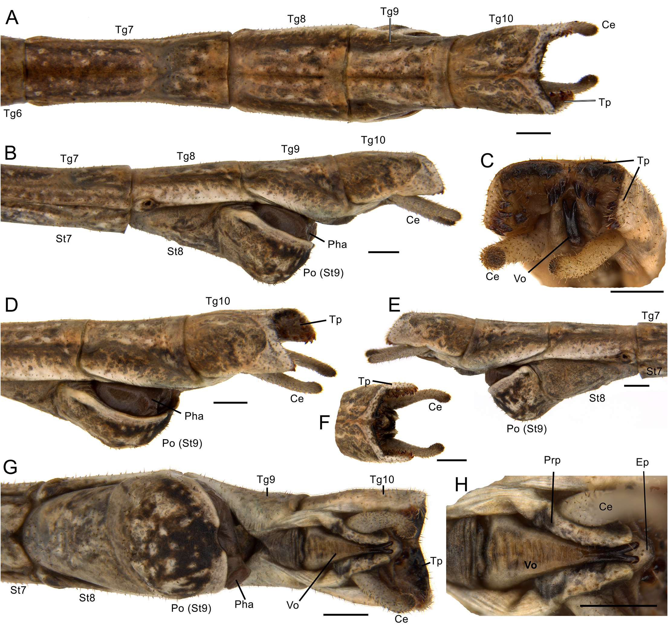
ABDOMEN ( Figs 34 View Fig , 36A View Fig , 37 View Fig ). External surface as in thorax but bearing slightly more scattered short setae across entire length. Median segment shorter than metanotum, anteriorly with two parasagittal ovoid stains ( Fig. 36A View Fig ). Combined length of segments II–X as long as combined length of thorax
and median segment. Terga II–VIII and sterna II–VII bearing lateral carinae near lateral margins. All segments longer than wide. Segment II shorter than III, segment III of same length as VII and shorter than IV, segments IV–VI of same length, tergum VIII significantly shorter than preceding segment and slightly longer than IX and X, tergum X slightly longer than IX. Tergum II widening towards posterior, tergum III widest, slightly wider than II and IV, terga IV and V about same width, tergum VI wider than preceding segment, with lateral margins convex in dorsal view, tergum VII narrower than all preceding segments, slightly wider than VIII, terga VIII–X around same width or very slightly tapering ( Fig. 37A View Fig ). Tergum X somewhat conical in lateral view ( Fig. 37B View Fig ) and in dorsal view almost parallel-sided but at ¾ the length tapering towards posterior, posterior margin emarginate with round lateral margins ( Fig. 37A View Fig ). Epiproct very prominent, elongate, lingulate in dorsal view, dorsoventrally flattened, visible dorsally and laterally ( Fig. 37A–B View Fig ). Paraprocts elongate, posteriorly fairly acute, straight, bearing setae on posterior margin, laterally bearing cerci ( Fig. 37B–C View Fig ). Cerci extremely elongate, straight, basally narrower fitting paraprocts, pointing to posterior, gradually tapering and slightly shorter than terga IX and X combined ( Fig. 37A–C View Fig ), bearing four types of setae. Praeopercular organ very prominent, lobed,
elevated and folded backwards, forming a round dorsoventrally flattened, centrally depressed projection ( Fig. 37B–E View Fig ). Subgenital plate roundly lanceolate, short, almost reaching ⅓ of length of tergum X. Subgenital plate bearing two parasagittal carinae beginning on anterior margin and running half length of segment before becoming flatter and more setose, tapering towards posterior and totally covering gonapophyses and gonoplac ( Fig. 37B–C View Fig ). Gonapophyses and gonoplac flattened, dorsoventrally for gonapophyses VIII, lateroventrally for IX and laterally for gonoplac ( Fig. 37F–G View Fig ). Gonapophyses VIII longer than IX, gonapophysis VIII reaching ca 0.8× length of tergum IX, gonapophysis IX reaching ca 0.6 × length of tergum IX. Gonapophysis VIII linear and gradually tapering, IX conical, tapering towards posterior, with lateral margin concave and inner margin straight ( Fig. 37F–G View Fig ). Gonapophyses IX ventrally folded to fit within gonapophyses VIII. Gonangulum distinctly reduced, flat, not lobed. Gonoplac lorate, somewhat wide in lateral view, bearing setae and slightly longer than gonapophyses VIII ( Fig. 37F–G View Fig ).
Other females
MEASUREMENTS (in mm, N = 8). Body 92–113.5, head 5.0–5.8, antennae 48.0–58.0, pronotum 3.3–4.1, mesonotum 20.0–25.5, metanotum 7.5–9.9, median segment 4.9–7.3, abdomen (excluding median segment) 44.0–54.7, cercus 4.4–6.0, profemur 21.4–24.7, protibia 20.8–26.0, mesofemur 16.5–19.5, mesotibia 16.3–20.3, metafemur 20.0–23.6, metatibia 20.6–28.7.
VARIATION. Some few specimens may present shorter cerci due to regeneration from breaking in previous instars (e.g., Fig. 45C View Fig ). Some specimens may present more elongate gonapophyses IX, longer than gonoplac (e.g., Fig. 45D View Fig ).
COLOUR ( Figs 34 View Fig , 44–46A View Fig View Fig View Fig ). Body generally light to mid green, beige, brown, grey or dark, with or without irregular stains of different tones.
Male paratype (MZUSP V0602)
MEASUREMENTS (in mm). Body 57.6, head 2.9, antennae at least 48.8, pronotum 2.3, mesonotum 14.0, metanotum 5.6, median segment 4.1, abdomen (excluding median segment) 28.7, cercus 1.1, profemur 19.2, protibia 21.9, mesofemur 15.3, mesotibia 17.0, metafemur 18.8, metatibia 22.1.
COLOUR ( Figs 38–40 View Fig View Fig View Fig , similar to paratype in Fig. 34C View Fig ). Entirely light to mid brownish with irregular stains of different tones and scattered whitish to creamy granules.
HEAD ( Fig. 38 View Fig ). Similar to female except: eyes, scapus and pedicel proportionally larger, head widening from posterior margin towards anterior in dorsal view, vertex very gently convex near anterior margin above eyes, frontal suture more acute ( Fig. 38A View Fig ). Paraglossa wider, inner margin straight ( Fig. 38E View Fig ). Galea thinner, more elongate, slightly constricted before apical portion, galealobulus wider, less prominent ( Fig. 38C View Fig ). Labial palpal segments slightly less widened ( Fig. 38F View Fig ). Antennae reaching at least tergum VI, scapus ca 1.7× as long as wide ( Fig. 38A View Fig ).
THORAX ( Figs 38A–B, F View Fig , 39A View Fig , similar to paratype in Fig. 34C View Fig ). Similar to female except: significantly thinner and more elongate than that of female. Meso-, metathorax and median segment with straight lateral margins in dorsal view, not convex ( Fig. 39A View Fig ). Mesothorax ca 6.5 × as long as prothorax. Mesocoxopleurite larger, triangular. Metanotum 1.3× as long as median segment ( Fig. 39A View Fig ). Metafurca Y-shaped.
LEGS ( Fig. 39B–G View Fig , similar to paratype in Fig. 34C View Fig ). Similar to female except: legs thinner than those of female, basitarsi relatively longer ( Fig. 39B, D, G View Fig ). All setae generally longer than those of female ( Fig. 39B–F View Fig ). Setae over carinae of femora slightly sparser, over carinae of tibiae longer ( Fig. 39B, D, G View Fig ). Carinae of femora, tibiae and tarsi with single row of setae except for ventral carinae of basitarsi
with two to three rows. Apical spine in ventral carinae of tibiae less developed, discrete. All basitarsi longer than respective following tarsomeres combined ( Fig. 39B, D, G View Fig ). Ventrolateral hairy setae of tarsomeres denser. Euplantulae slightly shorter, on tarsomeres V rougher ( Fig. 39E View Fig ).
ABDOMEN ( Figs 39A View Fig , 40 View Fig , 41A–B View Fig , similar to paratype in Fig. 34C View Fig ). Thinner than that of female. Lateral carinae absent in median segment. Segments II–VII slightly constricted medially from anterior to posterior regions. Terga II, III and VI about same length, terga IV and V about same length and slightly longer than III, tergum VII shorter than VI, tergum VIII shorter than preceding segment and slightly longer than IX, terga IX and X about same length. Terga II–V about same width, tergum VI slightly narrower than preceding segment, tergum VII as wide as VI but slightly wider posteriorly, VIII wider than VII, slightly widening towards posterior, IX slightly narrower than VIII, tapering towards posterior, X narrower anteriorly, slightly widened medially with maximum width around as wide as maximum width of VIII and IX ( Fig. 40A View Fig ). Tergum X presenting lateral flat flexing expansions bent downwards from half length to posterior margin, with somewhat acute ventrolateral edges directed posteriad ( Fig. 40A–G View Fig ). Paraprocts totally covered by these expansions. Dorsal posterior margin of tergum X widely v-shaped emarginate ( Fig. 40A, F View Fig ). Ventral area of posterior dorsal margin of tergum X strongly sclerotized, black ( Fig. 40C View Fig ). Thorn pads composed of ca 28–35 strong, inwardly curved sclerotized conical teeth, variable in size, with very few in sclerotized area on ventral surface of posterior dorsal margin of tergum X, with teeth present also anteroventrally (near apex of vomer) and laterally at ventral surface of posterior margin of lateral expansions ( Fig. 40C View Fig ). Tergum VIII and sternum VIII separated, not fused ( Fig. 40B, E View Fig ). Sternum VIII partly fused with sternum IX, border between both sterna shown as asymmetric sulcus curving towards posterior on right side ( Fig. 40G View Fig ). Sternum IX reduced with anterior portion indistinct; only shown as poculum ( Fig. 40B, E View Fig ). Poculum reaching around ¾ length of tergum IX. Due to asymmetry of sulcus, poculum shorter at right side than left side ( Fig. 40B, E View Fig ). Poculum fairly short, round in lateral view, about as long as tall or slightly longer than tall at left side, round in ventral view, posterior margin medially with wide emarginate short curvature towards anterior ( Fig. 40G View Fig ). Cerci slightly shorter than tergum X, inwardly curved and with round apex ( Fig. 40A–F View Fig ). Epiproct small, hidden in dorsal and lateral views ( Fig. 40H View Fig ). Vomer elongate, dorsoventrally flattened, terminal hook elongate and strongly sclerotized, symmetrically bifid near posterior margin with round apices gently curving upwards ( Fig. 40C, G–H View Fig ).
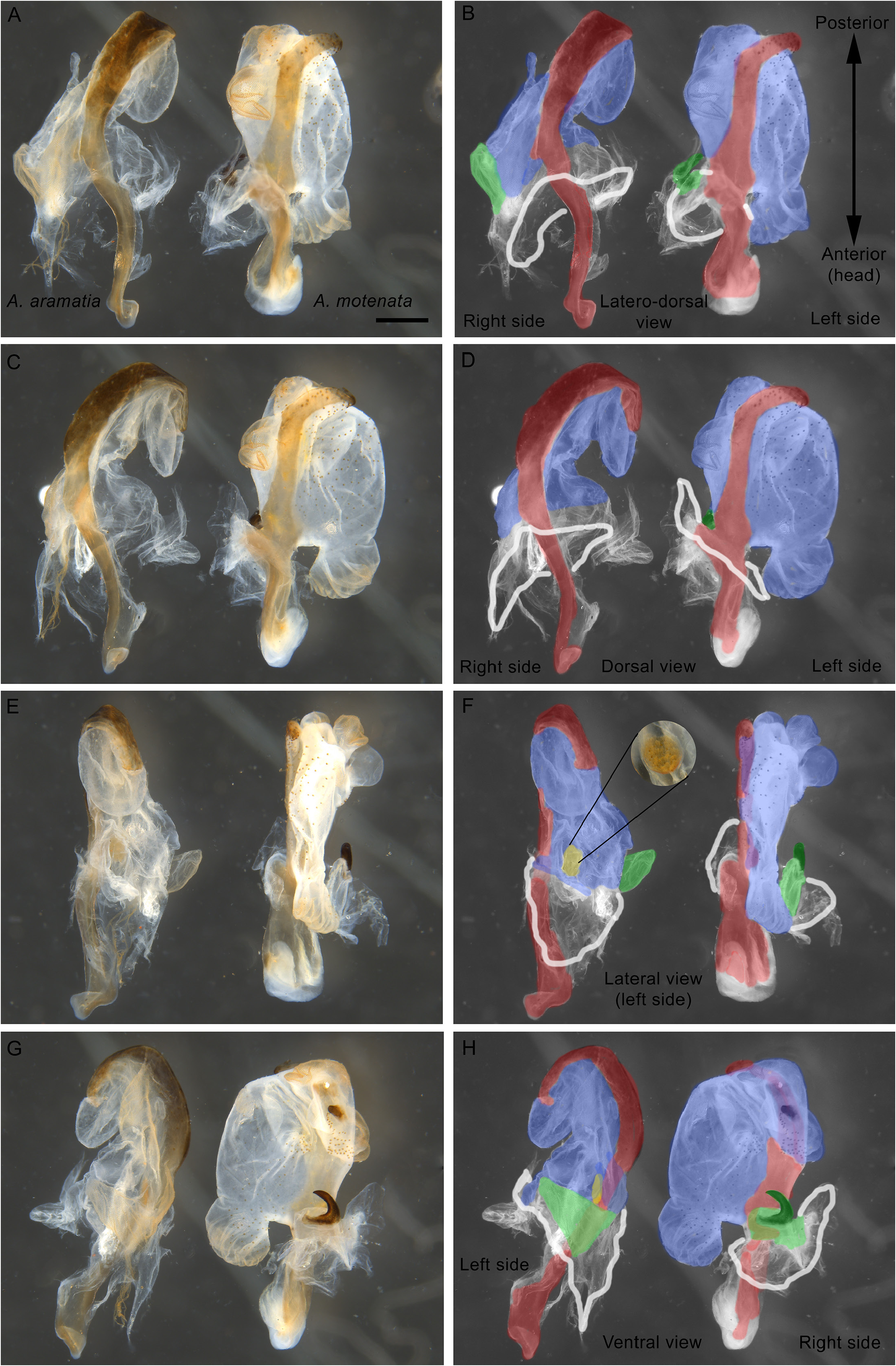
GENITALIA ( Figs 41 View Fig , 43 View Fig , right side). Male genitalia or phallic organ attached ventrally at area corresponding to asymmetric sulcus between sterna VIII and IX ( Fig. 41A–B View Fig ) by large muscles connected to anterior portion of dorsal sclerite ( Fig 41C–D View Fig ). Phallic organ composed of dorsalmost sclerite (dorsal sclerite), wide lobe with fine granulation originating ventrally from dorsal sclerite (longitudinal lobe), and smaller and shorter lobe basally, slightly ventrally, at right side, the (right) basal lobe. Dorsal sclerite very elongate and penetrating inside body cavity ( Fig. 43 View Fig ). Anterior portion penetrating body, wider and medially constricted. Short truncate branch at right side pointing towards posterior, connecting dorsal sclerite to body wall at end of anterior portion ( Fig. 43C–D View Fig ). Posterior portion narrower, slender, slightly tapering towards apex and bent to left near apex ( Figs 41D View Fig , 43A–D View Fig ). Longitudinal lobe wide, anteriorly to left branching in round lobule pointing towards anterior, with densely finely granulated round lobule dorsally before apex, originating near dorsal sclerite. Longitudinal lobe posteriorly with several folding and smaller lobules, with dense fine granulations ( Fig. 41F View Fig ), larger and sparser circular granulations, ventrally with weakly sclerotized area near apex and more posteriorly with strong, projecting, small and round sclerite ( Fig. 41C–F View Fig ). Basal lobe short and apically bearing claw-like sclerite, strongly sclerotized, with few sparse granules and with wide base ( Fig. 41E View Fig ).
Other males
MEASUREMENTS (paratypes, range in mm, N = 6). Body 61–70.1, head 2.7–3.2, antennae 50.0–57.0, pronotum 2.0–2.6, mesonotum 15.8–17.4, metanotum 6.3–7.5, median segment 4.4–5.2, abdomen
(excluding median segment) 29.8–34.2, cercus 0.9–1.2, profemur 20.5–22.7, protibia 22.4–24.8, mesofemur 16.0–17.8, mesotibia 17.9–19.1, metafemur 20.0–23.2, metatibia 22.4–25.2.
VARIATION. Thorn pad at ventral area of posterior dorsal margin of tergum X bearing 2–10 spines.
COLOUR ( Figs 34A, C View Fig , 44–45 View Fig View Fig ). Entirely light to dark brownish, light greenish, grey or dark with irregular stains of different tones and with or without stains of beige, dark red, black with scattered whitish to creamy granules.
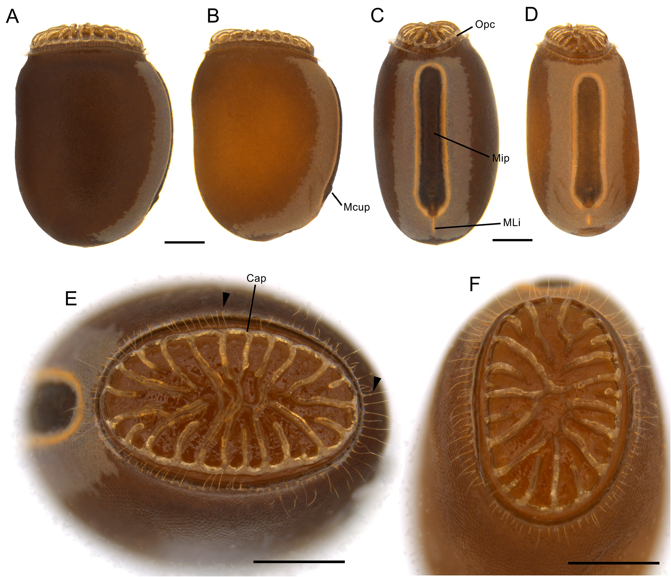
Egg ( Fig. 42 View Fig )
Measurements (in mm, N = 10): length 2.7–2.8, height 1.9–2.0, width 1.4–1.5. Relatively small,somewhat subrectangular in lateral view, slightly constricted at opercular collar, laterally compressed and ca 1.8 × as long as wide and 1.4× as long as tall ( Fig. 42A–D View Fig ). Capsule surface smooth but with finely punctuated appearance ( Fig. 42A–B View Fig ). Colour varying in shades of orangish or reddish brown with creamy lighter
band on dorsal surface extending in short posterior lateral patches around polar area. Micropylar plate very elongate, occupying large area of dorsal region, with round margins, almost parallel-sided with posterior portion gently widened ( Fig. 42C–D View Fig ). Micropylar plate with lighter round elevation delimiting inner flat region confluent with micropylar cup ( Fig. 42C–D View Fig ). Micropylar cup small, rounded and only slightly elevated. Median line short and of same colour and elevation as elevated margin of micropylar plate, almost reaching polar area, sometimes disjunct with micropylar plate ( Fig. 42C–D View Fig ). Opercular collar very slightly narrower (constricted) than rest of capsule, smooth, whitish with several minute and delicate long bristles surrounding edge ( Fig. 42E–F View Fig ). Operculum elliptical with marginally radial elevations with amber texture, irregular and less elevated at centre with some elevations fused centrally ( Fig. 42A–F View Fig ).
Distribution ( Fig. 52 View Fig )
Known only from the type locality, in the Serra do Cipó District, Santana do Riacho, Minas Gerais, Brazil. Serra do Cipó is part of the Serra do Espinhaço (Espinhaço mountain range), which is a wide and long mountain range in Brazil typically dominated by campos rupestres (“rupestrian grasslands”), a grassland to savannic rocky formation ( Fig. 53G–H View Fig ). All individuals of the species were found in campos rupestres vegetation near a road running through the Serra do Cipó ( Fig. 53G–H View Fig ). Certainly, the species also occurs in a nearby protected area continuous with the type locality, also in the Serra do Cipó formation, the Parque Nacional da Serra do Cipó, although it has not yet been recorded for the Park.
Biology
At the type locality at Serra do Cipó specimens were found in the rainy season at several stages from early nymphs to mating adult pairs. In two nights of searching, more than 20 specimens were seen over a few kilometres. Specimens were observed feeding on native locust berries ( Byrsonima Rich ex. Kunth, Malpighiaceae ) ( Fig. 44 View Fig ) and on guava trees alongside the road (native to Brazil but not to that particular environment; it is widely dispersed in urbanized areas). In captivity, they also fed on Brazilian cherry. Similar to Arumatia dubia gen. et comb. nov. both nymphs and adults are agitated when handled but in a somewhat lessened manner. Reared specimens were not observed to hide resting along a branch, but usually with mid and hindlegs keeping the body elevated. Eggs laid by adult females since collecting took 90–150 days to hatch but most of 10 controlled eggs hatched within 105 days. The first nymphs to hatch were adult males or pre-subadult females when totalling 60–73 days of development and females became adults at around 90–105 days of development.
In one of the mating attempts observed under captivity, the male repeatedly touched the female with his antennae while climbing on her, but the pair parted ways shortly after. Another two mating attempts resulted in a quick tapping of the antennae on the female while climbing on her and either only the male or the pair started to frenetically move their legs. The male quickly jumped on top of the female facing
the same direction with all tarsi touching her thorax. Then, while the female was shaking, the male positioned his abdomen beneath the abdomen of the female by the left side, tapped with the apex of his abdomen and then significantly bent his terga VIII to X anteriorly and slid the apex of his abdomen up and down the ventral surface of the abdomen of the female until the posterior margin of his tergum X attached to the female’s praeopercular organ ( Fig. 45A View Fig ). Then, the male pushed his abdomen backwards so the poculum contacted the female’s subgenital plate and copulation started ( Fig. 45A–B View Fig ). At this moment, the male’s abdomen curved upwards so the ventral side of the apex of his abdomen contacted the ventro-lateral area of the tip of the female’s abdomen ( Fig. 45B View Fig ). The poculum faced the right side of the female’s subgenital plate and attached that way. Later on, the male was seen to hang attached to the female only by the abdomen and eventually mounted on the top of the female again but with the abdomen now curving to the right side so that his abdomen curved downwards instead of upwards ( Fig. 45C View Fig ). They remained in copulation for around 50 hours. Males seen already in copulation with females were always positioned by the right side ( Figs 34A View Fig , 44 View Fig , 45 View Fig ).
Males attach to the female in three ways: by grasping the female’s subgenital plate with his curved cerci ( Fig. 45D–E View Fig ), by inserting his vomer underneath the praeopercular organ (hidden from view in the pictures) and by folding the lateral expansions of its tergum X, grasping the female praeopercular organ ( Fig. 45D–E View Fig ).


| MZUSP |
Museu de Zoologia da Universidade de Sao Paulo |
No known copyright restrictions apply. See Agosti, D., Egloff, W., 2009. Taxonomic information exchange and copyright: the Plazi approach. BMC Research Notes 2009, 2:53 for further explanation.
|
Kingdom |
|
|
Phylum |
|
|
Class |
|
|
Order |
|
|
Family |
|
|
Genus |